Association of body mass index with disease
severity and prognosis in patients with
non-cystic
fi
brosis bronchiectasis
Q. Qi
1, T. Li
1, J.C. Li
2and Y. Li
1 1Department of Respirology, Qilu Hospital of Shandong University, Jinan, Shandong Province, China2
Neurosurgical Intensive Care Unit, the First Affiliated Hospital, Sun Yat-sen University, Guangzhou, Guangdong Province, China
Abstract
The objective of this observational, multicenter study was to evaluate the association of body mass index (BMI) with disease severity and prognosis in patients with non-cystic fibrosis bronchiectasis. A total of 339 patients (197 females, 142 males) diagnosed with non-cystic fibrosis bronchiectasis by high-resolution computed tomography were classified into four groups: underweight (BMIo18.5 kg/m2), normal weight (18.5pBMIo25.0 kg/m2), overweight (25.0pBMIo30.0 kg/m2), and obese (BMIX30.0 kg/m2). Clinical variables expressing disease severity were recorded, and
acute exacerbations, hospitalizations, and survival rates were estimated during the follow-up period. The mean BMI was 21.90 kg/m2. The underweight group comprised 28.61% of all patients. BMI was negatively correlated with acute exacerbations, C-reactive protein, erythrocyte sedimentation rate, radiographic extent of bronchiectasis, and chronic colonization by
P. aeruginosaand positively correlated with pulmonary function indices. BMI was a significant predictor of hospitalization risk independent of relevant covariates. The 1-, 2-, 3-, and 4-year cumulative survival rates were 94%, 86%, 81%, and 73%, respectively. Survival rates decreased with decreasing BMI (w2=35.16, Po0.001). The arterial carbon dioxide partial pressure,
inspiratory capacity, age, BMI, and predicted percentage of forced expiratory volume in 1 s independently predicted survival in the Cox proportional hazard model. In conclusion, an underweight status was highly prevalent among patients with non-cystic
fibrosis bronchiectasis. Patients with a lower BMI were prone to developing more acute exacerbations, worse pulmonary function, amplified systemic inflammation, and chronic colonization by P. aeruginosa. BMI was a major determinant of hospitalization and death risks. BMI should be considered in the routine assessment of patients with non-cystic fibrosis bronchiectasis.
Key words: Bronchiectasis; Body mass index; Prognosis; Survival; Underweight
Introduction
Bronchiectasis is an abnormal, permanent dilatation of the bronchi and bronchioles caused by repeated cycles of airway infection and inflammation (1). Bronchiectasis is usually divided into non-cystic fibrosis (non-CF) bronch-iectasis, which affects a heterogeneous population and has various etiologies, and bronchiectasis due to cystic fibrosis (CF). CF is an autosomal recessive genetic disorder that not only affects the lungs, but also damages the pancreas, intestines, liver, sweat glands, and vas deferens. CF is rare in Asian races (2) and is considered to be a disease predominantly of Caucasian origin (3). Therefore, the present study only focused on patients with non-CF bronchiectasis.
Non-CF bronchiectasis is associated with chronic cough and expectoration, frequent respiratory infections, lung dysfunction, and advanced dyspnea. These symp-toms impose a significant burden on patients, resulting in worsening of quality of life and premature mortality (4). Extrapulmonary manifestations of non-CF bronchiectasis include muscle dysfunction, decreased exercise capa-city, fatigue, and a deteriorating health status (5). Clinically, some patients with non-CF bronchiectasis exhibit weight loss and nutritional depletion. A cross-sectional study of 93 patients with bronchiectasis found that 14% of patients presented with malnutrition as defined by a body mass index (BMI) ofo18.5 kg/m2(6).
Correspondence: Yu Li: <[email protected]>.
Another study found that the prevalence of malnutrition (defined as a BMI ofo20 kg/m2) was nearly 30% among
patients with bronchiectasis (7). A poor nutritional status was directly related to decreasing pulmonary function, and this link was a proposed predictive factor of morbidity and mortality in patients with chronic respiratory diseases (8). Based on our literature review, whether malnutrition accompanies non-CF bronchiectasis or is an important extrapulmonary feature of non-CF bronchiectasis remains unclear.
Measurement of BMI is a simple method for screening malnutrition. BMI has served as an indepen-dent prognostic factor for chronic obstructive pulmonary disease (COPD), with a clear association between a low BMI and increased mortality (9). In 2004, one study described a clear association between a low BMI and increased mortality in patients with end-stage respiratory disease (including 33 patients with bronch-iectasis) (7). Likewise, a Turkish study suggested that a high BMI was beneficial for survival in patients with bronchiectasis (10). Thus, in addition to the conven-tional treatment strategies for non-CF bronchiectasis, attention to the nutritional status may promote more favorable outcomes. However, there are no data regarding the association of BMI with disease severity and prognosis in patients with non-CF bronchiectasis in Asia.
In this study, we evaluated the relationships between BMI and clinical variables of disease severity in patients with non-CF bronchiectasis and explored the predictive factors for the risks of hospitalization and mortality in these patients.
Material and Methods
Patients
Four general hospitals (Qilu Hospital of Shandong University, Chest Hospital of Shandong Province, the Second Affiliated Hospital of Shandong University of Traditional Chinese Medicine, and Binzhou People’s Hospital of Shandong Province) in China participated in this observational, multicenter cohort study. Inpatients and outpatients were consecutively recruited from 1 January 2010 to 31 December 2013. Bronchiectasis was diagnosed by high-resolution computed tomography scans of the chest. The presence of bronchiectasis on high-resolution computed tomography images was based on criteria published by McShane et al. (1), including the following: the internal diameter of the bronchus was larger than that of its accompanying vessel (signet ring sign), the bronchus did not taper as it traveled to the periphery of the lung, or the bronchus terminated in a cyst. The underlying etiology of bronch-iectasis was determined after performing the tests recommended in the British Thoracic Society guideline for non-CF bronchiectasis (11). Patients were excluded if
they had a diagnosis of asthma, COPD, traction bronchiectasis due to lungfibrosis, or malignant tumors. In total, 339 Chinese patients were enrolled in the study and were followed until 1 April 2014. This observational study was approved by the Ethics Committee of Qilu Hospital of Shandong University, and all patients gave informed consent.
Basic data
The following basic data were recorded for each patient: age, gender, body weight, body height, and smoking history. Total symptom duration in years was calculated from the date of symptom onset to the date of recruitment into this study. BMI was calculated by dividing weight in kilograms by the square of height in meters. Patients were categorized into four groups according to the World Health Organization expert consultation on BMI criteria for Asian populations: underweight (BMIo18.5 kg/m2), normal
weight (18.5pBMIo25.0 kg/m2), overweight (25.0pBMI o30.0 kg/m2), and obese (BMIX30.0 kg/m2) (12).
Variables of disease severity
Pulmonary function. The pulmonary function indices measured in this study were the forced vital capacity (FVC), forced expiratory volume in 1 s (FEV1), FEV1/
FVC, predicted percentage of FVC, predicted percentage of FEV1, and inspiratory capacity using a MasterScreen
spirometer (Jaeger, Germany). The ventilatory func-tion of patients with non-CF bronchiectasis was classi-fied into one of four categories according to the American Thoracic Society COPD guidelines: normal ventilatory function, obstructive ventilatory dysfunction, restrictive ventilatory dysfunction, or mixed ventilatory dysfunction (13).
Arterial blood gas analyses. Arterial blood gas analyses were performed at rest and in room air with a blood gas analyzer (Radiometer Medical ApS, Denmark). Arterial oxygen tension (PaO2), arterial carbon dioxide
partial pressure (PaCO2), and arterial oxygen saturation
were measured while the patients breathed room air. According to the British Thoracic Society guideline for noninvasive ventilation in acute respiratory failure, respira-tory failure was defined as a PaO2 of o8.0 kPa
(60 mmHg), with or without a PaCO2 of 46.7 kPa
(50 mmHg) by arterial blood gas analyses while breathing air at sea level (14).
Acute exacerbations. According to the British Thoracic Society guideline for non-CF bronchiectasis, an acute exacerbation of bronchiectasis was defined as either a change in one or more of the common symptoms of bronchiectasis (sputum volume or purulence, dyspnea, cough, and fatigue/malaise) or the onset of new symptoms (fever, pleurisy, or hemoptysis) (11).
graded from 0 to 4 in accordance with the description of breathlessness.
Radiographic extent of bronchiectasis. The radio-graphic extent of bronchiectasis was determined accord-ing to established computed tomography criteria usaccord-ing the following scoring system: grade 1: localized bronchiecta-sis affecting one or part of one bronchopulmonary segment, grade 2: bronchiectasis in more than one bronchopulmonary segment (extensive), and grade 3: generalized cystic bronchiectasis (16).
Chronic colonization by P. aeruginosa. Gram-stained sputum samples with 425 polymorphonuclear leukocytes ando10 squamous cells perfield using a low-magnification lens were considered to be valid sputum samples. Valid sputum samples were obtained from all patients and processed for qualitative bacterial culture. Chronic coloniza-tion byP. aeruginosawas defined as at least three isolates of P. aeruginosa over a 3-month period and at least two isolates 3 months apart over a 1-year period (11).
Systemic inflammation. The peripheral blood C-reactive protein level was assessed by immunonephelom-etry (Cardio-Phase, Dade Behring Marburg GmbH, USA), and the erythrocyte sedimentation rate was measured using the Westergren method.
Follow-up study
The maximum follow-up duration was 51 months. Follow-up examinations were performed at 3-month intervals. During each follow-up, we recorded severity of respiratory symptoms, frequency of acute exacerbations, number of hospital admissions due to non-CF bronchiec-tasis, and information regarding survival. For patients who could not be followed up, an effort was made to contact the patient by telephone to obtain information. Survival rates were determined at 1 to 4 years.
Outcomes and prognosis
Two outcome parameters were prospectively recorded: the number of hospitalizations each year and the mortality rate. BMI was considered for its predictive value of outcomes, together with demographic data, pulmonary function, arterial blood gas analyses, C-reactive protein level, erythrocyte sedimentation rate, radiographic extent of bronchiectasis, and chronic coloni-zation byP. aeruginosa.
Statistical analysis
Descriptive data are reported as mean±SD or number (%). Analysis of variance was used to compare normally distributed variables among three or more groups; the Student-Newman-Keuls-q test was used for multiple comparisons. When data were not normally distributed, log transformation of the non-normal variables was performed before analysis of variance. Comparisons between qualitative variables were performed with the chi-squared test or Fisher’s exact test when necessary.
Spearman rank correlation analysis was performed to analyze whether there was a correlation between two ordered categorical variables. Univariate and multivariate regression analyses were used to study the determinants of the risk of hospitalization. For survival analysis, parameters with a significant impact on survival in a univariate Cox model analysis were tested in a multi-variate Cox proportional hazard model analysis. The survival process was described by Kaplan-Meier survival analysis. The log-rank test was used to test differences in the cumulative survival curves. Statistical analyses were performed using SPSS Statistics for Windows, Version 19.0 (SPSS Inc., USA). A P value of o0.05 was considered statistically significant.
Results
General characteristics
The baseline characteristics of the 339 patients with non-CF bronchiectasis are shown in Table 1. In total, 78.47% of the patients were lifetime nonsmokers. The mean BMI was 21.90 kg/m2among all patients, and the prevalence of underweight patients was high (28.61%). The mean MMRC dyspnea score of 1.95 demonstrated a moderate severity of breathlessness. Among all 339 patients, 118 patients’arterial blood gas analysis results met the diagnostic criteria for respiratory failure. Patients with normal ventilatory function only accounted for 29.20% of all patients. Obstructive ventilatory dysfunction was the most common pattern of pulmonary dysfunction (56.67%), followed by mixed ventilatory dysfunction (35.42%) and restrictive ventilatory dysfunction (7.91%). All patients underwent sputum sample collection and analysis. In total, 179 patients’sputum specimens tested positive (52.80%). The most common isolated pathogen wasP. aeruginosa
(77.09%). Other pathogens wereAcinetobacter baumannii
(5.59%), Haemophilus parainfluenzae (4.47%), Candida albicans (4.47%), Aspergillus spp. (3.35%), Klebsiella pneumoniae (2.23%), Escherichia coli (2.23%) and
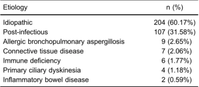
Staphylococcus haemolyticus (0.57%). The underlying etiologies of non-CF bronchiectasis among the patients in this study are listed in Table 2. Underlying causes were identified in 135 patients (39.83%). However, no cause could be established in 204 patients (60.17%); these patients were considered to have idiopathic non-CF bronchiectasis.
Comparison among the four study groups
acute exacerbations, FVC, FEV1/FVC, inspiratory
capa-city, PaO2, C-reactive protein level, erythrocyte
sedimen-tation rate, and chronic colonization byP. aeruginosa. In addition, acute exacerbations, the C-reactive protein level, and the erythrocyte sedimentation rate in the underweight group were significantly higher than those in the other three groups (Po0.05), while the inspiratory capacity in the underweight group was significantly lower than that in the other three groups (Po0.05). Total symptom duration in years and rate of chronic colonization byP. aeruginosa
in the underweight group were significantly higher than those in the normal weight and overweight groups (Po0.05), while FVC, FEV1/FVC, and PaO2 were significantly lower than
those in the normal weight and overweight groups (Po0.05). However, the differences in the symptom duration, FVC, FEV1/FVC, and PaO2between the underweight and obese
groups were not statistically significant. BMI was negatively correlated with the radiographic extent of bronchiectasis according to Spearman rank correlation analysis (rs=–0.312, Po0.001).
Determinants of hospitalization risk
As shown in Table 4, the factors associated with a risk of hospitalization in the univariate regression analysis were age, total symptom duration in years, MMRC dyspnea score, BMI, FVC, predicted percentage of FVC, FEV1, predicted percentage of FEV1, FEV1/FVC,
inspira-tory capacity, PaO2, PaCO2, arterial oxygen saturation,
C-reactive protein level, and erythrocyte sedimentation rate (Po0.05). Sex was not associated with the risk of hospitali-zation. Only the MMRC dyspnea score, BMI, erythrocyte sedimentation rate, C-reactive protein level, and total symptom duration in years appeared as independent predictors of hospitalization in the multivariate analysis. BMI was negatively correlated with the risk of hospitalization (standard regression coefficient=–0.26, Po0.001), while the MMRC dyspnea score, erythrocyte sedimentation rate, C-reactive protein level, and total symptom duration in years were positively correlated with the risk of hospitalization. In addition, the MMRC dyspnea score and BMI had more significant effects on the risk of hospitalization than did the erythrocyte sedimentation rate, C-reactive protein level, or total symptom duration.
Follow-up and survival
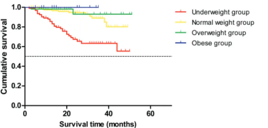
The minimum and maximum follow-up times were 2 and 51 months, respectively. Survival was recorded during a follow-up of 21.70±12.38 months. Forty-three patients died, and all died of respiratory and circulatory failure. The 1-, 2-, 3-, and 4-year cumulative survival rates were 94%, 86%, 81%, and 73%, respectively. As shown in Figure 1, the cumulative survival curves were statistically Table 2. Underlying etiology of 339 patients with non-cystic
fibrosis bronchiectasis.
Etiology n (%)
Idiopathic 204 (60.17%)
Post-infectious 107 (31.58%)
Allergic bronchopulmonary aspergillosis 9 (2.65%) Connective tissue disease 7 (2.06%)
Immune deficiency 6 (1.77%)
Primary ciliary dyskinesia 4 (1.18%) Inflammatory bowel disease 2 (0.59%) Table 1.General characteristics of 339 patients with non-cystic
fibrosis bronchiectasis.
Variables
Age (years) 56.00±13.52
Gender
Female 197 (58.11%)
Male 142 (41.89%)
Smoking status
Current smoker 48 (14.16%)
Ex-smoker 25 (7.37%)
Never smoked 266 (78.47%)
Smoking history (pack-years)* 6.37±14.08 Total symptom duration (in years) 16.80±10.00
BMI (kg/m2) 21.90±4.05
Acute exacerbations (times/year) 1.67±1.51
MMRC dyspnea score 1.95±1.40
FVC (L) 2.25±0.81
FVC, % predicted (%) 73.16±18.54
FEV1(L) 1.48±0.81
FEV1, % predicted (%) 58.51±26.02
FEV1/FVC (%) 62.96±17.39
Inspiratory capacity (L) 1.45±0.50
PaO2(mmHg) 72.88±14.83
PaCO2(mmHg) 43.10±12.13
Arterial oxygen saturation (%) 93.06±4.60
C-reactive protein (mg/L) 20.06±24.00 Erythrocyte sedimentation rate (mm/h) 27.89±25.33
Radiographic extent of bronchiectasisw
Grade 1 110 (32.45%)
Grade 2 167 (49.26%)
Grade 3 62 (18.29%)
Sputum culture positive 179 (52.80%)
Data are reported as mean ±SD or number (%). BMI, body mass index; MMRC, modified Medical Research Council dyspnea scale; FVC, forced vital capacity; FEV1, forced expiratory volume
in 1 s; PaO2, arterial oxygen tension; PaCO2, arterial carbon
different among the four groups (w2=31.67, Po0.001), and the underweight group had the lowest cumulative survival rate. Moreover, the mortality rate increased gradually as the BMI decreased according to a trend test (w2=35.16, Po0.001).
Predictive factors of mortality
Predictive factors of mortality according to Cox proportional hazard model analysis are reported in Table 5. Parameters with a significant impact on survival after univariate Cox model analysis were PaCO2, inspiratory
capacity, age, BMI, predicted percentage of FEV1, total
symptom duration in years, predicted percentage of FVC, FEV1/FVC, PaO2, C-reactive protein level, erythrocyte
sedimentation rate, radiographic extent of bronchiectasis, and chronic colonization byP. aeruginosa. However, when these parameters were tested in a multivariate Cox proportional hazard model, mortality was not influenced by the total symptom duration in years, predicted percentage of FVC, FEV1/FVC, PaO2, C-reactive protein
level, erythrocyte sedimentation rate, radiographic extent of bronchiectasis, or chronic colonization by P. aerugi-nosa. Five parameters were independently associated with survival in the multivariate analysis: PaCO2,
inspira-tory capacity, age, BMI, and predicted percentage of FEV1. Low values for BMI, inspiratory capacity, and
predicted percentage of FEV1and high values for PaCO2
and age were significantly associated with increased mortality.
Discussion
The mainfindings of the present study are as follows: an underweight status was highly prevalent among patients with non-CF bronchiectasis; BMI was associated with indicators reflecting disease severity, and patients with a lower BMI were prone to developing more acute exacerba-tions, worse pulmonary function, amplified systemic inflam-mation, and chronic colonization byP. aeruginosa; and BMI was a major determinant of hospitalization and death risks Table 3.Comparison of demographic data and clinical variables among the four groups.
Variables BMI categories P
Underweight (n=97)
Normal weight (n=173)
Overweight (n=55)
Obese (n=14)
Age (years) 56.19±13.73 54.69±13.98 58.85±11.27 59.64±13.22 NS
Female/male (n) 65/32 98/75 27/28 7/7 NS
Current smoker/ex-smoker/never smoked (n) 9/5/83 24/17/132 14/0/41 1/3/10 NS Smoking history (pack-years)* 4.58±13.50 6.25±13.02 9.18±17.31 9.29±15.92 NS
Total symptom duration (in years) 21.80±15.52 14.80±14.40 14.61±13.64 15.50±18.92 0.002= Acute exacerbations (times/year) 3.15±1.47 1.17±1.08 0.87±1.02 1.00±0.88 o0.001y
MMRC dyspnea score 2.91±1.13 1.53±1.31 1.44±1.32 2.50±0.94 NS
FVC (L) 1.88±0.66 2.39±0.81 2.55±0.87 2.00±0.62 o0.001y
FVC, % predicted (%) 64.75±15.25 76.70±18.40 79.00±19.61 64.79±15.28 NS
FEV1(L) 1.07±0.61 1.65±0.83 1.74±0.82 1.26±0.53 NS
FEV1, % predicted (%) 44.51±20.43 64.24±26.18 66.91±25.79 51.60±20.90 NS
FEV1/FVC (%) 54.72±15.62 66.15±17.14 67.49±16.66 62.84±17.12 o0.001y
Inspiratory capacity (L) 1.17±0.38 1.54±0.48 1.64±0.58 1.41±0.39 o0.001y
PaO2(mmHg) 66.34±14.43 75.47±14.58 76.72±1.77 71.25±13.40 o0.001y
PaCO2(mmHg) 44.80±12.44 41.96±11.13 41.77±10.55 50.71±21.66 NS
Arterial oxygen saturation (%) 91.16±4.99 93.73±4.38 94.47±3.66 92.3±3.76 NS C-reactive protein (mg/L) 37.25±29.31 14.00±17.64 11.29±17.49 10.43±10.61 o0.001y
Erythrocyte sedimentation rate (mm/h) 44.78±28.71 22.19±20.63 18.71±20.24 17.21±14.89 o0.001y Radiographic extent of bronchiectasisw
(grade 1/grade 2/grade 3) (n)
12/52/33 68/80/25 23/29/3 7/6/1 o0.001||
Chronic colonization byP. aeruginosa(n) 61 55 17 5 o0.001z
Data are reported as mean±SD or number. BMI, body mass index; MMRC, modified Medical Research Council dyspnea scale; FVC, forced vital capacity; FEV1, forced expiratory volume in 1 s; PaO2, arterial oxygen tension; PaCO2, arterial carbon dioxide partial
pressure; NS, not significant. *Pack-years were calculated by multiplying the average number of packs of cigarettes smoked per day by the number of years a person has smoked. wRadiographic extent of bronchiectasis was de
fined as follows: grade 1: localized bronchiectasis affecting one or part of one bronchopulmonary segment, grade 2: bronchiectasis in more than one bronchopulmonary segment (extensive), and grade 3: generalized cystic bronchiectasis.=Po0.05, analysis of variance.yPo0.001, analysis of variance.
||
Spearman rank correlation analysis (rs=–0.312,Po0.001). z
while a low BMI was associated with an unfavorable prognosis.
The prevalence of nutritional depletion in patients with non-CF bronchiectasis has not been fully studied. In the present study, we found a high prevalence of an under-weight status among patients with non-CF bronchiectasis (28.61%). This percentage is similar to those in other studies that reported nutritional depletion rates of 14% and 30% as analyzed by BMI (6,7). Therefore, weight loss was a frequently occurring phenomenon in patients with non-CF bronchiectasis.
The association between BMI and pulmonary function in patients with chronic respiratory diseases has been recognized for many years, but it has mainly been documented in patients with COPD. In one study of patients with COPD, BMI was positively associated with FEV1/FVC and the predicted percentage of FEV1(17). In
another study, the incidence of airflow obstruction (defined as an FEV1/FVC of o70%) in patients with COPD was
significantly higher in those with a BMI of o18.5 kg/m2
than in those with a BMI of X18.5 kg/m2 (18). Several recent studies have documented a clear association between a low BMI and poor pulmonary function in patients with bronchiectasis. A retrospective analysis of patients with bronchiectasis reported that BMI was positively correlated with FEV1 (7). In a multicenter
cross-sectional survey of outpatients on long-term oxygen therapy or home mechanical ventilation (including 39 patients with bronchiectasis), BMI was found to be positively associated with the predicted percentage of FVC and predicted percentage of FEV1(19). Additionally,
a low BMI was significantly associated with a low inspiratory capacity (20). The results of our study are in line with those of previous studies. We demonstrated that BMI was positively correlated with FVC, FEV1/FVC, and
inspiratory capacity. Our study has shown that patients with non-CF bronchiectasis with a lower BMI were prone to developing worse pulmonary function. The hypothesis was that weight loss (particularly loss of muscle mass) caused by malnutrition might promote a decrease in respiratory muscle strength, eventually leading to worse pulmonary function (21). Long-term longitudinal analyses are needed to better identify the effect of a low BMI on the reduction of pulmonary function.
Both the C-reactive protein level and erythrocyte sedimentation rate have known value as markers of systemic inflammation and are indirect markers of disease activity and quality of life for patients with non-CF bronchiectasis (11). The association between systemic inflammation and nutritional depletion in patients with chronic respiratory diseases has recently become an area of increasing research interest. In one study, the authors reported that an overflow of inflammatory cytokines might lead to malnutrition in patients with COPD (22). Moreover, in a cross-sectional study of patients with COPD, C-reactive protein levels were found to be significantly higher in patients with a low BMI (p21 kg/m2) than in those with a normal-to-high BMI (421 kg/m2), and an elevated C-reactive protein level was considered to be an indicator of malnutrition in patients with COPD (23). Consistent with other studies of chronic respiratory diseases, we found that BMI was negatively correlated with the C-reactive protein level and erythrocyte sedimentation rate in patients with non-CF bronchiectasis and that underweight patients had significantly higher C-reactive protein levels and erythrocyte sedimentation rates than did other patients. Our data suggest a link between a low BMI and increased systemic inflammation. Data from the current study support a previously proposed concept of disease-related malnutrition, in which disease is a major factor for malnutrition and the risk of malnutrition increases with disease severity (24). The mechanism of disease-related malnutrition is multifactorial, but the combination of decreased nutritional intake, increased energy and protein requirements, and the presence of inflammation probably plays the central role (25). A recently proposed hypothesis Table 4. Stepwise regression analysis to assess independent
predictors of risk of hospitalization.
Prediction variables* Effectw Cumulative R2= P
MMRC dyspnea score 0.44 0.41 o0.001 BMI (kg/m2)
–0.26 0.52 o0.001 Erythrocyte sedimentation
rate (mm/h)
0.15 0.56 0.007
C-reactive protein (mg/L) 0.14 0.57 0.014 Total symptom duration
(in years)
0.08 0.58 0.039
Age (years) NS
FVC (L) NS
FVC, % predicted (%) NS
FEV1(L) NS
FEV1, % predicted (%) NS
FEV1/FVC (%) NS
Inspiratory capacity (L) NS
PaO2(mmHg) NS
PaCO2(mmHg) NS
Arterial oxygen saturation (%) NS
MMRC, modified Medical Research Council dyspnea scale; BMI, body mass index; FVC, forced vital capacity; FEV1, forced
expiratory volume in 1 s; PaO2, arterial oxygen tension; PaCO2,
arterial carbon dioxide partial pressure; NS, not significant. *In univariate regression analysis, factors associated with the risk of hospitalization were the prediction variables shown in the table. Forward stepwise regression analysis was performed to seek the optimal standard regression equation: Ŷ = 0.443X1-0.255X2+
0.148X3+0.135X4+0.08X5, whereŶ represents hospitalization
and X1, X2, X3, X4, andX5 represent the MMRC score, BMI,
erythrocyte sedimentation rate, C-reactive protein level, and total symptom duration in years, respectively. wEffect indicates
standard regression coefficient. =R2
states that inflammation plays a key role in the pathogen-esis of disease-related malnutrition (26). Our findings do not allow for the establishment of a causal relationship between inflammation and weight loss in patients with non-CF bronchiectasis, but may provide insights into the cause of weight loss and malnutrition in these patients.
Lower respiratory tract infections repeatedly occur in patients with non-CF bronchiectasis. In one study, sputum
cultures tested positive forK. pneumoniaeorStreptococcus pneumoniaeduring the initial or stable phase of bronchiec-tasis. With disease progression, however, P. aeruginosa
replaced other pathogens and colonized the sputum (27). Very few studies have investigated the relationship between weight loss and chronic colonization byP. aeruginosa.One study of patients with COPD found that a low BMI was one of the independent determinants of a positive sputum culture Table 5.Predictive factors of mortality according to Cox proportional hazard model.
Variables Univariate analysis Multivariate analysis
HR=(95%CI) P HR=(95%CI) P
PaCO2(X50vso50 mmHg) 11.22 (5.95–21.18) o0.001 2.13 (0.99–4.59) 0.047 Inspiratory capacity (L) 0.02 (0.01–0.06) o0.001 0.18 (0.05–0.65) 0.009 Age (X55vso55 years) 13.73 (3.32–56.86) 0.001 7.70 (1.79–33.26) 0.006 BMI categories*
(group 4vsgroup 3vsgroup 2vsgroup 1)
0.27 (0.16–0.47) o0.001 0.48 (0.27–0.85) 0.011
FEV1, % predicted (%) 0.93 (0.91–0.95) o0.001 0.96 (0.93–1.00) 0.024 Total symptom duration in years
(X20vs10–20vsp10 years)
2.46 (1.71–3.54) o0.001 NS
FVC, % predicted (%) 0.93 (0.92–0.95) o0.001 NS
FEV1/FVC (%) 0.92 (0.90–0.94) o0.001 NS
PaO2(o60vsX60 mmHg) 7.65 (4.03–14.54) o0.001 NS
C-reactive protein (mg/L) 1.02 (1.01–1.02) 0.001 NS
Erythrocyte sedimentation rate (mm/h) 1.02 (1.01–1.03) o0.001 NS
Radiographic extent of bronchiectasisw
(grade 3vsgrade 2vsgrade 1)
3.42 (2.13–5.51) o0.001 NS
Chronic colonization byP. aeruginosa 4.79 (2.35–9.74) o0.001 NS
Gender (malevsfemale) NS
=
Data are reported as hazards ratio (HR), with 95% confidence intervals (CI) in parentheses. PaCO2, arterial carbon dioxide partial
pressure; BMI, body mass index; FEV1, forced expiratory volume in 1 s; FVC, forced vital capacity; PaO2, arterial oxygen tension;
HR, hazards ratio; CI, confidence intervals; NS, no significance. *Patients were categorized into 4 groups: group 1: underweight (BMIo18.5 kg/m2), group 2: normal weight (18.5pBMIo25 kg/m2), group 3: overweight (25pBMIo30 kg/m2), and group 4: obese (BMIX30 kg/m2).wRadiographic extent of bronchiectasis was defined as follows: grade 1: localized bronchiectasis affecting one or part of one bronchopulmonary segment, grade 2: bronchiectasis in more than one bronchopulmonary segment (extensive), and grade 3: generalized cystic bronchiectasis.
and that the most common pathogen isolated was
P. aeruginosa (28). The present study showed that BMI was negatively correlated with the rate of chronic coloniza-tion byP. aeruginosa, demonstrating that patients with non-CF bronchiectasis with a low BMI are more susceptible to chronic colonization by P. aeruginosa. Published data suggest that malnutrition is an independent factor associated with nosocomial infections (29). The causal relationship between weight loss and P. aeruginosa infection has recently been explored. Mouse models of chronic broncho-pulmonary infection withP. aeruginosaexhibited significant weight loss, and the weight loss was directly correlated with the degree of pulmonary inflammation (30). Using a mouse model ofP. aeruginosainfection, Kishta et al. (31) showed that nutritionally derived products with anti-inflammatory and antioxidant properties limited the bacterial burden and protein oxidation in P. aeruginosa lung infection. The hypothesis was that P. aeruginosa lung infection is associated with a marked inflammatory response and oxidative stress and that there is an intimate relationship among P. aeruginosa lung infection, inflammation, and weight loss.
One of the main concerns in patients with non-CF bronchiectasis is identification of the determinants of hospitalization because hospitalization is associated with a high mortality rate and is the main source of costs. In the present study, the number of hospitalizations was inde-pendently determined by a high MMRC dyspnea score, low BMI, elevated erythrocyte sedimentation rate, ele-vated C-reactive protein level, and rising total symptom duration in years. Dyspnea is one of the main symptoms of bronchiectasis, and Onen et al. (10) reported that dyspnea as measured by the MMRC dyspnea score was correlated with prognosis in patients with non-CF bronchi-ectasis. Additionally, a prospective study showed that a low BMI was associated with an increased risk of hospitalization in patients with non-CF bronchiectasis (7). Consistent with these results, our findings also demon-strated the relationship between a low BMI and risk of hospitalization. The hypothesized mechanism was the vicious circle of malnutrition and infection. A possible intermediate pathway could be immunodeficiency sec-ondary to malnutrition. It has been proposed that malnutrition might influence the organism’s defense processes by impairing the lymphohematopoietic organs and modifying the immune response (32). Therefore, malnourished individuals have a greater susceptibility to infection. In turn, repeated infections and frequent exacerbations lead to weight loss by reduced dietary intake and increased resting energy expenditure (33).
The influence of a high PaCO2on survival was clearly
demonstrated in a 4-year follow-up study of patients with bronchiectasis (10). The level of hypercapnia reflects the severity of the respiratory impairment. For this reason, patients with chronic hypercapnia during follow-up have a worse prognosis than do patients with normocapnia (10).
The predicted percentage of FEV1 also has significant
predictive power with respect to mortality, which confirms the recent hypothesis proposed by Martinez-Garcia et al. (4). An observational prospective study of hospitalized patients in Brazil showed that malnourished patients had a higher risk of death than did well-nourished patients (34). Furthermore, a prospective cohort study of patients with bronchiectasis showed that a low BMI was an indepen-dent predictor of long-term mortality (10). The mainfinding of our study is that nutritional depletion, as evaluated by BMI, not only correlated with the risk of hospitalization, but also appeared to be an independent predictor of mortality in patients with non-CF bronchiectasis. We found that, among patients with non-CF bronchiectasis, those with a low BMI had a worse prognosis than those with a normal-to-high BMI. The suggested mechanism for the higher mortality in patients with a lower BMI may be an impaired immune response with respiratory muscle weakness. Previous studies have found that maintaining an optimal nutritional status and achieving protein balance during routine care was important in the prevention of muscle loss and further improvements in the clinical and overall outcomes of patients with CF (35). Likewise, the Cochrane database of systemic reviews of randomized controlled trials suggested that nutritional support improves the prognosis of patients with COPD and is useful for their comprehensive care (36). Nutritional support as an additional therapy for non-CF bronchiectasis has often been neglected. Randomized controlled clinical trials are needed to explore the impact of nutritional manage-ment on clinical outcomes of patients with non-CF bronchiectasis.
tree-in-bud pattern, bronchiolectasis, and excess mucus (11). Imaging signs of small airway involvement are reportedly an important modality with which to monitor the progres-sion of bronchiectasis due to CF (39). Unfortunately, we did not evaluate the imaging signs of small airway involvement. Further studies will be conducted to evaluate the radiographic extent of bronchiectasis in patients with non-CF bronchiectasis in detail. Fourth, some examina-tions were only performed at baseline. We failed to record the changes in pulmonary function, arterial blood gas analyses, or high-resolution computed tomography scans of the chest from baseline into the follow-up period. Long-term longitudinal analyses are needed to demonstrate the relationship between the BMI and rate of lung function decline.
In the present study, an underweight status was highly prevalent among patients with non-CF bronchiectasis. Patients with a lower BMI were prone to increased acute exacerbations, worse pulmonary function, amplified systemic inflammation, and chronic colonization by
P. aeruginosa. BMI was one of the major determinants of hospitalization and death risks in patients with non-CF bronchiectasis. BMI should be considered in the routine assessment of patients with non-CF bronchiectasis.
Acknowledgments
Research supported by Science and Technology Planning Project of Shandong Province (#2012GSF11859), China.
References
1. McShane PJ, Naureckas ET, Tino G, Strek ME. Non-cystic
fibrosis bronchiectasis.Am J Respir Crit Care Med2013; 188: 647-656, doi: 10.1164/rccm.201303-0411CI.
2. Li N, Pei P, Bu DF, He B, Wang GF. A novel CFTR mutation found in a Chinese patient with cysticfibrosis.Chin Med J
2006; 119: 103-109.
3. Li W, Sun L, Corey M, Zou F, Lee S, Cojocaru AL, et al. Understanding the population structure of North American patients with cysticfibrosis.Clin Genet2011; 79: 136-146, doi: 10.1111/j.1399-0004.2010.01502.x.
4. Martinez-Garcia MA, de Gracia J, Vendrell Relat M, Giron RM, Maiz Carro L, de la Rosa Carrillo D, et al. Multi-dimensional approach to non-cysticfibrosis bronchiectasis: the FACED score.Eur Respir J2014; 43: 1357-1367, doi: 10.1183/09031936.00026313.
5. Ozalp O, Inal-Inc, Calik E, Vardar-Yagli N, Saglam M, Savci S, et al. Extrapulmonary features of bronchiectasis: muscle function, exercise capacity, fatigue, and health status. Multi-discip Respir Med2012; 7: 3, doi: 10.1186/2049-6958-7-3. 6. Olveira G, Olveira C, Gaspar I, Porras N, Martin-Nunez G,
Rubio E, et al. Fat-free mass depletion and inflammation in patients with bronchiectasis. J Acad Nutr Diet2012; 112: 1999-2006, doi: 10.1016/j.jand.2012.08.013.
7. Cano NJ, Pichard C, Roth H, Court-Fortune, Cynober L, Gerard-Boncompain M, et al. C-reactive protein and body mass index predict outcome in end-stage respiratory failure.
Chest2004; 126: 540-546, doi: 10.1378/chest.126.2.540. 8. Olveira G, Olveira C. [Nutrition, cystic fibrosis and the
digestive tract].Nutr Hosp2008; 23 (Suppl 2): 71-86. 9. Celli BR, Cote CG, Marin JM, Casanova C, Montes de Oca
M, Mendez RA, et al. The body-mass index, airflow obstruction, dyspnea, and exercise capacity index in chronic obstructive pulmonary disease.N Engl J Med 2004; 350: 1005-1012, doi: 10.1056/NEJMoa021322.
10. Onen ZP, Gulbay BE, Sen E, Yildiz OA, Saryal S, Acican T, et al. Analysis of the factors related to mortality in patients with bronchiectasis.Respir Med2007; 101: 1390-1397, doi: 10.1016/j.rmed.2007.02.002.
11. Pasteur MC, Bilton D, Hill AT. British Thoracic Society guideline for non-CF bronchiectasis.Thorax2010; 65 (Suppl 1): i1-i58, doi: 10.1136/thx.2010.136119.
12. WHO Expert Consultation. Appropriate body-mass index for Asian populations and its implications for policy and intervention strategies. Lancet 2004; 363: 157-163, doi: 10.1016/S0140-6736(03)15268-3.
13. Celli BR, MacNee W. Standards for the diagnosis and treatment of patients with COPD: a summary of the ATS/ ERS position paper.Eur Respir J2004; 23: 932-946, doi: 10.1183/09031936.04.00014304.
14. British Thoracic Society Standards of Care Committee. Non-invasive ventilation in acute respiratory failure.Thorax2002; 57: 192-211, doi: 10.1136/thorax.57.3.192.
15. Launois C, Barbe C, Bertin E, Nardi J, Perotin JM, Dury S, et al. The modified Medical Research Council scale for the assessment of dyspnea in daily living in obesity: a pilot study. BMC Pulm Med 2012; 12: 61, doi: 10.1186/1471-2466-12-61.
16. Grenier P, Cordeau MP, Beigelman C. High-resolution computed tomography of the airways. J Thorac Imaging
1993; 8: 213-229, doi: 10.1097/00005382-199322000-00006.
17. Qiu T, Tang YJ, Xu ZB, Xu D, Xiao J, Zhang MK, et al. Association between body mass index and pulmonary function of patients with chronic obstructive pulmonary disease.Chin Med J2009; 122: 1110-1111.
18. Chakrabarti B, Purkait S, Gun P, Moore VC, Choudhuri S, Zaman MJ, et al. Chronic airflow limitation in a rural Indian population: etiology and relationship to body mass index.Int J Chron Obstruct Pulmon Dis 2011; 6: 543-549, doi: 10.2147/COPD.
19. Cano NJ, Roth H, Court-Ortune, Cynober L, Gerard-Boncompain M, Cuvelier A, et al. Nutritional depletion in patients on long-term oxygen therapy and/or home mechan-ical ventilation.Eur Respir J2002; 20: 30-37, doi: 10.1183/ 09031936.02.01812001.
20. Tantucci C, Pinelli V, Cossi S, Guerini M, Donato F, Grassi V. Reference values and repeatability of inspiratory capacity for men and women aged 65–85.Respir Med2006; 100: 871-877, doi: 10.1016/j.rmed.2005.08.017.
quality assurance (CFQA) project.Thorax2002; 57: 596-601, doi: 10.1136/thorax.57.7.596.
22. Higashimoto Y, Yamagata T, Honda N, Satoh R, Sano H, Iwanaga T, et al. Clinical and inflammatory factors asso-ciated with body mass index in elderly patients with chronic obstructive pulmonary disease.Geriatr Gerontol Int 2011; 11: 32-38, doi: 10.1111/j.1447-0594.2010.00629.x. 23. Karadag F, Kirdar S, Karul AB, Ceylan E. The value of
C-reactive protein as a marker of systemic inflammation in stable chronic obstructive pulmonary disease.Eur J Intern Med2008; 19: 104-108, doi: 10.1016/j.ejim.2007.04.026. 24. Soeters PB, Reijven PL, van Bokhorst-de van der Schueren
MA, Schols JM, Halfens RJ, Meijers JM, et al. A rational approach to nutritional assessment.Clin Nutr2008; 27: 706-716, doi: 10.1016/j.clnu.2008.07.009.
25. Norman K, Pichard C, Lochs H, Pirlich M. Prognostic impact of disease-related malnutrition.Clin Nutr2008; 27: 5-15, doi: 10.1016/j.clnu.2007.10.007.
26. Jensen GL, Wheeler D. A new approach to defining and diagnosing malnutrition in adult critical illness.Curr Opin Crit Care 2012; 18: 206-211, doi: 10.1097/MCC.0b013e3283 51683a.
27. Caballero E, Drobnic ME, Perez MT, Manresa JM, Ferrer A, Orriols R. Anti-Pseudomonas aeruginosaantibody detection in patients with bronchiectasis without cysticfibrosis.Thorax
2001; 56: 669-674, doi: 10.1136/thorax.56.9.669.
28. Tsimogianni AM, Papiris SA, Kanavaki S, Stathopoulos GT, Sotiropoulou C, Manali ED, et al. Predictors of positive sputum cultures in exacerbations of chronic obstructive pulmonary disease.Respirology2009; 14: 1114-1120, doi: 10.1111/j.1440-1843.2009.01615.x.
29. Schneider SM, Veyres P, Pivot X, Soummer AM, Jambou P, Filippi J, et al. Malnutrition is an independent factor associated with nosocomial infections.Br J Nutr2004; 92: 105-111, doi: 10.1079/BJN20041152.
30. van Heeckeren AM, Tscheikuna J, Walenga RW, Konstan MW, Davis PB, Erokwu B, et al. Effect ofPseudomonas
infection on weight loss, lung mechanics, and cytokines in mice.Am J Respir Crit Care Med2000; 161: 271-279, doi: 10.1164/ajrccm.161.1.9903019.
31. Kishta OA, Iskandar M, Dauletbaev N, Kubow S, Lands LC. Pressurized whey protein can limit bacterial
burden and protein oxidation in Pseudomonas aeruginosa lung infection.Nutrition2013; 29: 918-924, doi: 10.1016/j. nut.2012.11.009.
32. Ortiz R, Cortes L, Cortes E, Medina H. Malnutrition alters the rates of apoptosis in splenocytes and thymocyte subpopula-tions of rats. Clin Exp Immunol 2009; 155: 96-106, doi: 10.1111/cei.2009.155.issue-1.
33. Hallin R, Koivisto-Hursti UK, Lindberg E, Janson C. Nutritional status, dietary energy intake and the risk of exacerbations in patients with chronic obstructive pulmonary disease (COPD). Respir Med 2006; 100: 561-567, doi: 10.1016/j.rmed.2005.05.020.
34. Pasquini TA, Neder HD, Araujo-Junqueira L, De-Souza DA. Clinical outcome of protein-energy malnourished patients in a Brazilian university hospital.Braz J Med Biol Res2012; 45: 1301-1307, doi: 10.1590/1414-431X20122586. 35. Engelen MP, Com G, Deutz NE. Protein is an important
but undervalued macronutrient in the nutritional care of patients with cysticfibrosis.Curr Opin Clin Nutr Metab Care2014; 17: 515-520, doi: 10.1097/MCO.00000000000 00100.
36. Ferreira IM, Brooks D, White J, Goldstein R. Nutritional supplementation for stable chronic obstructive pulmonary disease. Cochrane Database Syst Rev 2012; 12: CD00 0998.
37. Raslan M, Gonzalez MC, Torrinhas RS, Ravacci GR, Pereira JC, Waitzberg DL. Complementarity of Subjective Global Assessment (SGA) and Nutritional Risk Screening 2002 (NRS 2002) for predicting poor clinical outcomes in hospitalized patients. Clin Nutr 2011; 30: 49-53, doi: 10.1016/j.clnu.2010.07.002.
38. Vestbo J, Prescott E, Almdal T, Dahl M, Nordestgaard BG, Andersen T, et al. Body mass, fat-free body mass, and prognosis in patients with chronic obstructive pulmonary disease from a random population sample:findings from the Copenhagen City Heart Study.Am J Respir Crit Care Med
2006; 173: 79-83.