ISSN 1678-992X
ABSTRACT: The aims of this study were to determine the prevalence of viruses in 119 samples from 32 grapevine cultivars, collected from nine vineyards in a specific grape-growing area in southeastern Brazil, perform a partial molecular characterization of 14 isolates of Grapevine Syrah virus 1 (GSyV-1) and Grapevine leafroll-associated virus 3 (GLRaV-3) and assess the coat protein genetic variability of these viruses. The detection of viruses was implemented by real-time RT-PCR (reverse transcription polymerase chain reaction) aiming to detect seven viruses and one viroid. With the exception of the Grapevine Cabernet Sauvignon reovirus (GCSV), the viruses and viroid that were evaluated were widespread in the sampled areas, often in high prevalence and multiple infections, ranging from 15 % up to 76 %. Eight isolates of GSyV-1 and six of GLRaV-3, partially characterized by complete coat protein gene nucleotide sequencing and a variability study showed nucleotide identities ranging from 91 % to 99 % (GSyV-1) and from 98 % to 100 % (GLRaV-3) among themselves, respectively. Comparisons between conventional and real-time RT-PCR detections were implemented for GSyV-1 and GLRaV-3 infections. Analysis of genetic variability indicated molecular differences between GSyV-1 and GLRaV-3 isolates and negative selection acting on the coat protein gene of both viruses. This is the first report of GSyV-1 in commercial vineyards in Brazil. The survey revealed widespread infections of seven important pathogens in one prominent Brazilian grape-producing region implying contaminated grapevine cuttings in the spread of disease.
Keywords: Vitis, diagnosis, variability, incidence, leafroll
Molecular characterization of GSyV-1 and GLRaV-3 and prevalence of
Cátia Jacira Martins de Moura1, Thor Vinícius Martins Fajardo2*, Marcelo Eiras1, Fábio Nascimento da Silva3, Osmar Nickel2
1Biological Institute − Dept. of Plant Pathology − Lab. of Plant Virology, Av. Conselheiro Rodrigues Alves, 1252 − 04014-900 − São Paulo, SP − Brazil.
2Embrapa Grape & Wine, R. Livramento, 515 − 95701-008 − Bento Gonçalves, RS − Brazil.
3Santa Catarina State University/Centre of Agroveterinary Sciences, Av. Luiz de Camões, 2090 − 88520-000 − Lages, SC − Brazil.
*Corresponding author <[email protected]>
Edited by: Emerson Medeiros Del Ponte
Received August 19, 2016 Accepted November 08, 2016
Introduction
Grapevine (Vitis spp.) is one of the main fruit crops worldwide, in terms of its socioeconomic importance and area under cultivation. It is affected by several graft-transmissible agents that cause associated diseases. Its vegetative propagation has contributed to the worldwide spread of these pathogens and its perennial life cycle has accelerated the mixing of several pathogens in a single vine. These diseases cause crop losses and reduced plant vigor (Maliogkaet al., 2015).
In Brazil, the occurrence of 18 viruses has al-ready been reported in many viticultural areas (Basso et al., 2014). Grapevine yellow speckle viroid 1 (GYSVd-1, Apscaviroid) infects grapevines in Brazil, among other viroid species infecting this host elsewhere (Fajardo et al., 2016). Grapevine Syrah virus 1 (GSyV-1, Marafivirus) was first identified in the USA in connection with Syr-ah decline symptoms (Al RwSyr-ahnih et al., 2009). Since then, its presence has been reported in many coun-tries (Glasa et al., 2015). This virus is phylogenetically related to Grapevine fleck virus (GFkV, Maculavirus) which has spread worldwide and is a non-mechanical-ly transmissible virus associated with fleck symptoms (Sabanadzovic et al., 2001) that can be detected in la-tent infections in V. vinifera cultivars (Martelli, 2014). Grapevine leafroll disease (GLRD) is the most economi-cally damaging and widespread viral disease of grape-vine throughout the world (Almeida et al., 2013). It can cause up to 40 % yield losses (Naiduet al., 2015).
grapevine viruses in a grape-growing area
Several viral species (GLRaV-1, -2, -3, -4 and its strains and -7) designated Grapevine leafroll-associated virus (Closteroviridae) are related to GLRD, which can occur alone or as a viral complex, amongst which GLRaV-3 stands out (Maree et al., 2013). Grapevine rugose wood disease (GRWD) is a complex disease occurring in sev-eral grapevine cultivating regions. Some of the agents associated with GRWD are Grapevine rupestris stem pitting-associated virus (GRSPaV) and Grapevine virus A and B (GVA and GVB, Betaflexiviridae) (Martelli, 2014). Grapevine Cabernet Sauvignon reovirus (GCSV, Reo-viridae) was recently discovered infecting grapevines in the USA (Al Rwahnih et al., 2015).
There are few studies on the diversity and genetic variability of viruses and viroids in vineyards. Determin-ing which species are prevalent in the main producDetermin-ing regions, as well as the characterization and genetic vari-ability studies are important to understand how patho-gens evolve, predict epidemics and recommend control measures. The aims of this study were to investigate the occurrence of viruses and viroids in a survey of samples from Brazilian vineyards, perform molecular character-ization and molecular variability studies on the coat pro-tein gene of local isolates of two virus species.
Materials and Methods
Plant material and insect vectors
For the purpose of taking a survey, symptom-atic and asymptomsymptom-atic leaves and mature canes of 119
Plant Pathology
|
Resear
ch Ar
grapevine plants including 32 cultivars were collected at random during the middle summer season in 2016 from nine vineyards (13.2 sample/vineyard and 3.7 sample/ cv. in average) cultivated in the São Roque Municipal-ity, in the eastern region of the State of São Paulo, Brazil (Latitude 23º 31' 45" S, Longitude 47º 08' 07" W and altitude 771 m). Table and wine cultivars of grapevines sampled were: V. vinifera (Alfrocheiro, Alicante, Alvarin-ho, Aragonês, Arinto D’ouro, Cabernet Franc, C. Sau-vignon, Chardonnay, Fernão Dias, Malbec, Marselan, Moscato Setubal, Pinot Noir, Pinotage, Rebo, Sauvignon Blanc, Syrah, Tinta Cão, Tinta Roriz, Touriga Nacional, Verdelho), V. labrusca (Bordô, Bordô Grano D’oro, Con-cord, Isabel, Niagara Branca, N. Rosada) and hybrids (BRS cultivars: Carmem, Isis, Lorena, Margot, Violeta).
Twenty-eight compound samples of scale insects (11 samples) or mealybugs (17 samples) (ca. 20 adult individuals per sample) were collected inside the vine-yards sampled in São Roque. Mealybugs and soft scale insects were identified as Planococcus citri (Risso) and Pseudococcus longispinus (Targioni Tozzetti) (Hemiptera: Pseudococcidae) and Partenolecanium spp. (Hemiptera: Coccidae). Both healthy and infected mealybugs and soft scale insects were included in the assays as negative and positive controls, respectively. These insects were maintained in non-hosts of grapevine viruses and in vi-rus infected grapevines.
RNA extraction and quality
The total RNA extractions from 1 g of petioles, veins of leaves or scrapings of mature stems were ob-tained through the adsorption of nucleic acids on silica particles (Rott and Jelkmann, 2001), grinding plant tis-sues in liquid nitrogen, and following the prescribed protocol. Total RNA quality was monitored by evalua-tion using primers and probes for 18S rRNA (Osman et al., 2007). Total RNA was extracted from the mealybugs or scale insects using Trizol reagent (Invitrogen) accord-ing to the manufacturer's instructions.
Real time RT-PCR amplification
All plant samples were analyzed for the previ-ously mentioned viruses and the viroid (GCSV, GSyV-1, GRSPaV, GVA, GVB, GLRaV-3, GFkV and GYSVd-1) by real-time RT-PCR (RT-qPCR). In the performed analyses, RNAse-free water, healthy grapevines and positive con-trols from mother stock plants and the viral collection at Embrapa Grape and Wine, respectively, were included. Real-time RT-PCR reactions (One Step RT-PCR) were carried out in 96-well plates using the TaqMan Fast Vi-rus 1-Step Master Mix (Life Technologies) kit: 3 µL of the 4X TaqMan Fast Virus 1-Step Master Mix, 0.6 µL of the mixture of primers and probe (415 nM primer and 85 nM probe), 3 µL of total RNA (ca. 300 ng) to a final volume of 12 µL. Reactions were performed in a ther-mocycler StepOnePlus Real-time PCR System (Applied Biosystems) as follows: 45 °C for 35 min (for reverse transcription), 95 °C for 10 min, followed by 40 cycles
at 95 °C for 15 s (denaturation) and 60 °C for 1 min (an-nealing and extension). The reaction data were analyzed in terms of presence/absence assays and graphically, us-ing the StepOne Software program v.2.3 (Applied Bio-systems), by determining the Cq (quantification cycle). The primers and probes used for viruses and viroid tections by real-time RT-PCR have been previously de-scribed (Osman et al., 2007; Osman et al., 2008; Osman and Rowhani, 2008; Bianchi et al., 2015) or designed in this work for GCSV, primers Ctg 468F (5’ACGTTGGAT-CAACTAGCCGAAG3’) (Al Rwahnih et al., 2015) and GCSV-CS103r (5’ACCCATGTAAATTACACGCCTTC3’) and GCSV-CS103 probe (5’TGCTCCTATGTTCGTTAT-GCCATG3’). All probes were labeled with 6-FAM or VIC and TAMRA in the 5’ and 3’ ends, respectively. All insect samples were tested for Grapevine leafroll-associated virus 1 (GLRaV-1), GVA, GVB and GLRaV-3 infections by RT-qPCR.
Conventional RT-PCR amplification
For the molecular characterization of the coat protein (CP) genes, five (GSyV-1) and six (GLRaV-3) detected isolates were selected among infected plants. Additionally, other three isolates of GSyV-1 collected in an experimental vineyard in Bento Gonçalves, south-ern Brazil (Latitude 29º10'17" S, Longitude 51º31'09" W and altitude 691 m), were characterized. The prim-er pairs used to amplify GSyV-1 by one-step RT-PCR were GVQCP-R (5’GCATTGCTGCGCATTGGAGG3’), GVQCP-F (5’TCCCAGCTTCAGGGTGAATT3’) (En-gel et al., 2010) and GLRaV-3 were LR3-9445c (5’CTACTTCTTTTGCAATAGTT3’) and LR3-8504v (5’ATGGCATTTGAACTGAAATT3’) (Fajardo et al., 2007), complementary and viral sense, respectively. The RT-PCR in a single step was carried out using the One Step RT-PCR kit (Qiagen) and reactions were car-ried out according to the manufacturer with 4 µL of to-tal RNA (ca. 400 ng). The thermal amplification cycling was: 50 °C for 30 min, 95 °C for 15 min, 35 cycles: 94 °C for 50 s, 50 °C for 50 s, 72 °C for 1 min, and a final extension of 72 °C for 10 min. The RT-PCR prod-ucts were analyzed on 1 % agarose gels prepared in a TBE buffer pH 8.0, in the presence of ethidium bro-mide and visualized under UV light.
Comparison of diagnostic tests
To compare real time RT-PCR (RT-qPCR) and con-ventional RT-PCR for GSyV-1 and GLRaV-3 indexings, seventeen samples, randomly selected among those pre-viously determined as GSyV-1- or GLRaV-3-positive by RT-qPCR, were analyzed by conventional RT-PCR using aforementioned specific primers and reaction condi-tions.
Cloning and sequencing, nucleotide alignments and phylogenetic relationships
System kit (Promega). The eluted DNA fragments were ligated into pGEM-T Easy vector (Promega). The recom-binant plasmids were used to transform Escherichia coli DH5a competent cells by heat shock. The recombinant plasmids of transformed bacterial colonies were extract-ed using the Wizard Plus SV Minipreps DNA Purifica-tion System kit (Promega). The presence of the cloned viral fragment in the recombinant plasmids was con-firmed by digestion with the EcoRI restriction enzyme (Sambrook and Russell, 2001). The automatic nucleotide sequencing (Sanger method) was carried out with two clones per isolate. Multiple sequence alignments of nu-cleotides (nt) and deduced amino acids (daa) and the ma-trix generation of nt and daa identities were carried out using Clustal X 1.8 (Thompson et al., 1997) and BioEdit 7.2.5 softwares. The sequences obtained for GSyV-1 were aligned with the reference sequence of GSyV-1 (GenBank NC_012484) and all complete coat protein gene sequences available in GenBank. Also GLRaV-3 sequences were aligned with the reference sequence of GLRaV-3 (NC_004667) as well as with all CP genes of 13 GLRaV-3 isolates with complete genome available in the GenBank, and with six other Brazilian GLRaV-3 isolates that had been previously characterized. Phylogenetic re-lationships were determined from the aligned sequenc-es by using the maximum parsimony method (10,000 bootstrap replications) implemented in the MEGA 6.0 program (Tamura et al., 2013). The GenBank accession codes of the nucleotide sequences of the isolates used for phylogenetic analysis are presented in Table 1 and Figures 1 and 2. The molecular weight (MW) of the coat proteins of GSyV-1 and GLRaV-3 were calculated using the EXPASy software program (http://web.expasy.org/ compute_pi/).
Description of the coat protein genetic variability and selection analysis
The molecular variability descriptors [total num-ber of segregating sites (S), mean nucleotide differences
among sequences (K), nucleotide diversity (π), haplo-types number (H), haplotype diversity (Hd) and Wat-terson’s estimator for the population-scaled mutation rate] were estimated using the DnaSP software program v.5.10 (Rozas et al., 2003). The mean pairwise number of π per site was also calculated using a sliding window of 100 bases, with a step size of 25 bases across coat pro-tein genes of GSyV-1 and GLRaV-3. Coat propro-tein gene and site specific selection pressures were analyzed
us-Figure 1 −Phylogenetic tree based on the alignment of nucleotide
sequences of isolates REB-BR, MAR-BR, TR1-BR, TR2-BR and ALF-BR from São Roque and isolates MH, TRAJ-ALF-BR and VF-ALF-BR from Bento Gonçalves, southeastern and southern Brazil, respectively and other foreign isolates of Grapevine Syrah virus 1 (GSyV-1, Tymoviridae, Marafivirus). The tree was constructed by the maximum parsimony method, using the MEGA 6.0 program, and bootstrap support from 10,000 replications. Names of GSyV-1 isolates and origins were included according to GenBank accession codes, and specific clusters are indicated. Maize rayado fino virus
(MRFV), the type species of the genus Marafivirus (NC_002786) was used as outgroup. Bar = number of substitutions per site.
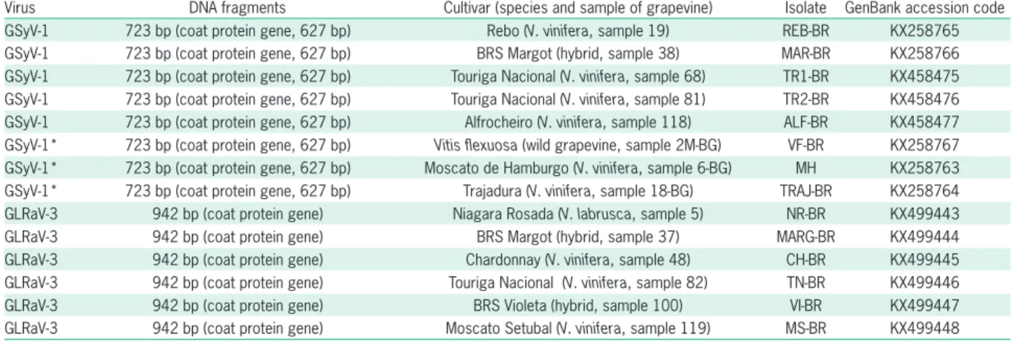
Table 1 − Grapevine Syrah virus 1 (GSyV-1) and Grapevine leafroll-associated virus 3 (GLRaV-3) molecularly characterized isolates.
ing four algorithms, Single Likelihood Ancestor (SLAC), Fixed Effects Likelihood (FEL), Random Effects Likeli-hood (REL) and Partitioning for Robust Inference of Selection (PARRIS) within the HyPhy software (http:// www.hyphy.org) implemented in the Datamonkey server (www.datamonkey.org) with default conditions. The nucleotide substitution model used was General re-versible substitution (REV). To avoid the recombination events effect on the selection analysis, recombination analysis was carried out using the RDP program v.3.44 (Martin et al., 2010) and the GARD method (available at the Datamonkey server).
Results and Discussion
Incidence of viruses and viroid in grapevines
The viruses and viroid infecting grapevines in the São Roque municipality were unknown and the knowl-edge of the phytosanitary status of a defined region is important for proposing management and control pro-cedures for grapevine diseases. Accordingly, 119
grape-vine accessions assayed by real time RT-PCR revealed a high level of infection in the analyzed samples: 0 % (GCSV), 15 % (GSyV-1), 28 % (GVB), 61 % (GRSPaV), 62 % (GVA), 65 % (GLRaV-3), 66 % (GYSVd-1) and 76 % (GFkV). The results also showed 11 %, 8 % and 81 % of incidence of healthy samples (without eight pathogens tested for), infected with only one pathogen and infect-ed with two or more viruses or viroid, respectively. To confirm the RT-qPCR results, two GYSVd-1 amplicons of 126 bp were sequenced resulting in 98 % nucleotide identities with other Brazilian isolates of this viroid (Fa-jardo et al., 2016).
In general, these pathogens were found widely distributed in the sampled vineyards. These results were supported by the fact that many grapevines (34 %) ex-hibited low vegetative vigor and general symptoms of virus infections [leafroll, unusual color (reddening or yellowing) or atypical appearance (coriaceous aspect) of the leaves, rugose wood (corky bark, grooving or pit-ting)] as a consequence of long-term maintenance (mean of 7 years) in vineyards. Grapevines infected by viruses may not exhibit noticeable symptoms because infections may be latent or asymptomatic in some host genotypes and commercial cultivars such as those usually observed in Vitis labrusca and hybrids; however, even in these cases, the virus infection can cause damage. Although crop losses were not assessed in the sampled vineyards, they probably occurred as has already been determined by other authors (Vega et al., 2011; Naidu et al., 2015). The correlation of virus and viroid infections and symp-tom expression might be related to other factors, such as temperature, certain virus isolates or strains, or multiple pathogen infections, making it impossible to diagnose precisely viruses and viroid infections based exclusively on symptomatology (Olmos et al., 2016). Despite these findings, there is a need for further evaluation of oth-er grape-growing areas to detoth-ermine the geographical distribution of these pathogens in Brazil. These results expand the knowledge about the incidence and virus distribution infecting grapevines in a representative Brazilian grape-growing region, and provide relevant in-formation for the development of control strategies and management of these diseases, with emphasis on the im-portance of the use of propagation material of virus-free vine in the establishment of new vineyards.
Detection of viruses and a viroid (GRSPaV, GVA, GVB, GLRaV-3, GFkV and GYSVd-1) infecting grapevine in Brazil has already been reported (Basso et al., 2014) as well as another viral species (GSyV-1), not previously reported in commercial Brazilian vineyards. In many grape-growing countries and regions, virus surveys have been conducted on grapevines, for example, in Argen-tina (Volpe et al., 2010), Chile (Fiore et al., 2011), China (Liu et al., 2013), the United States (Jones et al., 2015), Slovakia, the Czech Republic (Glasa et al., 2015) and in two regions of northeastern Brazil (Catarino et al., 2015), in which the incidence of viruses was highly variable, depending on the viral species, grapevine cultivars and
Figure 2 −Phylogenetic tree based on the alignment of nucleotide
sequences of isolates NR-BR, MARG-BR, CH-BR, TN-BR, VI-BR and MS-BR from São Roque; isolates RC-PE, Pet-1, Pet-2, Pet-3 and Pet-4 from Petrolina and isolate IS2 from Bento Gonçalves, southeastern, northeastern and southern Brazil, respectively and other foreign isolates of Grapevine leafroll-associated virus 3 (GLRaV-3). The tree was constructed by the maximum parsimony method, using the MEGA 6.0 program, and bootstrap support from 10,000 replications. Names of GLRaV-3 isolates and origins were included according to GenBank accession codes, and specific clusters are indicated as defined by Maree et al. (2015).
sampled regions. However, invariably, significant in-fection rates were reported, similar to the observation made in the present survey.
The presence of, at least, six different viruses and one viroid infecting vineyards in São Roque may be at-tributable to the redistribuition of infected propagative materials from other Brazilian infected grape-growing regions or from other countries through the planting of imported, infected propagative materials and cuttings. In conclusion, it was shown by indexing that viruses and one viroid were able to infect 106 out of 119 samples analyzed comprising 32 different grapevine genotypes that included cultivars of American (V. labrusca) and European (V. vinifera) grapes, interspecific hybrids and rootstocks (Table 1).
Mealybugs and soft scale insects as vectors
RT-qPCR analyses of potential vectors collected in the same vineyards showed that 0 % (GLRaV-1), 4 % (GVB), 18 % (GVA) and 61 % (GLRaV-3) out of 28 sam-ples of mealybugs and soft scales were infected. These rates were similar to those observed for the same vi-ruses in the analyzed grapevine samples, suggesting that mealybugs and soft scale insects are spreading these vi-rus species in the assayed vineyards. Another point to be considered is the availability of infected grapevines in-side and outin-side the vineyard (source of inoculum) from which viruliferous vectors would perform transmission. Thus, the high frequency of virus-infected vines empha-sizes the importance of clean plant materials as well as control of mealybugs (Jones et al., 2015).
Several mealybugs and soft scale species have already been reported as virus vectors in grapevine such as those mealybug species tested in this study and Partenolecanium corni (Almeida et al., 2013). GLRaV-1 and -3, GVA and GVB are transmitted by mealybugs and soft scale insects in a semipersistent manner (Tsai et al., 2010; Le Maguet et al., 2012), while GSyV-1 could be transmitted by leafhoppers (Al Rwahnih et al., 2009) and GCSV, GRSPaV, GFkV and GYSVd-1 have vectors that are still unknown or are not vector-transmissible. These viruses are transmitted by grafting with infected propagative materials (Martelli, 2014). In some cases, transmission modes may explain the observed incidence rates of viruses, in others there is no correlation. High incidence of GRSPaV, GFkV and GYSVd-1 has been found. However, since their vectors are not known, their spread is likely attributable to the use of infected cuttings or buds in the initial establishment of the sam-pled vineyards. Other cases, where high virus incidence rates were also observed (i.e. GLRaV-3 and GVA) could probably be attributed to mealybug transmissions in the sampled vineyards. Similar reasoning would be applied to the other assayed viruses (GCSV, GSyV-1 and GVB).
Comparison of diagnostic tests
In the assays carried out to compare RT-qPCR and conventional RT-PCR using specific primers to amplify
two viruses, we detected only 35 % (GLRaV-3) and 29 % (GSyV-1) by conventional RT-PCR, out of seventeen posi-tive samples (100 %) determined by RT-qPCR to both viruses. Divergences in nucleotide identities among iso-lates of these viral species have been related previously (Fajardo et al., 2007; Glasa et al., 2015), resulting from absence or mismatch of pairing of primers and probes with the virus sequence. This result highlights the rel-evance of knowledge of sequence variability when de-signing primers and probes for the reliable detection of a wide range of isolates of target viruses. Besides con-ventional RT-PCR, RT-qPCR was also used in this work to evaluate its capacity to detect a range of GSyV-1 and GLRaV-3 isolates, regardless of their variabilities. Based on the results obtained, it was demonstrated that prim-ers and probes defined by Bianchi et al. (2015) and Os-man et al. (2007) were suitable for a wide diagnosis of GSyV-1 and GLRaV-3, respectively. The GSyV-1 and GLRaV-3 sequence variabilities observed in the molecu-larly characterized samples could explain that the prim-ers used for conventional RT-PCR were not suitable for accurately covering the sequences analyzed.
Sequencing, nucleotide alignments and phylogenetic relationships
DNA fragments of 723 bp comprising the com-plete gene of GSyV-1 capsid protein (627 nt and 208 daa) were amplified from five isolates collected in São Roque and three from Bento Gonçalves by RT-PCR us-ing specific primers, and their nucleotide sequences were submitted to the GenBank (Table 1). The mul-tiple alignment of the CP sequences of the Brazilian isolates of GSyV-1 revealed expressive nucleotide and amino acid divergences among certain isolates, sug-gesting high variability among them. The nucleotide sequences of the eight Brazilian isolates clustered in groups I (MH, VF-BR, TRAJ-BR, ALF-BR and MAR-BR isolates), II (REB-MAR-BR) and III (TR1-MAR-BR and TR2-MAR-BR) (Figure 1), showing nt and daa identities ranging from 91 % to 99 % and from 96 % to 100 % among them-selves, respectively. The nt and daa identities between the type-isolate of GSyV-1 (NC_012484) and the Brazil-ian isolates ranged from 92 % to 98 % and from 97 % to 99 %, respectively, clustering this North American isolate in the group III together with other North Amer-ican isolates. All Brazilian isolates clustered closer to North and South American isolates than to European isolates (Figure 1). Comparisons among Slovak, Czech and Hungarian strains of GSyV-1 also suggest high variability of European GSyV-1 strains (Czotter et al., 2015; Glasa et al., 2015). Variability among Chilean and foreign GSyV-1 isolates has already been reported by Engel et al. (2010). The calculated MW of the CPs of GSyV-1 Brazilian isolates was ca. 22 kDa, similar to that reported by Al Rwahnih et al. (2009).
RT-PCR using specific primers, and their nucleotide se-quences were submitted to GenBank (Table 1). The mul-tiple alignment of the CP sequences of Brazilian isolates of GLRaV-3 revealed restricted nucleotide and amino acid divergences among isolates, suggesting there is low variability among the analyzed isolates. The nucleotide sequences of the six Brazilian isolates (NR-BR, CH-BR, VI-BR, MARG-BR, TN-BR and MS-BR) clustered to-gether (Figure 2), in group I as defined by Maree et al. (2015), and showed nt and daa identities ranging from 98 % to 100 % and from 98 % to 100 % among them-selves, respectively. The nt and daa identities between the type-isolate of GLRaV-3 (NC_004667) and the Bra-zilian isolates ranged from 98 to 99 % and from 97 % to 99 %, respectively, placing this North American iso-late in group I together with all GLRaV-3 isoiso-lates from São Roque, Brazil (Figure 2). The calculated MW of the CPs of Brazilian isolates of GLRaV-3 was ca. 35 kDa, similar to that reported by Fajardo et al. (2007). Vari-ability among GLRaV-3 isolates had already been veri-fied (Maree et al., 2015) including some isolates from southern and northeastern Brazil (Fajardo et al., 2007; Catarino et al., 2015). Other previously molecularly characterized Brazilian isolates of GLRaV-3 (Fajardo et al., 2007; Basso et al., 2010; Catarino et al., 2015) also clustered in group I (Pet-1, Pet-2, Pet-3 and RC-PE) and group II (Pet-4 and IS2) (Figure 2), suggesting that the re-stricted variability observed in São Roque, SP was wider with isolates from grape-growing areas in southern and northeastern Brazil, two Brazilian viticultural regions far apart from each other.
With regard to the genetic variability of the two viruses analyzed, a high level of deduced amino acid identities was found among isolates collected from dif-ferent grape-growing regions in Brazil, emphasizing the validity of the information about possible common sources of infected propagative vines used in the coun-try. Five GSyV-1 isolates from São Roque (southeastern Brazil) shared 96-100 % daa identities with three isolates from Bento Gonçalves (southern Brazil) (GenBank aces-sion codes in Table 1). Six GLRaV-3 isolates from São Roque (Table 1) shared 94-100 % daa identities with five isolates from Pernambuco State (northeastern Brazil, GenBank KJ704369, DQ680141, DQ680142, DQ062152, AY753208) and one from Bento Gonçalves (GenBank HM059034) (data not shown).
Variability of the CP gene of GSyV-1 and GLRaV-3 isolates infecting a broad range of grapevine cultivars was demonstrated with expressive differences among themselves such as REB-BR, TR1-BR and TR2-BR iso-lates of GSyV-1. Thus, reliable detection methods for viruses and viroids, besides determining pathogen iden-tity are important not only for taxonomical purposes, but also for constituting an essential requirement for the development of broader spectrum and precise detection methods (Olmos et al., 2016).
Coat protein genetic variability and selection analysis
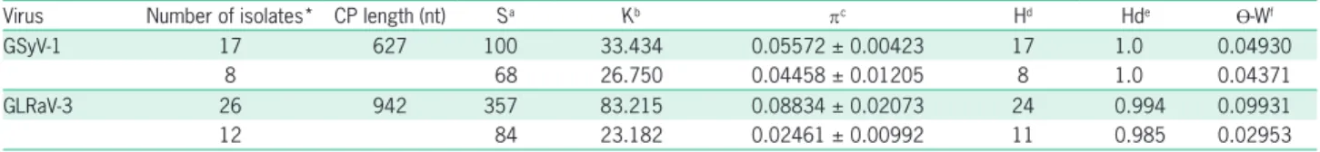
The coat protein gene molecular variability of GSyV-1 and GLRaV-3 was evaluated using two datasets. The first consisted of both Brazilian and foreign isolates (the same isolates used in phylogenetic analysis for GSyV-1 and GLRaV-3, excluding the outgroup) and the second consisted of Brazilian isolates only. Descriptors for dataset consisting of both Brazilian and foreign isolates indicated higher genetic variability than the dataset consisting of Brazilian isolates only for both viruses. However, the dif-ferences verified between the two datasets were lower for GSyV-1 (Table 2). This was probably a consequence of the isolates that were analyzed together originating from different countries. These results corroborate the lowest nt and daa identities observed in the majority of compari-sons between Brazilian and foreign isolates. The higher genetic variability is represented in turn by a higher num-ber of segregating sites (S), nucleotide diversity (π), haplo-type number (H) and haplohaplo-type diversity (Hd) (Table 2).
Nucleotide diversity (π) was lower than 0.089 for both viruses in the two datasets analyzed (Table 2). The GLRaV-3 virus (dataset with both Brazilian and foreign isolates) had the highest π value (0.08834 ± 0.02073), and the lowest π value was from the dataset with Brazilian isolates (0.02461 ± 0.00992). In general, when the vari-ability descriptors for the dataset consisting of Brazilian isolates only are analyzed, the GSyV-1 virus had greater genetic variability. The π value detected in GSyV-1 and GLRaV-3 (for both datasets) is in concordance with the
π values found by García-Arenal et al. (2001). These au-thors presented π values in the range of 0.002 to 0.224 for the coat protein gene of Cotton leaf curl virus (CLCuV), Citrus tristeza virus (CTV), Cucurbit yellow stunting disor-der virus (CYSDV), Groundnut rosette assistor virus (GRAV),
Table 2 − Descriptors of molecular variability for Grapevine Syrah virus 1 (GSyV-1) and Grapevine leafroll-associated virus 3 (GLRaV-3) coat
protein (CP) genes from Brazil and other foreign isolates.
Virus Number of isolates* CP length (nt) Sa Kb πc Hd Hde ϴ-Wf
GSyV-1 17 627 100 33.434 0.05572 ± 0.00423 17 1.0 0.04930
8 68 26.750 0.04458 ± 0.01205 8 1.0 0.04371
GLRaV-3 26 942 357 83.215 0.08834 ± 0.02073 24 0.994 0.09931
12 84 23.182 0.02461 ± 0.00992 11 0.985 0.02953
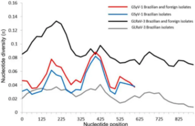
Rice tungro spherical virus (RTSV), Rice yellow mottle virus (RYMV), Sweet potato chlorotic stunt virus (SPCSV) and Yam mosaic virus (YMV). Nucleotide diversity (π) was also analyzed throughout the length of the coat protein gene of GSyV-1 and GLRaV-3, using the two previously described datasets. The tendency for π values in the graphic was similar for the two datasets in each virus, differing only in absolute values of π (Figure 3). The GSyV-1 virus showed the greatest nucleotide diversity at positions 175 and 400, and the GLRaV-3 virus presented greater nucleotide di-versity close to the N-terminal region (Figure 3). The Wat-terson’s estimator for the population-scaled mutation rate (ϴ-W) for the two viruses (both datasets) was in the order of 10−2 (Table 2).
Recombination events interfere with the selection analysis. Thus recombination was analyzed and no re-combination events were detected in datasets used for site-specific selection analysis (data not shown). In this analysis we used only the datasets consisting of Brazilian isolates, which represent a population for each virus. The presence of positive and negative selection at each site of the coat protein gene was evaluated. The coat protein genes of GSyV-1 and GLRaV-3 showed dN/dS ratios (not synonymous/synonymous substitution ratios) lower than 1.0, indicating negative or purifying selection (Table 3). The coat protein gene of GLRaV-3 showed a lower dN/dS ratio [GLRaV-3 (0.050) and GSyV-1 (0.182)], suggesting it was more constrained. No sites under positive selection in all the methods used have been detected. The number
Table 3 − Selection analysis for coat protein genes of Brazilian isolates of Grapevine Syrah virus 1 (GSyV-1) and Grapevine leafroll-associated
virus 3 (GLRaV-3).
Virus Number of isolates dN/dS SLAC
a FELb RELc PARRISd
PS NS PS NS PS NS PS
GSyV-1 8 0.182 - 2 - 27 - 57
-GLRaV-3 12 0.050 - - - 34 - 1
-PS = number of positive selection sites; NS = number of negative selection sites; (-) no site under selection; aSingle likelihood ancestor counting (SLAC); bFixed effects likelihood (FEL); cRandom effects likelihood (REL); dPartitioning for robust inference of selection (PARRIS); dN/dS = not synonymous/synonymous substitutions ratios.
of sites under negative selection varies according to the method employed (Table 3). Selection pressures can be associated with the maintenance of structural features of the virus (García-Arenal et al., 2001). For example, amino acids that are important in the assembly and stabiliza-tion of the coat protein are conserved in tobamoviruses (Altschuh et al., 1987). Viral proteins are multifunc-tional and may be involved in other processes such as replication, cell-to-cell, and long distance movement and transmission (García-Arenal et al., 2001). Therefore, in general, negative selection is predominant in the coding regions of viral proteins. The results presented here are consistent with the fact that the coat protein gene of ar-thropod-vectored viruses, such as GLRaV-3 and possibly GSyV-1, is the region under strongest negative selection and is the most constrained (Chare and Holmes, 2004; Zanardo et al., 2014).
The results of this study revealed genetic variability in these two viruses in Brazilian grapevines that should be taken into consideration in symptomatological assess-ments, and biological and molecular indexing. Although the most remarkable changes were observed in the nu-cleotide sequences, the deduced amino acid sequences of coat proteins have a propensity for less variation among isolates from different origins (geographical regions, hosts). The capsid protein is structural. Thus evolutionary factors could restrict changes that might be deleterious for the virus as was observed for GSyV-1 and GLRaV-3. For example, changes in capsid proteins could result in loss of interactions with vectors or plant host factors. In addition, GSyV-1 and GLRaV-3 have grapevine as a single natural host, which would further restrict these changes (Catarino et al., 2015).
A survey and molecular characterization based on the methods used here allowed detection and further evaluation of the genetic variability and geographical distribution of viral and viroidal pathogens in a Brazilian grape-growing area. This provides valuable information for the implementation of regional management practices to control virus diseases, such as the monitoring of vector transmission in vineyards and establishment of new vine-yards with pathogen-free propagative materials.
Acknowledgements
This research was financially supported by Em-brapa (Project 02.13.14.002). The authors would like to acknowledge Marcos Fernando Vanni (Embrapa
Figure 3 − Mean pairwise number of nucleotide differences per site
Grape & Wine) for his technical support, Márcio Pereira (Federal Institute of Education, Science and Technology of São Paulo, São Roque) for his help in the sampling and Vitor Cezar Pacheco da Silva for tax-onomic identification of the mealybugs. Marcelo Eiras is supported by a CNPq (Brazilian National Council for Scientific and Technological Development) re-search fellowship.
References
Al Rwahnih, M.; Daubert, S.; Golino, D.; Rowhani, A. 2009. Deep sequencing analysis of RNAs from a grapevine showing Syrah decline symptoms reveals a multiple virus infection that includes a novel virus. Virology 387: 395-401.
Al Rwahnih, M.; Daubert, S.; Golino, D.; Islas, C.; Rowhani, A. 2015. Comparison of next-generation sequencing versus biological indexing for the optimal detection of viral pathogens in grapevine. Phytopathology 105: 758-763.
Almeida, R.P.P.; Daane, K.M.; Bell, V.A.; Blaisdell, G.K.; Cooper, M.L.; Herrbach, E.; Pietersen, G. 2013. Ecology and management of grapevine leafroll disease. Frontiers in Microbiology 4: article 94.
Altschuh, D.; Lesk, A.M.; Bloomer, A.C.; Klug, A. 1987. Correlation of co-ordinated amino acid substitutions with function in viruses related to tobacco mosaic virus. Journal of Molecular Biology 193: 693-707.
Basso, M.F.; Fajardo, T.V.M.; Eiras, M.; Ayub, R.A.; Nickel, O. 2010. Molecular detection and identification of virus associated with symptomatic and symptomless grapevines. Ciência Rural 40: 2249-2255 (in Portuguese, with abstract in English).
Basso, M.F.; Fajardo, T.V.M.; Pio-Ribeiro, G.; Eiras, M.; Zerbini, F.M. 2014. Advances and prospects in the study of viral and subviral diseases in grapevine with emphasis on the situation in Brazil. Revisão Anual de Patologia de Plantas 22: 160-207 (in Portuguese, with abstract in English).
Bianchi, G.L.; Amicis, F.; Sabbata, L.; Bernardo, N.; Governatori, G.; Nonino, F.; Prete, G.; Marrazzo, T.; Versolatto, S.; Frausin, C. 2015. Occurrence of Grapevine Pinot gris virus
in Friuli Venezia Giulia (Italy): field monitoring and virus quantification by real-time RT-PCR. EPPO Bulletin 45: 22-32.
Catarino, A.M.; Fajardo, T.V.M.; Pio-Ribeiro, G.; Eiras, M.; Nickel, O. 2015. Incidence of viruses in grapevines in the Brazilian Northeast and partial molecular characterization of local virus isolates. Ciência Rural 45: 379-385 (in Portuguese, with abstract in English).
Chare, E.R.; Holmes, E.C. 2004. Selection pressures in the capsid genes of plant RNA viruses reflect mode of transmission. Journal of General Virology 85: 3149-3157.
Czotter, N.; Szabó, E.; Molnar, J.; Kocsis, L.; Deák, T.; Bisztray, Gy.; Tusnády, G.E.; Burgyán, J.; Várallyay, E. 2015. First description of Grapevine Syrah virus 1 in vineyards of Hungary. Journal of Plant Pathology 97: S74.
Engel, E.A.; Rivera, P.A.; Valenzuela, P.D.T. 2010. First report of
Grapevine Syrah virus-1 in Chilean grapevines. Plant Disease
94: 633.
Fajardo, T.V.M.; Dianese, E.C.; Eiras, M.; Cerqueira, D.M.; Lopes, D.B.; Ferreira, M.A.S.V.; Martins, C.R.F. 2007. Variability of the coat protein gene of Grapevine leafroll-associated virus 3 in Brazil. Fitopatologia Brasileira 32: 335-340.
Fajardo, T.V.M.; Eiras, M.; Nickel, O. 2016. Detection and molecular characterization of Grapevine yellow speckle viroid 1
isolates infecting grapevines in Brazil. Tropical Plant Pathology 41: 246-253.
Fiore, N.; Zamorano, A.; Rivera, L.; González, F.; Aballay, E.; Montealegre, J.; Pino, A.M. 2011. Grapevine viruses in the Atacama region of Chile. Journal of Phytopathology 159: 743-750.
Garcia-Arenal, F.; Fraile, A.; Malpica, J.M. 2001. Variability and genetic structure of plant virus populations. Annual Review of Phytopathology 39: 157-186.
Glasa, M.; Predajna, L.; Soltys, K.; Sabanadzovic, S.; Olmos, A. 2015. Detection and molecular characterisation of Grapevine
Syrah virus-1 isolates from Central Europe. Virus Genes 51:
112-121.
Jones, T.J.; Rayapati, N.A.; Nita, M. 2015. Occurrence of
Grapevine leafroll associated virus-2, -3 and Grapevine fleck virus
in Virginia, U.S.A., and factors affecting virus infected vines. European Journal of Plant Pathology 142: 209-222.
Le Maguet, J.; Beuve, M.; Herrbach, E.; Lemaire, O. 2012. Transmission of six ampeloviruses and two vitiviruses to grapevine by Phenacoccus aceris (Signoret). Phytopathology 102: 717-723.
Liu, M.H.; Li, M.J.; Qi, H.H.; Guo, R.; Liu, X.M.; Wang, Q.; Cheng, Y.Q. 2013. Occurrence of grapevine leafroll-associated viruses in China. Plant Disease 97: 1339-1345.
Maliogka, V.I.; Martelli, G.P.; Fuchs, M.; Katis, N.I. 2015. Control of viruses infecting grapevine. p. 175-227. In: Loebenstein, G.; Katis, N.I., eds. Advances in Virus Research. Academic Press, Burlington, USA.
Maree, H.J.; Almeida, R.P.P.; Bester, R.; Chooi, K.M.; Cohen, D.; Dolja, V.V.; Fuchs, M.F.; Golino, D.A.; Jooste, A.E.C.; Martelli, G.P.; Naidu, R.A.; Rowhani, A.; Saldarelli, P.; Burger, J.T. 2013.
Grapevine leafroll-associated virus 3. Frontiers in Microbiology
4: 82 doi: 10.3389/fmicb.2013.00082.
Maree, H.J.; Pirie, M.D.; Bester, R.; Oosthuizen, K.; Burger, J.T. 2015. Phylogenomic analysis reveals deep divergence and recombination in an economically important grapevine virus. Plos One 10: e0126819.
Martin, D.P.; Lemey, P.; Lott, M.; Moulton, V.; Posada, D.; Lefeuvre, P. 2010. RDP3: a flexible and fast computer program for analyzing recombination. Bioinformatics 26: 2462-2463.
Martelli, G.P. 2014. Directory of virus and virus-like diseases of the grapevine and their agents. Journal of Plant Pathology 96-1S: 1-136.
Naidu, R.A.; Marre, H.J.; Burger, J.T. 2015. Grapevine leafroll disease and associated viruses: a unique pathosystem. Annual Review of Phytopathology 53: 613-634.
Osman, F.; Leutenegger, C.; Golino, D.; Rowhani, A. 2007. Real-time RT-PCR (TaqMan) assays for the detection of Grapevine
leafroll associated viruses 1-5 and 9. Journal of Virological
Methods 141: 22-29.
Osman, F.; Leutenegger, C.; Golino, D.; Rowhani, A. 2008. Comparison of low-density arrays, RT-PCR and real-time TaqMan RT-PCR in detection of grapevine viruses. Journal of Virological Methods 149: 292-299.
Osman, F.; Rowhani, A. 2008. Real-time RT-PCR (TaqMan) assays for the detection of viruses associated with Rugose wood complex of grapevine. Journal of Virological Methods 154: 69-75.
Rott, M.E.; Jelkmann, W. 2001. Characterization and detection of several filamentous viruses of cherry: adaptation of an alternative cloning method (DOP-PCR) and modification of an RNA extraction protocol. European Journal of Plant Pathology 107: 411-420.
Rozas, J.; Sanchez-Del Barrio, J.C.; Messeguer, X.; Rozas, R. 2003. DnaSP, DNA polymorphism analyses by the coalescent and other methods. Bioinformatics 19: 2496-2497.
Sabanadzovic, S.; Ghanem-Sabanadzovic, N.A.; Saldarelli, P.; Martelli, G.P. 2001. Complete nucleotide sequence and genome organization of Grapevine fleck virus. Journal of General Virology 82: 2009-2015.
Sambrook, J.; Russell, D. 2001. Molecular Cloning: A Laboratory Manual. 3ed. CSHL Press, New York, NY, USA.
Tamura, K.; Stecher, G.; Peterson, D.; Filipski, A.; Kumar, S. 2013. MEGA6: molecular evolutionary genetics analysis version 6.0. Molecular Biology and Evolution 30: 2725-2729.
Thompson, J.D.; Gibson, T.J.; Plewniak, F.; Jeanmougin, F.; Higgins, D.G. 1997. The Clustal X windows interface: flexible strategies for multiple sequence alignment aided by quality tools. Nucleic Acids Research 24: 4876-4882.
Tsai, C.W.; Rowhani, A.; Golino, D.A.; Daane, K.M.; Almeida, R.P.P. 2010. Mealybug transmission of Grapevine leafroll viruses: an analysis of virus-vector specificity. Phytopathology 100: 830-834.
Vega, A.; Gutiérrez, R.A.; Peña-Neira, A.; Cramer, G.R.; Arce-Johnson, P. 2011. Compatible GLRaV-3 viral infections affect berry ripening decreasing sugar accumulation and anthocyanin biosynthesis in Vitis vinifera. Plant Molecular Biology 77: 261-274. Volpe, M.L.; Talquenca, S.G.; Engel, E.A.; Gracia, O. 2010.
Incidence of Grapevine leafroll associated viruses -1, -2, and -3 in Mendoza vineyards. Tropical Plant Pathology 35: 377-380. Zanardo, L.G.; Silva, F.N.; Lima, A.T.; Milanesi, D.F.;