Gale ctins: a ke y inte rse ctio n
be twe e n glyco bio lo gy and
im m uno lo gy
Inmunología, Departamento de Bioquímica Clínica, Facultad de Ciencias Q uímicas,
Universidad Nacional de Córdoba, Córdoba, Argentina G.A. Rabinovich,
C.M. Riera, C.A. Landa and C.E. Sotomayor
Abstract
Galectins are a family of evolutionarily conserved animal lectins, widely distributed from lower invertebrates to mammals. They share sequence and structure similarities in the carbohydrate recognition domain and specificity for polylactosamine-enriched glycoconjugates. In the last few years significant experimental data have been accumu-lated concerning their participation in different biological processes requiring carbohydrate recognition such as cell adhesion, cell growth regulation, inflammation, immunomodulation, apoptosis and metas-tasis. In the present review we will discuss some exciting questions and advances in galectin research, highlighting the significance of these proteins in immunological processes and their implications in biomedical research, disease diagnosis and clinical intervention. De-signing novel therapeutic strategies based on carbohydrate recogni-tion will provide answers for the treatment of autoimmune disorders, inflammatory processes, allergic reactions and tumor spreading.
Co rre spo nde nce G.A. Rabinovich Inmunología
Departamento de Bioquímica Clínica Facultad de Ciencias Q uímicas Universidad Nacional de Córdoba Ciudad Universitaria
Pabellón Argentina CC61, Agencia Postal 4 5000 Córdoba Argentina
Fax: + 54-351-433-4174 E-mail: gabyrabi@ onenet.com.ar
Presented at the XXVII Annual Meeting of the Brazilian Society of Biochemistry and Molecular Biology, Caxambu, MG, Brasil, May 23-26, 1998.
G.A. Rabinovich received the “Young Talent in Life-Sciences-98" award for this article from the Sociedade Brasileira de Bioquímica e Biologia Molecular and Amersham Pharmacia Biotech.
Research supported in part by CO NICET, CO NICO R, Fundación Antorchas and British Council.
Received December 2, 1998 Accepted February 1, 1999
Ke y wo rds ·Galectins
·Immunomodulation ·Apoptosis
·Adhesion ·Embryogenesis ·Metastasis
Intro ductio n
What are gale ctins?
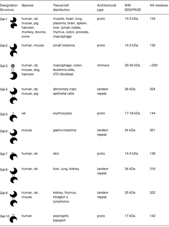
Galectins are a family of evolutionarily conserved proteins widely distributed in na-ture from lower invertebrates to mammals (1-3). They are defined by their sequence and structure similarities in the carbohydrate recognition domain (CRD) and their speci-ficity for polylactosamine-enriched glyco-conjugates (1,2). Ten mammalian galectins have been well identified in a wide variety of tissues of several species (4).
Ho w are gale ctins de signe d?
According to their architecture,
mam-Species Tissue/cell Architectural M M AA residues
distribution type SDS/PAGE
human, rat, muscle, heart, lung, proto 14.5 kDa 134
mouse, pig, placenta, brain, spleen, hamster, liver, lymph nodes, monkey, bovine, thymus, colon, prostate,
ovine macrophage
human, mouse small intestine proto 14.5 kDa 132
human, rat, macrophage, colon, chimera 29-35 kDa >250
mouse, dog, leukemia cells,
hamster 3T3 fibroblast
human, rat, alimentary tract, tandem 36 kDa 324
mouse, pig epithelial cells repeat
rat erythrocytes proto 17-18 kDa 144
mouse gastro-intestine tandem 34 kDa 301
repeat
human, rat skin proto 14.5 kDa 136
human, rat liver, lung, kidney tandem 34 kDa 316
repeat
human, rat, kidney, thymus, tandem 35 kDa 322
mouse Hodgkin’s repeat
lymphoma
human eosinophil, proto 17 kDa 142
basophil Designation
Structure
Gal-1
Gal-2
Gal-3
Gal-4
Gal-5
Gal-6
Gal-7
Gal-8
Gal-9
Gal-10
Table 1 - Galectin family: properties and tissue distribution.
M M , M olecular mass; AA residues, amino acid residues.
mals, although it has also been detected in chickens (19). The third family of tandem-repeat galectins includes proteins with two distinct CRDs, such as galectins-4 (20), -6
charac-terized and found to be associated with mul-tiple regulatory elements.
Ho w we re gale ctins ide ntifie d?
Although the first galectins were identi-fied by their binding to ß-galactoside-sugars (5), the most recent galectins, such as galectins-5 and -8, were detected by immu-noscreening cDNA libraries (9,22) or by crossreactivity with other galectins identi-fied so far (24). In this context galectin-9 was found by screening tumor cDNA librar-ies with sera from tumor-bearing patients (23).
Why are gale ctins fo und in the e xtrace llular m ilie u if the y are de signe d as intrace llular pro te ins?
From the viewpoint of protein structure, galectins have been designed to play key roles inside the cells with an acetylated N-terminus, lack of signal sequences and bio-synthesis in free ribosomes. Although novel intracellular functions have been reported in the last few years, such as mRNA processing (25), most of the functions assigned to ß-galactoside-binding proteins are confined to the cell surface or extracellular milieu (26), suggesting that galectins are externalized by a non-classical secretory mechanism (27). The intracellular concentration of galec-tins under physiological conditions is as high as 0.01 mM in mammalian cells, but it can reach levels of approximately 0.1 mM in critical situations. Galectin expression was found to be modulated during embryogen-esis (28), being a typical hallmark of specific developmental stages. Moreover, galectin expression and subcellular distribution were reported to be highly susceptible to modula-tion by diverse stimuli such as sodium bu-tyrate (29), viral infections (30), tumor sup-pressor genes (31) or inflammatory agents (18). In the present paper we discuss in detail the regulated expression of RMGal, a rat
macrophage galectin-1-like protein, isolated and characterized in our laboratory, which showed immunomodulatory properties (24, 32). Gillenwater and colleagues (29) dem-onstrated that sodium butyrate, a known dif-ferentiating agent, was able to modulate ga-lectin-1 content in human head and neck squamous carcinoma cells by a combination of transcriptional regulation and inhibition of histone deacetylation.
What are the m e chanism s invo lve d in gale ctin se cre tio n?
This question still remains to be ascer-tained. However, two hypotheses have been considered regarding the externalization of galectin-1. The first involves the utilization of specific transmembrane carriers, like those used to export some bacterial toxins (33). The second considers the possibility that, after synthesis, galectin-1 becomes concen-trated in plasma membrane evaginations prior to secretion and further externalized to form galectin-enriched extracellular vesicles (27). A kind of infrequent mechanism of external-ization is also used by many cytokines and growth factors (34). Nevertheless, it is clear that galectins can be specifically targeted and secreted to exert their functions by inter-acting with intracellular or extracellular gly-coconjugates.
Functio ns o f gale ctins
What is the functio nal significance o f this e vo lutio narily co nse rve d family o f animal le ctins?
cru-cial for life. In this context, it may be pro-posed that other members of this family could potentially compensate for the absence of these proteins, as suggested for targeted disruption of other important genes (37).
Galectins have been involved in several
in vitro physiopathological processes requir-ing carbohydrate recognition, such as cell adhesion (26), cell growth regulation (38), immunomodulation (39,40), apoptosis (32, 41), inflammation (42), embryogenesis (28), reproduction (43,44), tumor spreading (45) and pre-mRNA splicing (25). It should be emphasized that the vast majority of ga-lectin functions have been assigned to galec-tins-1 and -3. Hence, functions correspond-ing to other members of this protein family remain to be elucidated in future work.
What are the m o le cular m e chanism s invo lve d in gale ctin functio ns?
Although galectins are supposed to exert their biological roles by crosslinking glyco-conjugate ligands, the precise mechanisms of action of these proteins and their signal transduction pathways remain largely un-known.
Gale ctins in ce ll adhe sio n
What e xpe rim e ntal e vide nce sugge sts that gale ctins co uld be invo lve d in ce ll adhe sio n?
Polylactosamine residues in extracellu-lar matrix (ECM) glycoproteins such as la-minin (46) and fibronectin (47) have been proposed to be candidate ligands for galec-tin-1. In view of this specific recognition, galectins have been postulated as powerful modulators of cell-cell and cell-ECM inter-actions.
In this co nte xt, are gale ctins pro -adhe sive o r anti-adhe sive pro te ins?
Controversial results have been reported
as to whether galectin-1 exerts a positive or a negative effect on cell adhesion to ECM glycoconjugates, raising the possibility that this dimeric protein could promote cell at-tachment or deat-tachment according to cell type or cell developmental stage (26). At first sight, galectin-1 promoted cell adhesion in various cell types, such as melanoma cell lines (48), F9 teratocarcinoma cells (46), olfactory neurons (49), rhabdomyosarcoma cells (47) and CHO fibroblasts (46), by bridg-ing oligosaccharides between specific cell surface glycoconjugates and ECM compo-nents. On the other hand, the presence of galectin-1 inhibited myoblast interaction with laminin by sterically blocking the laminin receptor a7ß1 integrin from recognizing la-minin, thus allowing myoblasts to fuse into myotubes (50).
Similarly, the effects of galectin-3 on cell adhesion were antagonic, depending on the physiopathological environment. While ga-lectin-3 promoted neutrophil adhesion to la-minin (51) in the context of inflammation, it showed a dramatic inhibitory effect on mela-noma cell adhesion to ECM in metastases (52). In this context, expression of galectins-1 and -3 in tumor cells was found to be correlated with high metastatic potential and low survival (53).
Gale ctins in ce ll gro wth re gulatio n
What is the ro le o f gale ctins in ce ll gro wth re gulatio n and diffe re ntiatio n?
Ho w can it be e xplaine d that antago nic stim ulato ry and inhibito ry pro pe rtie s co uld be pre se nt within the sam e m o le cule ?
To answer this question, an interesting study reports a biphasic modulation of cell growth by recombinant galectin-1 (55). While this ß-galactoside-binding protein was found to be mitogenic at low physiological concentrations, the growth-inhibitory prop-erties were apparent in a higher concentra-tion range, when tested on human cells in vitro. Moreover, galectin-1 may trigger ei-ther proliferation or cell growth arrest de-pending on the presence of concomitant en-vironmental signals, cell cycle stages, or the expression of its carbohydrate receptors on the cell surface (55). Other signaling mol-ecules, such as TGF ß (56) and FasL (57), have been shown to trigger different effects depending on the type and functional state of the cells.
Gale ctins in the im m une syste m
The ultimate goal of immunologists is to manipulate the immune response, so that deficient responses can be potentiated, while harmful responses are suppressed.
Are gale ctins impo rtant in the co nte xt o f innate immunity?
The innate immune system is important because it provides the early phases of host defense to protect the organism. Galectins have been implicated in different functions of innate immunity such as the respiratory burst (58), chemotaxis of eosinophils (59) and neutrophil migration during inflamma-tory episodes (51).
What was the first clue sugge sting that gale ctins co uld play a ro le in the re gulatio n o f the adaptive im m une re spo nse ?
That galectins could be implicated in the
regulation of the immune response was first inferred from galectin-1 localization in im-mune privileged sites of the body, such as the placenta (6,43,44) and the eye (60). In immune privileged sites multiple factors in-teract to prevent immunopathogenic pro-cesses from disrupting the critical functions of these particularly vulnerable organs. It has been recently demonstrated that FasL expression in immune privileged sites may have a key role in preserving tolerance by selectively killing inflammatory T cells by apoptosis (61). Accordingly, galectin-1 could be proposed as an alternative regulatory sig-nal to regulate immune privilege. Expres-sion of this protein in the first term gestation placenta would prevent inflammatory T cells from harming the fetus (43). Furthermore, galectin-1 expression in the eye would pro-tect this sensory organ from the devastating effects of an inflammatory response (61,62). In agreement, a protein related to the galec-tin family called GRIFIN (galecgalec-tin-related interfiber protein) was recently identified in the lens and in simple cellular structures of the eye (60).
Are gale ctins also lo calize d within the immune syste m?
the level of the S and G2/M stages (66).
What is the asso ciatio n be twe e n gale ctins and ato pic dise ase s?
Recent studies have shown that galectin-3, formerly defined as an Ig E-binding pro-tein, was able to inhibit IL-5 gene transcrip-tion and protein release from human eosino-phils and allergen-specific T cell lines (67). The possibility to shut down the IL-5 path-way could be determinant in the regulation of the T helper 2-dependent allergic reac-tions. Further investigations will be needed to clarify the mechanisms involved in this process.
What is the asso ciatio n be twe e n gale ctins and auto immunity?
Galectin-1 has shown immunosuppres-sive properties in two experimental models of autoimmunity. Administration of recom-binant galectin-1 prevented clinical and histopathological manifestations of experi-mental autoimmune encephalomyelitis (EAE) in susceptible Lewis rats (39) and autoimmune myasthenia gravis (EAMG) in rabbits (40).
What kind o f m o le cular m e chanism s are invo lve d in the se immuno mo dulato ry pro pe rtie s?
Although the precise mechanisms in-volved in these properties in vivo still remain to be elucidated, hypotheses have been raised concerning the ability of galectin-1 to affect processes in T cell suppressor commitment and in sensitization or deletion of antigen-specific T cells (39,40). Interestingly, in the last few years evidence has been obtained concerning the implications of galectin-1 in apoptosis of mature T cells (31,41,43) and particular subsets of immature cortical thy-mocytes (59), thus providing a breakthrough in glycoimmunology.
What was the first clue indicating that gale ctins co uld be implicate d in pro gramme d ce ll de ath?
The first clue was raised when Goldstone and Lavin (68) reported in 1991 that the galectin-1 gene was overexpressed during glucocorticoid-induced cell death.
What is the ro le o f gale ctin-1 in ce ntral to le rance ?
Concerning the immune system, cell death programs promote tolerance by deletion of autoreactive lymphocytes in the central lym-phoid organs and termination of immune responses in the peripheral immune system. At the central level, lymphocytes are suscep-tible to positive (69) and negative (70) selec-tion. Immature T cells that recognize self-peptides with high affinity in the context of autologous major histocompatibility com-plex (MHC) molecules undergo negative selection by programmed cell death (70). T lymphocytes that fail to receive antigen stim-ulation (neglected cells) also undergo a de-fault apoptotic program (69). The remaining T cells, which bind to peptide MHC com-plexes with low affinity, are positively se-lected to survive. In the context of new mol-ecules involved in the machinery of central death, galectin-1 expressed by human thy-mic epithelial cells (64) has been recently shown to induce apoptosis of cortical thy-mocytes (71). The model of galectin-1 gene knock-out mice (35) will be useful to eluci-date the precise role of galectin-1 in the elimination of non-selected and negatively selected cells during thymocyte development.
What is the ro le o f gale ctin-1 in pe riphe ral to le rance ?
context, galectin-1 has also been reported to induce apoptosis of mature T cells (32,41,43). The apoptotic effect of soluble galectin-1 depended upon the activation state of T cells. Perillo et al. (41) clearly showed that galec-tin-1 induced apoptosis of mature T cells bearing the CD45RO splicing product, which was found to be differentially glycosylated (73). This provides an alternative mechanism for explaining the immunosuppressive prop-erties exhibited by this highly conserved pro-tein family.
What is the link be twe e n apo pto sis and auto im m unity?
The importance of decreased apoptosis in the etiology of autoimmune disorders has been highlighted by the occurrence of lupus-like disorders in MRL-lpr/lpr or C3H-gld/ gld mouse strains, carrying spontaneous mutations in Fas or FasL genes (74), and in particular by the dominant Fas gene muta-tion associated with Canale-Smith syndrome, a human autoimmune lymphoproliferative disorder (75).
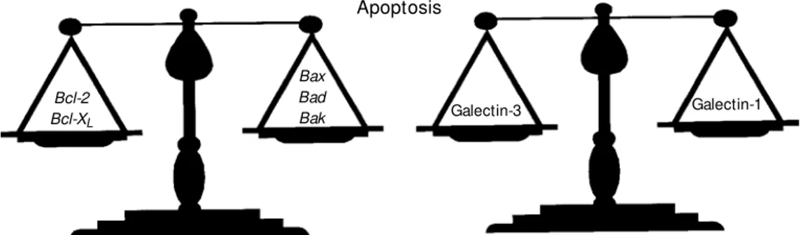
Gale ctins-1 and -3: why a no ve l paradigm?
Antagonic functions have been assigned to galectin-1 and galectin-3, providing clues for a novel paradigm. While galectin-1 has been shown to induce T cell apoptosis (32,41,43), galectin-3 has been conversely shown to prevent cell death (76). Similarly to members of the Bcl-2 family, galectins-1 and -3 belong to an additional family of
proteins with high sequence homologies but opposite effects on cell survival (Figure 1). As clearly shown in the Figure, Bcl-2 and
Bcl-XL are negative regulators of apoptosis, whereas Bax, Bad, and Bak contribute to a positive signal in the regulation of cell death (77). Transfection of galectin-3 cDNA into leukemia T cells conferred resistance to ap-optosis induced by Fas ligation and stauro-sporine (76). Of particular interest, galectin-3 showed a significant sequence similarity to
Bcl-2, mainly concentrated in the functional BH1 (NWGR) domain. Although this para-digm between galectins-1 and -3 seems to be attractive, it should not be conceived as a general principle. Future studies should chal-lenge this paradigm in the context of other physiological systems. Consistently, expres-sion of galectins-1 and -3 was found to be differentially regulated during mammalian gestation (43,44). Iglesias et al. (44) reported the coexistence of mitogenic galectin-3 and apoptotic galectin-1 (43) in ovine placenta. Interestingly, galectin-3 expression was found to be decreased in term ovine pla-centa, in comparison with the middle of the gestation period. In contrast, no significant decrease was observed in galectin-1 expres-sion. In this context, one might speculate that differential expression of both ß-galac-toside-binding lectins in ovine placenta could be associated with selective requirements at different developmental stages of gestation. Furthermore, the participation of other members of the galectin family in the control of programmed cell death should also be considered. In this sense, galectin-9, a novel
Bcl-2 Bcl-XL
Bax Bad
Bak Galectin-3 Galectin-1
Figure 1 - Galectins-1 and -3: a novel paradigm in the regulation of programmed cell death. Ga-lectins-1 and -3 represent an ad-dit ional f am ily of prot eins similar to the Bcl-2 family, w here different members exhibit se-quence and structure similari-ties, but opposite effects on cell survival.
tandem repeat lectin, has recently shown strong apoptotic activity toward murine thy-mocytes (78).
What is the functio nal significance o f gale ctin-1 in activate d m acro phage s?
Macrophages (Møs) play important roles in a wide spectrum of events from innate immunity to adaptive immune response (24). Since galectin-1 had shown such immuno-modulatory properties, we wondered whether it could be possible to identify its presence in key immunoregulatory cells such as Mfs and further demonstrate its functional mean-ing for target cells.
By means of immunochemical and im-munocytochemical studies, we have identi-fied the presence of a 15-kDa soluble protein with key features of mammalian galectin-1 in rat peritoneal Møs by using a polyclonal Ab raised against the extensively studied C-16 chicken isolectin (24). Rat macrophage galectin (RMGal) was purified from chemi-cally activated macrophages by a single step affinity chromatography on a lactosyl-Sepharose matrix, resulting in the isolation of a sharp protein band of 15 kDa, an iso-electric point of 4.8 and high hemagglutinat-ing activity specifically inhibited by saccha-rides bearing a ß-D-galactoside configura-tion (32). This protein behaved as a dimer under nondenaturing conditions, and inter-nal amino acid sequencing revealed exten-sive homologies (80-90%) with other mem-bers of the galectin-1 subfamily. Taken to-gether, all the properties described resembled those exhibited by mammalian galectin-1 (32). Resident, inflammatory or chemically ac-tivated Møs must be viewed as distinct cell subpopulations, since they show different phenotypes according to their activation sta-tus (24). In light of this observation, the next question we tackled was related to the regu-lation of total and surface RMGal expression in different Mø subpopulations. By Western blot analysis and further densitometric
quantitation, we found a 5-fold increase in total galectin expression in chemically acti-vated Møs and a 2-fold increase in inflam-matory Møs in comparison to peritoneal resi-dent cells (24). Moreover, modulation of RMGal expression was accompanied by an increase in cell surface immunostaining as shown by FACS analysis of non-permeabil-ized cells (24). Modulation of galectin ex-pression and subcellular distribution strongly suggested that it could be implicated in cru-cial immunological processes mediated by Møs. By using current techniques to evalu-ate apoptosis, such as TUNEL assay, trans-mission electron microscopy and DNA frag-mentation assay, we concluded that RMGal was clearly involved in apoptosis of mature T-cells and that this effect was dependent on its carbohydrate recognition domain. These results were more significant than those ob-tained using a heterologous system (79). Con-sistently, it has been reported that mono-cytes and dendritic cells from peripheral blood of healthy individuals can induce ap-optosis of mitogen-activated T-cells (80).
Co ncluding re m arks and future pe rspe ctive s
Elucidation of the molecular mechanisms involved in galectin functions will provide new insights in biomedical research, disease diagnosis, prognosis and clinical therapy.
At the level of disease diagnosis, Lutomski et al. (81) reported the presence of autoanti-bodies against galectin-1 in sera of patients with neurological disorders, particularly multiple sclerosis. Moreover, aberrant ex-pression of galectins on the surface of tumor cells seems to be indicative of a worse prog-nosis (53).
possibility to regulate the apoptotic thresh-old of autoreactive T cells using galectin-1 would be beneficial in autoimmunity. Sec-ond, down-regulation of the IL-5 gene by galectin-3 would be important in the treat-ment of allergic processes. Finally, neutral-izing antibodies or antisense oligonucleotides aimed at inhibiting galectins-1 and -3 ex-pression would potentially decrease the meta-static potential of tumor cells
Ackno wle dgm e nts
We would like to give special thanks to all the people who contributed to our work during the last few years, especially to Drs. Mercedes Iglesias, Nidia Modesti, Carlota Wolfenstein-Todel, Yuti Chernajovsky, Ofer Lider, Hanna Dreja, Gordon Daily, José Luis Bocco and Leonardo Castagna.
Re fe re nce s
1. Barondes SH, Cooper DNW, Gitt M A & Leffler H (1994). Galectins: structure and function of a large family of animal lectins. Journal of Biological Chem istry, 269: 20807-20810.
2. Barondes SH, Cast ronovo V, Cooper DNW, Cummings RD, Drickamer K, Feizi T, Gitt M A, Hirabayashi J, Hughes C, Kasai K, Leffler H, Liu F, Lotan R, M ercurio AM , M onsigni M , Pillai S, Poirer F, Raz A, Rigby PW J, Rini JM & W ang JL (1994). Galectins: a family of animal galactoside-binding lectins. Cell, 76: 597-598. 3. Kasi K & Hirabayashi J (1996). Galectins: a
family of animal lectins that decipher glycocodes. Journal of Biochemistry, 119: 1-8.
4. Leffler H (1997). Introduction to galectins. Trends in Glycoscience and Glycotechnol-ogy, 45: 9-19.
5. Hirabayashi J & Kasai K (1993). The family of metazoan metal-independent ß-galac-toside-binding lectins: structure, function and molecular evolution. Glycobiology, 3: 297-304.
6. Hirabayashi J & Kasai K (1984). Human placenta ß-galactoside-binding lectin. Pu-rification and some properties. Biochemi-cal and BiophysiBiochemi-cal Research Communi-cations, 122: 938-944.
7. Gitt M A & Barondes SH (1991). Genomic sequence and organization of tw o mem-bers of a human lectin gene family. Bio-chemistry, 30: 82-89.
8. Gitt M A, M assa SM , Leffler H & Barondes SH (1992). Isolation and expression of a gene encoding L-14-II, a new human soluble lactose-binding lectin. Journal of Biological Chemistry, 267: 10601-10606. 9. Gitt M A, Wisers M F, Leffler H, Herrmann
J, Xia Y-R, M assa SM , Cooper DNW, Luis AJ & Barondes SH (1995). Sequence and
mapping of galectin-5, a ß-galactoside-binding lectin, found in rat erythrocytes. Journal of Biological Chem istry, 270: 5032-5038.
10. M agnaldo T, Bernerd F & Darmon M (1995). Galectin-7, a human 14-kDa S-lec-tin, specifically expressed in keratinocytes and sensitive to retinoic acid. Develop-mental Biology, 168: 259-271.
11. Ackerman SJ, Corrette SE, Rosenberg HF, Bennet JC, M astrianni DM , Nicholson-Weller A, Nicholson-Weller PF, Chin DT & Tenen DG (1993). M olecular cloning and character-ization of human eosinophil Charcot-Ley-den cristal protein (lysophospholipase). Journal of Immunology, 150: 456-468. 12. Vast a GR, Ahm ed H, Am zel LM &
Bianchet M A (1997). Galectins from am-phibian species: carbohydrate specificity, molecular structure and evolution. Trends in Glycoscience and Glycotechnology, 9: 131-144.
13. Paroutaud P, Levi G, Teichberg VI & Strosberg AD (1987). Extensive amino acid homologies betw een animal lectins. Proceedings of the National Academy of Sciences, USA, 84: 6345-6348.
14. Hirabayashi J, Ubukata T & Kasai K (1996). Purification and molecular characterization of a novel 16-kDa galectin from the nema-tode Caenorhabditis elegans. Journal of Biological Chemistry, 271: 2497-2505. 15. Ohyama Y, Hirabayashi J, Oda Y, Oono S,
Kaw asaki H, Suzuki K & Kasai K (1986). Nucleotide sequence of chick 14 K ß-ga-lactoside-binding lectin mRNA. Biochemi-cal and BiophysiBiochemi-cal Research Communi-cations, 134: 51-56.
16. Sakakura Y, Hirabayashi J, Oda Y, Ohyama Y & Kasai K (1990). Structure of chicken 16-kDa ß-galactoside-binding lectin: com-plete amino acid sequence, cloning of
cDNA and production. Journal of Biologi-cal Chemistry, 265: 21573-21579. 17. Pfeifer K, Haasemann M , Gamulin V,
Bretting H, Fahrenholz F & M uller WE (1993). S-type lectins occur also in inver-tebrates: high conservation of the carbo-hydrate recognition domain in the lectin genes from the marine sponge Geodia cydonium. Glycobiology, 3: 179-184. 18. Sato S & Hughes RC (1994). Regulation of
secretion and surface expression of M ac-2, a galactoside-binding protein of macro-phages. Journal of Biological Chemistry, 269: 4424-4430.
19. Nurminskaya M & Linsenmayer TF (1996). Identification and characterization of up-regulated genes during chondrocyte hy-pert rophy. Developm ent al Dynam ics, 206: 260-271.
20. Oda Y, Herrmann J, Gitt M A, Turck CW, Burlingame AL, Barondes SH & Leffler H (1993). Soluble lactose-binding lectin from rat intestine w ith tw o different carbohy-drate-binding domains in the same chain. Journal of Biological Chem istry, 268: 5929-5939.
21. Gitt M A, Colnot C, Poirier F, Nani KJ, Barondes SH & Leffler H (1998). Galectin-4 and galectin-6 are tw o closely related lectins expressed in mouse gastrointesti-nal tract. Jourgastrointesti-nal of Biological Chemistry, 273: 2954-2960.
22. Hadari YR, Paz K, Dekel R, M estrovic T, Accili D & Zick Y (1995). Galectin-8: a new rat lectin related to galectin-4. Journal of Biological Chemistry, 270: 3447-3453. 23. Wada J & Kanw ar YS (1997).
Identifica-tion and characterizaIdentifica-tion of galectin-9, a novel ß-galactoside-binding mammalian lectin. Journal of Biological Chemistry, 272: 6078-6086.
Riera CM & Sotomayor CE (1996). Regu-lated expression of a 16-kd galectin-like protein in activated rat macrophages. Journal of Leukocyte Biology, 59: 363-370.
25. Dagher SF, Wang JL & Patterson RJ (1995). Identification of galectin-3 as a fac-tor in pre-mRNA splicing. Proceedings of the National Academy of Sciences, USA, 92: 1213-1217.
26. Cooper DNW (1997). Galectin-1: secre-tion and modulasecre-tion of cell interacsecre-tions w ith laminin. Trends in Glycoscience and Glycotechnology, 9: 57-67.
27. Cooper DNW & Barondes SH (1990). Evi-dence for export of a muscle lectin from cytosol to extracellular matrix and for novel secretory mechanism. Journal of Cell Biology, 110: 1681-1691.
28. Poirier F, Timmons PM , Chan C-T, Guenet J-L & Rigby P (1992). Expression of the L14 lectin during mouse embryogenesis suggests multiple roles during pre- and post-implantation development. Develop-ment, 115: 143-155.
29. Gillenw ater A, Xu XC, Estrov Y, Sacks PG, Lotan D & Lotan R (1998). M odulation of galectin-1 content in human head and neck squamous carcinoma cells by so-dium butyrate. International Journal of Cancer, 75: 217-224.
30. Hsu DK, Hammes SR, Kuw abara I, Greene WC & Liu FT (1996) Human T lymphotro-pic virus-I infection of human T lympho-cytes induces expression of the beta-ga-lactoside binding lectin, galectin-3. Jour-nal of Biological Chemistry, 148: 1661-1670.
31. Gaudin JC, Arar C, M onsigny M & Legrand A (1997). M odulation of the expression of the rabbit galectin-3 gene by p53 and c-Ha-ras proteins and PM A. Glycobiology, 7: 1089-1098.
32. Rabinovich GA, Iglesias M M , M odesti NM , Castagna LF, Wolfenstein-Todel C, Riera CM & Sotomayor CE (1998). Acti-vated rat macrophages produce a galec-tin-1-like protein that induces apoptosis of T cells: biochemical and functional charac-terization. Journal of Immunology, 160: 4831-4840.
33. Cleves AE, Cooper DN, Barondes SH & Kelly RB (1996). A new pathw ay for pro-tein export in Saccharomyces cerevisiae. Journal of Cell Biology, 133: 1017-1026. 34. Rubart elli A, Bajet t o A, Allavena G,
Wollman E & Sitia R (1992). Secretion of thioredoxin by normal and neoplastic cells through leaderless secretory pathw ay. Journal of Biological Chem istry, 267: 24161-24164.
35. Poirrier F & Robertson EJ (1993). Normal development of mice carrying a null mu-tation in the gene encoding the L-14 S-type lectin. Development, 119: 1229-1236.
36. Colnot C, Fow lis D, Ripoche M A, Bouchaert I & Poirier F (1998). Embryonic im plant at ion in galect in-1/galect in-3 double mutant mice. Developmental Dy-namics, 211: 306-313.
37. Colnot C, Ripoche M A, Fow lis D, Cannon V, Scaerou F, Cooper DNW & Poirier F (1997). The role of galectins in mouse development. Trends in Glycoscience and Glycotechnology, 9: 31-40.
38. Wells V & M allucci L (1991). Identification of an autocrine negative grow th factor: mouse ß-galactoside-binding protein is a cytostatic factor and cell grow th regula-tor. Cell, 64: 91-97.
39. Offner H, Celnik B, Bringman T, Casentini-Borocz D, Nedw in GE & Vandebark A (1990). Recombinant human ß-galacto-side-binding lectin suppresses clinical and histological signs of experimental autoim-mune encephalomyelitis. Journal of Neu-roimmunology, 28: 177-184.
40. Levy G, Tarrab-Hazdai R & Teichberg VI (1983). Prevent ion and t herapy w it h electrolectin of experimental autoimmune myasthenia gravis in rabbits. European Journal of Immunology, 13: 500-507. 41. Perillo NL, Pace KE, Seilhamer JJ & Baum
LG (1995). Apoptosis of T-cells mediated by galectin-1. Nature, 378: 736-739. 42. Yamaoka A, Kuw abara I, Frigeri LG & Liu
FT (1995). A human lectin, galectin-3 (ep-silon BP/M ac-2), stimulates superoxide production by neutrophils. Journal of Im-munology, 154: 3479-3487.
43. Iglesias M M , Rabinovich GA, Ivanovic V, Sotomayor CE & Wolfenstein-Todel C (1998). Galectin-1 from ovine placenta: amino-acid sequence, physicochemical properties and implications in T-cell death. European Journal of Biochemistry, 252: 400-407.
44. Iglesias M M , Rabinovich GA, Ambrosio AL, Cast agna LF, Sot om ayor CE & Wolfenstein-Todel CW (1998). Purification of galectin-3 from ovine placenta: devel-opmentally regulated expression and im-munological relevance. Glycobiology, 8: 59-65.
45. Raz A & Lotan R (1987). Endogenous ga-lactoside-binding lectins: a new class of functional tumor cell surface molecules related to metastasis. Cancer and M etas-tasis Review s, 6: 433-452.
46. Zhou Q & Cummings RD (1993). L-14 lec-tin recognition of laminin and its
promo-tion of in vitro cell adhesion. Archives of Biochemistry and Biophysics, 300: 6-17. 47. Ozeki Y, M at sui T, Yam am ot o Y,
Funahashi M , Hamako J & Titani K (1995). Tissue f ibronect in is an endogenous ligand for galectin-1. Glycobiology, 5: 255-261.
48. Van den Brüle FA, Buicu C, Baldet M , Sobel M E, Cooper DNW, M arschal P & Castronovo V (1995). Galectin-1 modu-lates human melanoma cell adhesion to laminin. Biochemical and Biophysical Re-search Communications, 209: 760-767. 49. M ahant happa NK, Cooper DNW ,
Barondes SH & Schw arting GA (1994). Rat olfactory neurons can utilize the en-dogenous lectin L-14, in a novel adhesion mechanism. Development, 120: 1373-1384.
50. Cooper DNW, M assa SM & Barondes SH (1991). Endogenous muscle lectin inhib-its myoblast adhesion to laminin. Journal of Cell Biology, 115: 1437-1448. 51. Kuw abara I & Liu F-T (1996). Galectin-3
promotes adhesion of human neutrophils to laminin. Journal of Immunology, 156: 3939-3944.
52. Ochieng J, Leite-Brow ning M L & Warfield P (1998). Regulation of cellular adhesion to extracellular matrix proteins by galec-tin-3. Biochemical and Biophysical Re-search Communications, 246: 788-791. 53. Bresalier RS, M azurek N, Sternberg LR,
Byrd JC, Yunker CK, M akker PN & Raz A (1998). M etastasis of human colon cancer is altered by modifying expression of the ß-galactoside binding protein galectin-3. Gastroenterology, 115: 287-296. 54. Sanford GL & Harris-Hooker S (1990).
Stimulation of vascular cell proliferation by ß-galactoside-binding lectins. FASEB Journal, 4: 2912-2918.
55. Adams L, Kenneth Scott G & Weinberg C (1996). Biphasic modulation of cell grow th by recombinant human galectin-1. Bio-chimica et Biophysica Acta, 1312: 137-144.
56. Roberts AB, Anzano M A, Wakefield LM , Roche NS, Stern DF & Sporn M B (1985). Type beta transforming grow th factor: a bifunctional regulator of cell grow th. Pro-ceedings of the National Academy of Sci-ences, USA, 82: 119-123.
57. Lynch DH, Ramsdell F & Alderson M R (1995). Fas and Fas L in the homeostatic regulation of immune responses. Immu-nology Today, 16: 569-574.
59. M atsumoto R, M atsumoto H, Seki M , Hata M , Asano Y, Kanegasaki S, Stevens RL & Hirashim a M (1998). Hum an ecalectin, a variant of human galectin-9, is a novel eosinophil chemoattractant pro-duced by T lymphocytes. Journal of Bio-logical Chemistry, 273: 16976-16984. 60. Ogden AT, Nunes I, Ko K, Wu S, Hines
CS, Wang AF, Hegde RS & Lang RA (1998). GRIFIN, a novel lens-specific pro-tein related to the galectin family. Journal of Biological Chem istry, 273: 28889-28896.
61. Griffith TS & Ferguson TA (1997). The role of Fas L-induced apoptosis in immune privilege. Immunology Today, 18: 240-244.
62. Gold R, Hartung HP & Lassman H (1997). T-cell apoptosis in autoimmune diseases: termination of inflammation in the ner-vous system and other sites w ith special-ized im m une-def ense m echanism . Trends in Neurosciences, 20: 399-404. 63. Baum LG, Seilhamer JJ, Pang M , Levine
WB, Beynon D & Berliner JA (1995). Syn-thesis of an endogenous lectin, galectin-1 by human endothelial cells is up-regulated by endothelial cell activation. Glycoconju-gate Journal, 12: 63-68.
64. Baum LG, Pang M , Perillo NL, Wu T, Delegaene A, Uittenbogaart CH, Fukuda M & Seilhamer JJ (1995). Human thymic epithelial cells express an endogenous lectin, galectin-1, w hich binds to core 2 O-glycans on thymocytes and T lymphoblas-toid cells. Journal of Experimental M edi-cine, 181: 877-887.
65. Blaser C, Kauf m ann M , M uller C, Zimmerman C, Wells V, M allucci L & Pircher H (1998). ß-galactoside-binding protein secreted by activated T cells in-hibits antigen-induced proliferation of T cells. European Journal of Immunology,
28: 2311-2319.
66. Allione A, Wells V, Forni G, M allucci L & Novelli F (1998). ß-galactoside-binding protein (ß-GBP) alters the cell cycle, up-regulates expression of the a- and ß-chains of the IFN-g receptor, and triggers IFN-g-mediated apoptosis of activated hu-man T lymphocytes. Journal of Immunol-ogy, 161: 2114-2119.
67. Cortegano I, del Pozo V, Cárdaba B, de Andres B, Gallardo S, del Amo A, Arrieta I, Jurado A, Palomino P, Liu F-T & Lahoz C (1998). Galectin-3 dow n-regulates IL-5 gene expression on different cell types. Journal of Immunology, 161: 385-389. 68. Goldstone SD & Lavin M F (1991).
Isola-tion of a cDNA clone, encoding a human ß-galact oside-binding prot ein overex-pressed during glucocorticoid-induced cell death. Biochemical and Biophysical Re-search Communications, 178: 746-750. 69. von Boehmer H (1994). Positive selection
of lymphocytes. Cell, 76: 219-228. 70. Nossal GJV (1994). Negative selection of
lymphocytes. Cell, 76: 229-239. 71. Perillo NL, Uittenbogaart CH, Nguyen JT
& Baum LG (1997). Galectin-1, an endog-enous lectin produced by thymic epitheli-al cells, induces apoptosis of human thy-mocytes. Journal of Experimental M edi-cine, 97: 1851-1858.
72. M cFarland HL, Critchfield JM , Racke M K, M ueller JP, Nye SH, Boehm e SA & Lenardo M J (1995). Amelioration of au-toimmune reactions by antigen-induced apoptosis of T cells. Advances in Experi-mental and M edical Biology, 383: 157-166.
73. Cyster JG, Fow ell D & Barclay AN (1994). Antigen determinants encoded by alter-natively spliced exons of CD45 are deter-mined by the polypeptide but influenced by glycosylation. International
Immunol-ogy, 6: 1875-1881.
74. Singer GG, Carrera AC, M arshak-Rothstein A, M artínez AC & Abbas AK (1994). Apoptosis, Fas and systemic au-toimmunity: the M RL-lpr/lpr model. Cur-rent Opinion in Immunology, 6: 913-920. 75. Fisher GH, Rosenberg FJ, Straus SE, Dale JK, M iddelton LA, Yin AY, Strober W, Lenardo M J & Puck JM (1995). Dominant interfering Fas gene mutations impair ap-optosis in a human autoimmune lympho-proliferative syndrome. Cell, 81: 935-946. 76. Yang RY, Hsu DK & Liu FT (1996). Expres-sion of galectin-3 modulates T cell grow th and apoptosis. Proceedings of the Na-tional Academy of Sciences, USA, 93: 6737-6742.
77. Kroemer G (1997). The proto-oncogene Bcl-2 and its role in regulating apoptosis. Nature M edicine, 3: 614-620.
78. Wada J, Ota K, Kumar A, Wallner EI & Kanw ar Y (1997). Developmental regula-tion, expression and apoptotic potential of galectin-9, a ß-galactoside-binding lec-tin. Journal of Clinical Investigation, 99: 2452-2461.
79. Rabinovich GA, M odesti NM , Castagna LF, Landa CA, Riera CM & Sotomayor CE (1997). Specific inhibition of lymphocyte proliferation and induction of apoptosis by CLL-I, a ß-galactoside-binding lectin. Jour-nal of Biochemistry, 122: 365-373. 80. Revillard JP, Adorini L, Goldm an M ,
Kabelitz D & Waldmann H (1998). Apopto-sis: potential for disease therapies. Im-munology Today, 19: 291-293.