Gale ctin-1 , an alte rnative signal
fo r T ce ll de ath, is incre ase d in
activate d m acro phage s
Laboratorio de Inmunología, Departamento de Bioquímica Clínica, Facultad de Ciencias Q uímicas, Universidad Nacional de Córdoba, Córdoba, Argentina
G.A. Rabinovich, C.M. Riera and C.E. Sotomayor
Abstract
Galectin-1 belongs to an evolutionarily conserved family of animal ß-galactoside-binding proteins, which exert their functions by crosslinking the oligosaccharides of specific glycoconjugate ligands. During the past decade, attempts to identify the functional role of galectin-1 suggested participation in the regulation of the immune response. Only in the last few years has the molecular mechanism involved in these properties been clearly elucidated, revealing a critical role for galectin-1 as an alternative signal in the generation of T cell death. In the present study we will discuss the latest advances in galectin research in the context of the regulation of the immune response, not only at the central level but also at the periphery. Moreover, we will review the purification, biochemical properties and functional signif-icance of a novel galectin-1-like protein from activated rat macro-phages, whose expression is differentially regulated according to the activation state of the cells. The novel role of a carbohydrate-binding protein in the regulation of apoptosis is providing a breakthrough in galectin research and extending the interface between immunology, glycobiology and clinical medicine.
Co rre spo nde nce C.E. Sotomayor
Laboratorio de Inmunología Departamento de Bioquímica Clínica Facultad de Ciencias Q uímicas Universidad Nacional de Córdoba Ciudad Universitaria
Pabellón Argentina 5000 Córdoba Argentina
Fax: + 54-351-433-4174 E-mail:
csotomay@ bioclin.fcq.unc.edu.ar
Presented at the 5th Brazilian Symposium on Extracellular Matrix - SIMEC, Angra dos Reis, RJ, Brasil, September 7-10, 1998.
Research supported by the Consejo Nacional de Investigaciones Científicas y Técnicas (CO NICET), Consejo de Investigaciones Científicas y Tecnológicas de la Provincia de Córdoba (CO NICO R), and Secretaría de Ciencia y Técnica de la UNC (SeCyT-UNC).
Received November 17, 1998 Accepted February 10, 1999
Ke y wo rds ·Galectin-1 ·Apoptosis
·Immunomodulation ·Macrophage
Intro ductio n
Galectins are a family of evolutionarily preserved proteins widely distributed in spe-cies ranging from fungus to man (1). The systematic name galectins has been re-cently proposed, referring to proteins which are characterized by a conserved carbohy-drate recognition domain (CRD), binding specificity for ß-galactoside-related sugars and conserved sequence motifs (2). Figure 1 illustrates a structural diagram for the classi-fication of mammalian and non-mammalian
galectins. Despite the fact that galectins ex-hibit classical features of cytosolic proteins, they are definitely exported into the extracel-lular milieu by a non-classical secretory path-way (3), where they interact with specific extracellular matrix ligands such as laminin and fibronectin (3,4). They may be targeted alternatively to the nucleus or sub-cytosolic compartments (5). Extracellularly, they have been implicated in the modulation of cell-cell and cell-cell-matrix interactions through glycoconjugate-mediated recognition.
galectins have been classified into three groups, namely proto-, chimera- and tandem repeat-types. This classification proposed by Hirabayashi and Kasai (6) was defined mainly according to the architectural features of this protein family, without any functional or evolutionary connotation. Proto-type galec-tins function as homobifunctional cross-link-ers able to dimerize in a non-covalent fash-ion. On the other hand, chimera- and tandem repeat-types act as heterobifunctional cross-linkers of glycoconjugates. Chimera-type galectins are designed to link carbohydrate and non-carbohydrate biomolecules (e.g., polynucleotides and polypeptides). The tan-dem repeat-type can cross-link different types of glycoconjugates (Figure 1).
Galectin-1 belongs to the proto-type fam-ily and was the first mammalian galectin identified. Despite considerable information
obtained at the biochemical and molecular level, its precise physiological functions af-ter more than 20 years of active research still remain unclear. Table 1 shows the distribu-tion of some galectin-1 in various tissues of diverse species. Studies of spatio-temporal distribution of this lectin have shown a highly conserved pattern throughout evolution. Its developmentally regulated expression strongly suggests that it could play a key role in some relevant physiological processes re-quiring protein-carbohydrate interactions (1,7,8). In this context, functions have been assigned to galectin-1 in development, mi-gration, cell adhesion and tumor metastasis (9-11).
Galectin-1 has been shown to exhibit specific immunomodulatory properties. Ad-ministration of exogenous galectin-1 showed therapeutic activity against the induction and progression of disease in two experimental animal models of autoimmunity, as described by Levi et al. (12) for myasthenia gravis and by Offner et al. (13) for autoimmune en-cephalomyelitis. Clear-cut evidence obtained from mouse strains which spontaneously develop autoimmune diseases and from more recent studies performed with transgenic or knockout mice clearly demonstrates that al-teration of the cell death program of autore-active lymphocytes is involved in the etiol-ogy of autoimmune diseases (14,15).
In this context, recent investigations high-lighted an additional role for galectin-1 as a mediator of T cell apoptosis, the physiologic cell death of immature cortical thymocytes in the thymus (16), and also at the periphery at the level of mature T cells (17-19).
Apo pto sis in immune syste m ho me o stasis
In multicellular organisms, homeostasis is maintained by an equilibrium between cell proliferation and cell death (20,21). Cell proliferation is a highly regulated process with numerous check points. While growth
Figure 1 - Galectin family. Three architectural types and distribu-tion in m am m alian and non-mammalian tissues.
ARCHITECTURAL TYPES
Proto Chimera Tandem repeat
BINDING FEATURES
Homologous carbohydrate
ligands
Carbohydrate and non-carbohydrate
ligands
Tw o distinct carbohydrate
ligands
DESIGNATIONS (mammalian)
Galectin-1 Galectin-2 Galectin-5 Galectin-7 Galectin-10
Galectin-4 Galectin-6 Galectin-8 Galectin-9 Galectin-3
DESIGNATIONS (non-mammalian)
Birds Chick 14 kDa Chick 16 kDa Amphibians
Xenopus 16 kDa Bufo 15 kDa Fish
Electrolectin Congerin Nematodes
Nematode 16 kDa Sponges
Gel 1/2 Fungi
Cgl-l/II
Birds Chick 30 kDa
Nematodes Nematode 32 kDa
factors and proto-oncogenes are positive regulators of the cell cycle, tumor suppres-sor genes such as p53 and Rb act to oppose uncontrolled proliferation by arresting cell cycle progression (20,22). Physiologic cell death takes place primarily through an evo-lutionarily conserved form of cell suicide termed apoptosis (23,24). The decision of a cell to undergo apoptosis can be influenced by a wide variety of extrinsic and intrinsic regulatory stimuli (25). This type of regula-tion allows the eliminaregula-tion of cells that have been produced in excess, have developed improperly, or have sustained genetic dam-age (23,25). Although diverse signals can induce apoptosis in a wide variety of cell
types, a number of evolutionarily conserved genes regulate a final common cell death pathway that is preserved from invertebrates to humans (26,27).
In the immune system, a self-destructive process such as apoptosis must be under tight control to avoid unwanted effects, such as potentially autoreactive lymphocytes and excess cells after the completion of an im-mune response (28,29). The presence of ga-lectin-1 in primary and secondary lymphoid organs such as thymus, lymph nodes and spleen, in connection with the ability of this lectin to induce apoptosis of immature and mature T cells, strongly suggests that this galectin may play an important role in the
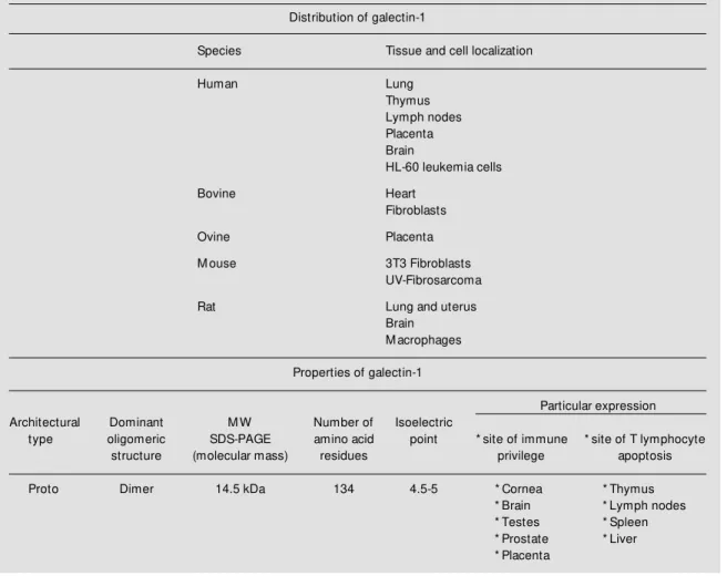
Table 1 - Distributions and properties of galectin-1.
Reference 67 summarizes all the members of the galectin family so far identified.
Distribution of galectin-1
Species Tissue and cell localization
Human Lung
Thymus Lymph nodes Placenta Brain
HL-60 leukemia cells
Bovine Heart
Fibroblasts
Ovine Placenta
M ouse 3T3 Fibroblasts
UV-Fibrosarcoma
Rat Lung and uterus
Brain M acrophages
Properties of galectin-1
Particular expression
Architectural Dominant M W Number of Isoelectric
type oligomeric SDS-PAGE amino acid point * site of immune * site of T lymphocyte
structure (molecular mass) residues privilege apoptosis
Proto Dimer 14.5 kDa 134 4.5-5 * Cornea * Thymus
* Brain * Lymph nodes
* Testes * Spleen
* Prostate * Liver
regulation of the immune response.
Life and de ath in the thymus
Apoptosis is a common event during T lymphocyte development for the production of immunocompetent T cells (29). Large numbers of precursor cells migrate into the thymus daily, where they are subjected to selection in a critical process of thymic edu-cation. The majority of these cells die as result of neglect, since they are neither posi-tively nor negaposi-tively selected (30). Those cells bearing T cell receptors (TCRs) that recognize self major histocompatibility com-plex proteins (MHC) are positively selected. Moreover, a subset of these cells recogniz-ing MHC with the high affinity is subjected to negative selection and consequently de-leted by apoptosis.
Death by neglect is definitely apoptotic (30) and occurs most probably via exposure to endogenous glucocorticoids (31). Thy-mocytes, in addition to the T cell line, are highly sensitive to apoptosis induced by glu-cocorticoids in an active process, requiring
de novo gene expression (32). In this respect, Ashwell and colleagues (33,34) performed
in vivo experiments demonstrating that a subset of thymic epithelial cells are ste-roidogenic and that inhibition of steroid syn-thesis modified the profile of lymphoid thy-mic populations. Furthermore, data obtained by the creation of transgenic lines of mice carrying an antisense glucocorticoid recep-tor (34) indicated that thymocytes that do not bind to MHC are deleted by endogenous steroids.
Elimination of autoreactive lymphocytes may occur via activation-induced cell death: the same signals that trigger activation of peripheral mature T cells induce apoptosis of thymocytes (35). The difference between positive selection of normally functional immature T cells and elimination of autore-active cells may lie in the affinity of their TCRs for self antigens and increasing
evi-dence suggests that co-stimulatory signals could play an important role in this process (35-37). Furthermore, positive selection might also result from antagonism between glucocorticoid and activation-induced death. This model clearly suggests that thymocytes unable to bind to the MHC at all receive no stimulation through the T cell receptor com-plex and are eliminated via glucocorticoid signals, whereas thymocytes able to trans-duce TCR signals of sufficient strength to overwhelm the glucocorticoid pathway are eliminated via activation-induced cell death. In connection with these findings, an inter-esting study reports the overexpression of the human galectin-1 gene during glucocor-ticoid-induced cell death (38).
It is well known that thymocyte matura-tion requires the participamatura-tion of thymic ep-ithelial cells and extracellular matrix com-ponents (39,40). In this context, Baum et al. (41) reported the expression of galectin-1 by human thymic epithelial (TE) cells and dem-onstrated that this endogenous lectin medi-ates the adhesion of thymocytes to TE cells. This observation suggested that specific oli-gosaccharide sequences on immature thy-mocytes might be candidate ligands for ga-lectin-1 synthesized by thymic stromal cells. Sensitivity of T cells to the lectin was found to be modulated by the expression of glyco-syltransferase enzymes that may modify the availability of oligosaccharide ligands for galectin-1. Elucidation of the functional sig-nificance of this interaction provided con-cluding evidence that galectin-1 induced ap-optosis of two distinct populations of non-selected and negatively non-selected CD4low
, CD8low
immature cortical thymocytes (16).
D e ath at the pe riphe ry
cell death. Activation-induced apoptosis of mature T cells occurs via Fas and Fas ligand (FasL) interactions. This has been clearly demonstrated in vitro using cell lines or T cell hybrids, which die in response to TCR binding (42-44) as well as in vivo (45,46) in spontaneous mutations in particular strains of MLR lpr/lpr or gld/gld mice, defective in Fas or FasL, respectively. In agreement, Nagata and colleagues (47) created Fas-/-mice by targeted deletion of the Fas gene. These mice displayed enhanced and acceler-ated lymphoproliferation in comparison to
lpr/lpr mice (48). Following administration of Staphylococcus enterotoxin B to these null-mutant mice, dramatically impaired de-letion occurred due to the absence of Fas antigen.
Fas is expressed on a wide variety of cell types including hematopoietic and epithelial cells. Expression of Fas on T and B lympho-cytes increases after antigen receptor-medi-ated activation (49). In contrast to the wide-spread distribution of Fas, its ligand exhibits a highly restricted pattern of expression. FasL expression is induced on mature CD4+ and CD8+ T lymphocytes following activation but is not expressed by other hematopoietic cells (50,51). FasL has also been reported to be constitutively expressed in two immuno-logically privileged tissues, such as the eye and the testis. Such expression could prevent damage inflicted by activated T cells to these tissues. Although most tissues can tolerate the nonspecific damage caused by inflam-matory responses, delicate tissues such as the eyes and the testis are susceptible to suffering irreparable damage after an in-flammatory episode. Hence, expression of high levels of FasL represents a defensive mechanism to prevent damage caused by inflammation through an induction of apop-tosis of activated cells expressing elevated levels of Fas antigen (52).
Despite striking similarities in their lo-calization, critical differences should be high-lighted between galectin-1 and FasL-induced
apoptosis. At first sight, FasL triggers apop-tosis by binding to Fas (CD95) through pro-tein-protein interaction, whereas galectin-1 binds to cell surface glycoconjugates on thy-mocytes (16) and mature T cells (18,19). Besides, galectin-1 and FasL apparently use different signal transduction pathways to engage the apoptotic program of the cell. Recently, Su et al. (53) and Perillo et al. (18) showed that the T lymphoblastoid cell line MOLT-4, which was insensitive to FasL-induced apoptosis, was susceptible to galec-tin-1. In contrast, a T lymphoblastoid cell line, which was sensitive to FasL, was resist-ant to the apoptotic effect of galectin-1. Moreover, since galectin-1 was capable of inducing apoptosis in the CD3-negative T lymphoblastoid cell lines, one should con-clude that galectin-1 does not trigger apopto-sis by cross-linking of the T cell receptor complex. These data strongly suggest that the mechanisms by which galectin-1 induced apoptosis are clearly distinct from those im-plicated in FasL- or T cell receptor-induced apoptosis.
Expression of galectin-1 in privileged immune sites, such as placenta, cornea and prostate (8,54), may contribute to the main-tenance of tolerance by inducing apoptosis of activated T cells responding to an injury, autoimmune damage or infection. Galectin-1 was found to be upregulated by metastatic rather than noninvasive tumors. In a way, tumors could also be considered to be privi-leged immune tissues and several mecha-nisms for tumor evasion of immune recogni-tion have been proposed, such as down-regulation of MHC class I expression. In this context, one may suspect that galectins in tumor cells can trigger apoptosis of activated T cells, thus allowing the tumor to escape immune attack.
Macrop hages: key im m unoregulatory cells at the p erip hery
immuno-regulatory cells implicated in critical func-tions at the periphery, such as initiation of the immune response and regulation of the activities of other immune cell populations. These highly adaptive cells are able to modify their behavior in response to different envi-ronmental signals. In this sense, the perito-neal cavity has been a useful system for studying phenotypic differences between Mø populations (55). Resident, inflammatory and activated Møs show different profiles of en-zymes and receptors which can be up- or down-regulated, and are closely related to the functional competence of these cells (55). Since galectin-1 demonstrated immuno-modulatory properties, we wondered whether this highly conserved structure could be pres-ent in Møs. If so, critical questions remain to be addressed: which is its precise biological function? Which is the cellular and molecu-lar target of this function?
In an attempt to address this question, we performed immunochemical and immuno-cytochemical studies using a polyclonal an-tibody raised against the galectin. Mø-en-riched populations were purified by plastic adherence from rat peritoneal cells and three different Mø subpopulations were defined. Resident Møs were obtained by washing the peritoneal cavity. Inflammatory Møs were recruited 3 days after intraperitoneal injec-tion with proteose-peptone and activated Møs were obtained by in vivo or in vitro treat-ments, such as administration of Bacillus Calmette-Guerin and activation by LPS or by chemical agents such as PMA (phorbol ester) or chemoattractants such as fMLP (56). Western blot analysis of total cell lysates obtained from the different stimulated Møs using the anti-galectin antibody revealed a single immunoreactive protein band corre-sponding to a molecular mass of 15 kDa in all purified Mø population. By densitomet-ric quantification we concluded that total expression of the ß-galactoside-binding pro-tein was increased about 5-fold in phorbol ester (PMA)- and chemotactic peptide
(fMLP)-activated Møs, and 2-fold in pep-tone-elicited inflammatory Møs. We per-formed flow cytometry experiments on the different Mø populations using fluorescein-labeled ED1 mAb for the detection of mon-ocyte macrophage lineage and phycoerythrin-conjugated galectin antibody. Dual param-eters counter plots showed a high proportion of double positive cells after exposure of cells to chemical agents such as PMA and fMLP (56). These results validate the con-cept that activation stimuli and different en-vironmental signals can modulate the ex-pression of this protein and suggest that this molecule could be involved in critical im-munological processes mediated by Møs.
critical residues found in other members of the mammalian galectin-1 subfamily. How-ever, we do not rule out the possibility that it may be an alternative isoform of galectin-1, as described for chicken isolectins (62).
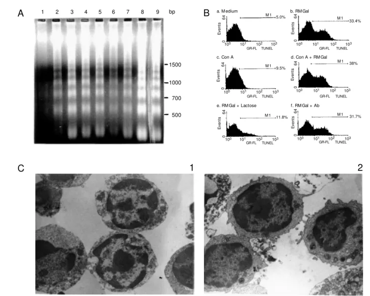
Since RMGal was overexpressed in acti-vated immunoregulatory cells, the next issue we attempted to investigate was related to the role of this galectin in T cell death. Hence, mitogenically stimulated and non-stimulated spleen mononuclear cells were cultured in the presence of optimal concen-trations of RMGal and then processed for DNA fragmentation, TUNEL assay and trans-mission electron microscopy. The electro-phoretic pattern of genomic DNA extracted after 6 h of cell culture is shown in Figure 2A. The typical DNA ladder of oligonucleo-some-sized fragments of ~180-200 bp was intensified in SpMs stimulated with Con A and exposed to RMGal (lane 3), and in SpMs incubated with RMGal alone at two different concentrations (lanes 4 and 5, respectively). In contrast, ladder type DNA fragmentation was almost absent in DNA extracted from SpMs cultured in medium alone (lane 1). Cells stimulated with Con A but not treated with RMGal showed the typical low inten-sity pattern of fragmentation characteristic of cell death following activation with mito-genic stimuli (lane 2). The same pattern was clearly observed when the T cell population was purified from total SpMs. Genomic DNA fragmentation was found to be particularly increased when RMGal was added to stimu-lated (lane 8), and nonstimustimu-lated (lane 9) T cells, in comparison to control of T cells in medium alone (lane 6) and T cells stimulated with Con A (lane 7). Thus, it should be emphasized that in our experimental condi-tions cell stimulation was not an essential step for RMGal-induced apoptosis.
In an attempt to quantify galectin-induced apoptosis, cells were cultured under the same conditions and processed for TUNEL detec-tion using flow cytometry. As clearly shown in Figure 2B, 33% of cells exposed to RMGal
were apoptotic, as demonstrated by specific incorporation of biotinylated dUTP into DNA breaks, whereas control samples cultured in medium alone showed 5% spontaneous ap-optosis. The proportion of TUNEL-positive cells increased to 38% when the cells were simultaneously incubated with Con A and the lectin in comparison to cells cultured in the presence of Con A but not exposed to RMGal. The carbohydrate recognition do-main of RMGal was involved in this func-tion since a ß-galactoside-related sugar, lac-tose, was able to induce a 33 to 11% de-crease in levels of TUNEL-positive cells. In contrast, these effects were not significantly blocked when cells were exposed to the lectin in the presence of the specific anti-body. Thus, induction of apoptosis by RMGal was highly specific and related to its carbo-hydrate-binding properties. Finally, ultra-structural studies of morphological changes induced by exposure to RMGal revealed the typical features of apoptosis (29) including reduction of the cytoplasmic volume, loss of surface microvilli, chromatin condensation and margination along the inner surface of the nuclear envelope (Figure 2C, panel 1 in comparison to panel 2). The results pre-sented here provide clear-cut evidence indi-cating that the purified Mø galectin is associ-ated with a positive control of the apoptotic threshold of T cells.
In the galectin family, galectin-1 pro-motes T cell apoptosis (16-19) and con-versely, galectin-3 has been recently reported to be involved in the inhibition of the apoptotic cell program through an interac-tion with the Bcl-2 proto-oncogene (63). The family of Bcl-2-related proteins constitutes one of the most relevant apoptotic regulatory gene products acting on the effector stages of apoptosis (64). Within the Bcl-2 family members, the ratio of death antagonists such as Bcl-2 and Bcl-XLto agonists such as Bax
9 8 7 6 5 4 3 2 1
1500
1000
700
500
A
C
1
2
Figure 2 - Rat macrophage galectin induces apoptosis of T cells. A, Electrophoretic analysis of internucleosomal DNA fragmentation induced by RM Gal. Spleen mononuclear cells w ere cultured in 24-w ell microtiter plates at a density of 2 x 10⁷
cells/w ell for 6 h in medium alone (lane 1), in medium containing Con A, 2.5 µg/ml (lane 2), and in medium containing Con A, 2.5 µg/ml, plus the addition of RM Gal at a concentration of 4 µg/ml (lane 3). Cells w ere also cultured w ith RM Gal (4 and 6 µg/ ml) in the absence of a mitogenic stimulus (lanes 4 and 5, respectively). The T cell-enriched population w as purified and cultured under identical conditions in medium alone (lane 6), in medium containing Con A (lane 7), in the presence of Con A plus RM Gal, 4 µg/ml (lane 8), and in the presence of RM Gal alone, 4 µg/ml (lane 9). Cells w ere then harvested and genomic DNA w as extracted. Samples w ere diluted in loading buffer and resolved on 1.8% agarose gel. The relative mobility of oligonucleosome-length DNA fragments reflects integer multiples of ~180-200 bp. M olecular standards (100 bp DNA ladder) are indicated on the right. B, Incorporation of biotinylated dUPT by exogenous TdT into DNA strand breaks generated after RM Gal treatment. T cells (2 x 10⁷ cells/w ell) w ere exposed to medium alone (a), 4 µg/ml RM Gal (b), 2.5 µg/ml Con A (c), 2.5
µg/ml Con A plus 4 µg/ml RM Gal (d), 4 µg/ml RM Gal in the presence of 100 mM lactose (e), or 4 µg/ml RM Gal in the presence of galectin Ab (f) for 6 h at 37oC in 5% CO
2. Samples w ere harvested, fixed, and permeabilized and the percentage of
apoptotic cells in each sample w as determined by flow cytometry analysis after TUNEL labeling. Cells treated w ith DNAse I w ere used as positive controls. C, Transmission electron microscopy examination of ultrastructural changes induced by RM Gal. T cells w ere purified and cultured in 24-w ell plates at a density of 2 x 10⁷ cells/w ell, in the presence (1), or in the
absence (2) of RM Gal (4 µg/ml). After 6 h, cells w ere harvested, w ashed and processed for transmission electron microscopy. Galectin-treated cells displayed the typical ultrastructural features compatible w ith apoptosis. M agnification: X12,000. (From Journal of Immunology, 160: 4831-4840, 1998. Copyright 1998, The American Society of Immunologists).
a. M edium b. RM Gal
6
4
E
v
e
n
ts
0
100 101 102 103
TUNEL GR-FL
5.0% M 1
6
4
E
v
e
n
ts
0
100 101 102 103
TUNEL GR-FL
6
4
E
v
e
n
ts
0
100 101 102 103
TUNEL GR-FL
6
4
E
v
e
n
ts
0
100 101 102 103
TUNEL GR-FL
6
4
E
v
e
n
ts
0
100 101 102 103
TUNEL GR-FL
6
4
E
v
e
n
ts
0
100 101 102 103
TUNEL GR-FL
33.4% M 1
38% M 1
31.7% M 1 11.8%
M 1 9.5% M 1
c. Con A d. Con A + RM Gal
e. RM Gal + Lactose f. RM Gal + Ab
galectins-1 and -3 could also represent an alternative pathway in the normal control of life, that is regulated by a delicate balance between cell proliferation, differentiation and death. In agreement with the results pre-sented for RMGal, we also provided evi-dence that the chicken galectin CLL-I, which has ~50% sequence similarities with galec-tin-1, inhibits growth of rat T cells via apop-tosis, which proved to be controlled in a time-, lectin concentration- and a saccha-ride-dependent manner (65).
Co ncluding re marks
On the basis of the results reviewed herein, one would expect knockout mice for galec-tin-1 to show autoimmune manifestations such as lupus-like disorders, as observed for spontaneous mutations in Fas and FasL in MLR lpr/lpr or gld/gld mice, respectively. However, no important phenotypic changes could be detected in null mutant-mice as regards the galectin-1 gene. By contrast, these mice were found to be completely vital and
proliferative (66). An exhaustive examina-tion of the immunological system is impera-tive in these genetically modified mice not only at the central level but also at the pe-riphery to search for potentially harmful autoaggressive clones and signs of disregu-lated apoptosis. Moreover, the possibility that other galectins identified thus far could compensate for the absence of this proto-type galectin-1 protein should also be con-sidered in future experimental work. Never-theless, the novel role of a carbohydrate-binding protein in the regulation of apopto-sis provided a breakthrough in galectin re-search and widened the interphase between immunology, glycobiology and clinical medi-cine.
Ackno wle dgm e nts
We thank Drs. Carlos Landa, Leonardo Castagna, Mercedes Iglesias, Nidia Modesti and Carlota-Wolfenstein Todel for their con-tribution.
Re fe re nce s
1. Barondes SH, Cooper DNW, Gitt M A & Leffler H (1994). Galectins: Structure and function of a large family of animal lectins. Journal of Biological Chem istry, 269: 20807-20810.
2. Barondes SH, Cast ronovo V, Cooper DNW, Cummings RD, Drickamer K, Feizi T, Gitt M A, Hirabayashi J, Hughes C, Kasai K, Leffler H, Liu F, Lotan R, M ercurio AM , M onsigni M , Pillai S, Poirer F, Raz A, Rigby PW J, Rini JM & W ang JL (1994). Galectins: a family of animal beta galacto-side-binding lectins. Cell, 76: 597-598. 3. M oonjae C & Cummings RD (1997).
Ga-lectin-1: Oligomeric structure and interac-tions w ith polylactosamine. Trends in Glycoscience and Glycotechnology, 45: 47-56.
4. Cooper DNW (1997). Galectin-1: Secre-tion and modulaSecre-tion of cell interacSecre-tions w ith laminin. Trends in Glycoscience and Glycotechnology, 45: 57-67.
5. Wang JL, Laing JG & Anderson RL (1991). Lectin in the cell nucleus. Glycobiology, 1:
243-252.
6. Hirabayashi J & Kasai K (1993). The family of metazoan metal-independent ß-galac-toside-binding lectins: structure, function and molecular evolution. Glycobiology, 3: 297-304.
7. Barondes SH (1986). Vertebrate lectins: properties and functions. In: Lienner I, Sharon N & Goldstein I (Editors), The Lec-tins: Properties, Functions and Applica-tions inBiology and M edicine. Academic Press, Orlando, FL, 447-465.
8. Kasai K & Hirabayashi J (1996). Galectins: a family of animal lectins that decipher glycocodes. Journal of Biochemistry, 119: 1-8.
9. Adams L, Kenneth Scott G & Weinberg CS (1996). Biphasic modulation of cell grow th by recombinant human galectin-1. Biochimica et Biophysica Acta,1312: 137-144.
10. Hughes RC (1992). Lectins as cell adhe-sion molecules. Current Opinion in Struc-tural Biology,2: 687-692.
11. Raz A & Lotan R (1987). Endogenous ga-lactoside-binding lectins: a new class of functional tumor cell surface molecules related to metastasis. Cancer and M etas-tasis Review s,6: 433-452.
12. Levi G, Tarrab-Hazdai R & Teichberg VI (1983). Prevent ion and t herapy w it h electrolectin of experimental autoimmune myasthenia gravis in rabbits. European Journal of Immunology, 13: 500-507. 13. Of f ner H, Celnik B, Bringm an TS,
Casent ini-Borocz D, Nedw in GE & Vandenbark A (1990). Recombinant hu-m an-galact oside-binding lect in sup-presses clinical and histological signs of experimental autoimmune encephalomy-elitis. Journal of Neuroimmunology, 28: 177-184.
Copeland NG, Jenkins A & Nagata S (1992). Lymphoproliferative disorder in mice explained by defects in Fas antigen that mediates apoptosis. Nature, 356: 314-317.
16. Perillo NL, Oittenbogaart CH, Nguyen JT & Baum LG (1997). Galectin-1, an endog-enous lectin produced by thymic epitheli-al cells, induces apoptosis of human thy-mocytes. Journal of Experimental M edi-cine, 97: 1851-1859.
17. Iglesias M M , Rabinovich GA, Ivanovic V, Sotomayor CE & Wolfenstein-Todel C (1998). Galectin-1 from ovine placenta: com plet e prim ary st ruct ure, physico-chemical properties and implications in the T cell death. EuropeanJournal of Bio-chemistry, 252: 400-407.
18. Perillo NL, Pace KE, Seilhamer JJ & Baum LG (1995). Apoptosis of T cells mediated by galectin-1. Nature,378: 736-739. 19. Rabinovich GA, Iglesias M M , M odesti
NM , Castagna L, Wolfenstein-Todel C, Riera CM & Sotomayor CE (1998). Rat activated macrophages produce a galec-tin-1-like protein w hich induces apoptosis of T cells: Biochemical and functional char-acterization. Journal of Immunology,160: 4831-4840.
20. Evan GI, Brow n L, Whyte M & Harrington E (1995). Apoptosis and the cell cycle. Current Opinion in Cell Biology, 7: 825-834.
21. Grassilli E, Carcereri de Prati A, M onti D, Troiano L, M enegazzi M , Barbieri D, Franceschi C & Suzuki H (1992). Studies of the relationship betw een cell prolifera-tion and cell death. II. Early gene expres-sion during concanavalin A-induced prolif-eration or dexamethasone-induced apop-tosis of rat thymocytes. Biochemical and Biophysical Research Communications, 188: 1261-1266.
22. Hartw ell LH & Kastan M B (1994). Cell cycle control and cancer. Science,266: 1821-1828.
23. Schw artzman RA & Cidlow ski JA (1993). Apoptosis: the biochemistry and molecu-lar biology of programmed cell death. En-docrine Review s, 14: 133-151.
24. Steller H (1995). M echanisms and genes of cellular suicide. Science, 267: 1445-1449.
25. Thompson CB (1995). Apoptosis in the pathogenesis and treatment of disease. Science, 267: 1456-1462.
26. Vaux DL, Haecker G & Strasser A (1994). An evolutionary perspective on apopto-sis. Cell, 76: 777-779.
27. Raff M C (1992). Social controls on cell sur-vival and cell death. Nature, 356: 397-400.
28. Squier M KT, Sehnert AJ & Cohen JJ (1995). Apoptosis in leukocytes. Journal of Leukocyte Biology,57: 2-10.
29. Cohen JJ, Duke RC, Fadok VA & Sellins KS (1992). Apoptosis and programmed cell death in immunity. Annual Review of Immunology, 10: 267-293.
30. Surh CD & Sprent J (1994). T-cell apopto-sis detected in situ during positive and negative selection in the thymus. Nature, 372: 100-103.
31. Sprent J, Lo D, Gao EK & Ron Y (1988). T cell selection in the thymus. Immunologi-cal Review s, 101: 172-190.
32. Wyllie AH (1980). Glucocorticoid induced thymocyte apoptosis is associated w ith endogenous endonuclease activation. Na-ture, 284: 555-556.
33. Vacchio M S, Papadopulous V & Ashw ell JD (1994). Steroid production in the thy-mus: implications for thymocyte selec-tion. Journal of Experimental M edicine, 179: 1835-1846.
34. King LB, Vacchio M S, Hunziker R, M argulies DH & Ashw ell JD (1995). A targeted of glucocorticoid receptor anti-sense transgene increases thymocyte ap-optosis and alters thymocyte develop-ment. Immunity, 5: 647-656.
35. Takayama H & Sitkovsky M V (1989). Po-tential use of an antagonist of cAM P-de-pendent protein kinase to block inhibition and modulate T-cell receptor-triggered ac-tivation of cytotoxic T-lymphocytes. Jour-nal of Pharmaceutical Sciences, 78: 8-10. 36. Iw ata M , M ukai M , Nakai Y & Iseki R (1992). Retinoic acids inhibit activation-induced apoptosis in T cell hybridomas and thymocytes. Journal of Immunology, 149: 3302-3308.
37. M igliorati G, Nicoletti I, Pagliacci M C & Riccardi C (1992). Glucocorticoid-induced thymocyte apoptosis: inhibition by inter-leukin-2 and interleukin-4. Pharmacologi-cal Research,25: 15-16.
38. Goldstone SD & Lavin M F (1991). Isola-tion of a cDNA clone, encoding a human-galact oside-binding prot ein overex-pressed during glucocorticoid-induced cell death. Biochemical and Biophysical Re-search Communications, 178: 746-750. 39. Anderson G, Ow en JJT, M oore NC &
Jenkinson EJ (1994). Thymic epithelial cells provide unique signals for positive selection of CD4+CD8+ thymocytes in vitro. Journal of Experimental M edicine, 179: 2027-2031.
40. Anderson G, M oore NC, Ow en JJT & Jenkinson EJ (1996). Cellular interactions in thymocyte development. Annual Re-view of Immunology, 14: 73-99.
41. Baum LG, Pang M , Perillo NL, Wu T, Delegeane A, Uittenbogaart CH, Fukuda M & Seilhamer JJ (1995). Human thymic epithelial cells express an endogenous lectin, galectin-1, w hich binds to core 2 O-glycans on thymocytes and T lymphoblas-toid cells. Journal of Experimental M edi-cine, 181: 877-887.
42. Dhein J, Walczak H, Baumler C, Debatin KM & Kramer PH (1995). Autocrine T-cell suicide mediated by APO-1 (Fas/CD95). Nature, 373: 438-441.
43. Bruner T, M ogil RJ, LaFace D, Yoo NJ, M ahoubl A, Echeverri F, M artin SJ, Force WR, Lynch DH, Ware CF & Green DR (1995). Cell-autonomous Fas (CD95)/Fas-ligand interaction mediates activation-in-duced apoptosis in T-cell hybridomas. Na-ture, 373: 441-444.
44. Ju S-T, Panka DJ, Cul H, Ettinger R, El-Khatib M , Sherr DH, Stanger BZ & M arsak-Rothstein A (1995). Fas (CD95)/FasL in-teractions required for programmed cell death after T-cell activation. Nature,373: 444-448.
45. Russell JH, Rush B, Weaver C & Wang R (1993). M ature T cells of autoimmune lpr/ lpr mice have a defect in antigen-stimulat-ed suicide. Proceedings of the National Academy of Sciences, USA, 90: 4409-4413.
46. Russell JH & Wang R (1993). Autoim-mune gld mutation uncouples suicide and cytokine/proliferation pathw ay in activated mature T cells. European Journal of Im-munology, 23: 2379-2382.
47. Adachi M , Suem at su S, Kondo T, Ogasaw ara J, Tanaka T, Yoshida N & Nagata S (1995). Targeted mutation in the Fas gene causes hyperplasia in peripheral lymphoid organs and liver. Nature Genet-ics, 11: 294-300.
48. Adachi M , Suematsu S, Suda T, Watanabe D, Fukuyama H, Ogasaw ara J, Tanaka T, Yoshida N & Nagata S (1996). Enhanced and accelerated lymphoproliferation in Fas-null-mice. Proceedings of the National Academy of Sciences, USA, 93: 2131-2136.
49. Nishim ura Y, Ishií A, Kobayashi Y, Yamasaki Y & Yonehara S (1995). Expres-sion and function of mouse Fas antigen on immature and mature T cells. Journal of Immunology,154: 4395-4403. 50. Tanaka M , Suda T, Takanahashi T &
Nagata S (1995). Expression of the func-tional soluble forms of the human Fas ligand in activated lymphocytes. EM BO Journal, 14: 1129-1135.
Expression of the Fas ligand in cells of the T cell lineage. Journal of Immunology, 154: 3806-3813.
52. Osborne BA (1996). Apoptosis and the maintenance of homeostasis in the im-mune system. Current Opinion in Immu-nology,8: 245-254.
53. Su ZZ, Lin J, Shen R, Fisher PE, Goldstein NI & Fisher PB (1996). Surface-epitope masking and expression cloning identifies the human prostate carcinoma tumor an-tigen gene PCTA-1, a member of the ga-lectin gene family. Proceedings of the Na-tional Academy of Sciences, USA, 93: 7252-7257.
54. Allen HJ, Sucato D, Gottstine S, Kisailus E, Nava H, Petrelli N, Castillo N & Wilson D (1991). Localisation of endogenous beta-galactoside-binding lectin in human cells and tissues. Tumor Biology, 12: 52-60.
55. Adams D & Hamilton TA (1984). The cell biology of macrophage activation. Annual Review of Immunology,2: 283-318. 56. Rabinovich GA, Castagna LF, Landa CA,
Riera CM & Sotomayor CE (1996). Regu-lated expression of a 16-kd galectin-like protein in activated rat macrophages. Journal of Leukocyte Biology, 59:
363-370.
57. Clerch LB, Whitney P, Hass M , Brew K, M iller T, Werner R & M assaro D (1988). Sequence of a full-length cDNA for rat lung ß-galactoside-binding protein: Pri-mary and secondary structure of the lec-tin. Biochemistry, 27: 692-699.
58. W ilson TJG, Firt h M N, Pow ell JT & Harrison FL (1989). The sequence of the mouse 14 kDa ß-galactoside-binding lec-tin and evidence for its synthesis on free cytoplasmic ribosomes. Biochemical Jour-nal, 261: 847-852.
59. Abbott WM , M ellor A, Edw ards Y & Feizi T (1989). Soluble bovine galactoside-bind-ing lectin cDNA reveals the complete amino acid sequence and an antigenic re-lationship w ith the major encephalitoge-nic domain of myelin basic protein. Bio-chemical Journal, 259: 283-290. 60. Hirabayashi J & Kasai K (1988). Complete
amino acid sequence of a ß-galactoside-binding lectin from human placenta. Jour-nal of Biochemistry, 104: 1-4.
61. Bladier D, Le Caer JP, Joubert R, Caron M & Rossier J (1991). ß-galactoside soluble lectin from human brain: complete amino acid sequence. Neurochemistry Interna-tional, 18: 275-283.
62. Beyer EC, Zw eig SE & Barondes SH (1980). Tw o lactose-binding lectins from chicken tissues. Purified lectin from intes-tine is different from those in liver and muscle. Journal of Biological Chemistry, 255: 4236-4239.
63. Yang RY, Hsu DK & Liu FT (1996). Expres-sion of galectin-3 modulates T cell grow th and apoptosis. Proceedings of the Na-tional Academy of Sciences, USA, 93: 6737-6742.
64. Kroemer G (1997). The proto-oncogene Bcl-2 and its role in regulating apoptosis. Nature M edicine, 6: 614-620.
65. Rabinovich GA, M odesti N, Castagna LF, Landa C, Riera CM & Sotomayor CE (1997). Specific inhibition of lymphocyte proliferation and induction of apoptosis by CLL-I, a ß-galactoside-binding lectin. Jour-nal of Biochemistry, 122: 365-373. 66. Poirrier F & Robertson EJ (1993). Normal
development of mice carrying a null mu-tation in the gene encoding the L14 S-type lectin. Development, 119: 1229-1236.