Topical HPMC/S-Nitrosoglutathione Solution
Decreases Inflammation and Bone
Resorption in Experimental Periodontal
Disease in Rats
Conceição S. Martins1, Renata F. C. Leitão1, Deiziane V. S. Costa1, Iracema M. Melo2,
Glaylton S. Santos1, Vilma Lima3, Victor Baldim4, Deysi V. T. Wong3, Luana E. Bonfim1, Cíntia B. Melo1, Marcelo G. de Oliveira4
*, Gerly A. C. Brito1
*
1Postgraduate Program in Morphofunctional Sciences, Department of Morphology, School of Medicine, Federal University of Ceará, Fortaleza, Ceará, Brazil,2Department of Clinical Dentistry, Faculty of Pharmacy, Dentistry and Nursing, School of Dentistry. Federal University of Ceará, Fortaleza, Ceará, Brazil,
3Department of Physiology and Pharmacology, School of Medicine, Federal University of Ceará, Fortaleza, Ceará, Brazil,4Institute of Chemistry, University of Campinas, UNICAMP, Campinas, São Paulo, Brazil
*[email protected](MGO);[email protected](GACB)
Abstract
S-nitrosoglutathione (GSNO) is a nitric oxide (NO) donor, which exerts antioxidant, anti-inflammatory, and microbicidal actions. Intragingival application of GSNO was already shown to decrease alveolar bone loss, inflammation and oxidative stress in an experimental periodontal disease (EPD) model. In the present study, we evaluated the potential thera-peutic effect of topical applications of hydroxypropylmethylcellulose (HPMC)/GSNO solu-tions on EPD in Wistar rats. EPD was induced by placing a sterilized nylon (3.0) thread ligature around the cervix of the second left upper molar of the animals, which received topi-cal applications of a HPMC solutions containing GSNO 2 or 10 mM or vehicle (HPMC solu-tion), 1 h prior to the placement of the ligature and then twice daily until sacrifice on day 11. Treatment with HPMC/GSNO 10 mM solution significantly reduced alveolar bone loss, oxi-dative stress and TNF-αe IL-1βlevels in the surrounding gingival tissue, and led to a decreased transcription of RANK and TNF-αgenes and elevated bone alkaline phospha-tase, compared to the HPMC group. In conclusion, topical application of HPMC/GSNO solu-tion is a potential treatment to reduce inflammasolu-tion and bone loss in periodontal disease.
Introduction
Periodontal diseases, including gingivitis, are highly prevalent [1] and characterized by the inflammation of the periodontal connective tissue with influx of inflammatory cells, especially polymorphonuclear leukocytes, monocytes and macrophages from the peripheral blood into the periodontal connective tissue. While gingivitis does not affect the underlying supporting structures of the teeth, periodontitis results in loss of connective tissue and bone support [2,3]
a11111
OPEN ACCESS
Citation:Martins CS, Leitão RFC, Costa DVS, Melo IM, Santos GS, Lima V, et al. (2016) Topical HPMC/ S-Nitrosoglutathione Solution Decreases Inflammation and Bone Resorption in Experimental Periodontal Disease in Rats. PLoS ONE 11(4): e0153716. doi:10.1371/journal.pone.0153716
Editor:Xing-Ming Shi, Georgia Regents University, UNITED STATES
Received:October 30, 2015
Accepted:April 1, 2016
Published:April 26, 2016
Copyright:© 2016 Martins et al. This is an open access article distributed under the terms of the Creative Commons Attribution License, which permits unrestricted use, distribution, and reproduction in any medium, provided the original author and source are credited.
Data Availability Statement:All relevant data are within the paper.
Funding:Leitao RFC and de Oliveira MG would like to acknowledge grants from The National Council for Scientific and Technological Development - CNPq (http://www.cnpq.br/) (projects #478380/2011-9 and 309390/2011-7, respectively). The funders had no role in study design, data collection and analysis, decision to publish, or preparation of the manuscript.
and can progress to bone destruction, tooth mobility and finally, tooth loss. In turn, the bacte-rial pathogens in the dental plaque stimulate host cells to release various pro-inflammatory cytokines such as IL-1 and TNF-α, which attract polymorphonucleocytes (PMNs) to the site of infection. In addition, the activation of neutrophils by periodontopathogenic microorganisms with the consequent production of reactive oxygen species (ROS) may induce periodontal tis-sue breakdown [4].
Endogenously produced nitric oxide (NO) is a mediator of several physiological and patho-physiological processes whose actions include the inactivation of ROS and the blocking of radi-cal propagation reactions [5,6]. It has also been demonstrated that NO has a variety of effects on bone, suppressing osteoclast bone resorption and promoting the growth of osteoblasts [7–13].
It has been demonstrated that intragingival administration of S-nitrosoglutathione (GSNO), an endogenously found NO carrier and donor decreases bone resorptionin vivoin a animal model of periodontitis [14], similarly to what has been shownin vitrowith L-arginine supple-mentation [15]. GSNO has already displayed microbicidal actions [16–18] as well as wound healing action [19] in several other studies. These results encouraged us to investigate the potential beneficial actions that could be obtained from the topical application of GSNO for treating periodontitis, avoiding thus an invasive approach. For this purpose, we incorporated GSNO in hydroxypropylmethylcellulose (HPMC) solution. HPMC is widely used as a hydro-philic pharmaceutical vehicle for drug delivery systems and due to its mucoadhesive property, comprises an adequate matrix for the topical NO delivery from GSNO in the oral cavity.
Materials and Methods
Reagents and drugs
Hydroxypropylmethylcellulose (HPMC; Mn 90000), glutathione (GSH), sodium nitrite (NaNO2), phosphate-buffered saline (PBS) pH 7.4, acetone and hydrochloric acid (HCl) were
purchased from Sigma-Aldrich (St. Louis, MO, USA). All reagents were analytical grade and used as received. All experiments were performed using deionized water from a Millipore Milli-Q gradient filtration system (Billerica, MA, USA).
S-nitrosoglutathione synthesis
S-nitrosoglutathione (GSNO) was synthesized by reacting GSH with NaNO2in equimolar
amounts in acidic solution (HCl, pH 2.0) as described elsewhere [19]. Solid, dry GSNO was stored protected from light at -20°C for further use.
Preparation of HPMC/GSNO solutions
Animals
Male Wistar rats from the Federal University of Ceará, weighting 200 to 250 g were housed in temperature-controlled rooms under 12 hour light-dark cycles and received water and foodad libitum. Surgical procedures and animal treatments were conducted in accordance with the guidelines of the Animal Research Ethics Committee (Comissão de Ética em Pesquisa Animal—
CEPA/DFF/FAMED/UFC—Permit number: 10/2013), of the Federal University of Ceará, Bra-zil, who approved all the procedures involving the animals. All efforts were made to minimize suffering.
Experimental Periodontal Disease Protocol
Experimental Periodontal Disease (EPD) was induced as previously described [20–21].Briefly, the animals were anesthetized with ketamine (90 mg/kg intraperitoneally) and xylazine (10 mg/kg intraperitoneally) and a sterilized nylon (0.3) thread ligature was placed around the cer-vix of the second left maxillary molar. The ligature was then knotted on the vestibular side of the tooth, so it remained subgingival on the palatal side and supragingival on the buccal side. The animals were euthanized with an overdose of ketamine and xylazine (300/30mg/kg; i.p.) on day 11th.
Topical application of HPMC/GSNO solutions
The animals were divided into groups of 6 animals each and received topical applications of HPMC/GSNO 2.0 mM or HPMC/GSNO 10.0 mM. The solutions were applied using small cot-ton swabs with a gentle massage to both palatal and vestibular sides of the left maxillary molars gingiva, starting 1 hour before the placement of the ligature and twice daily until euthanasia on the 11thday. Control groups consisted of animals subjected to the surgical procedure that received only the vehicle (HPMC) and a group of animals not subjected to experimental peri-odontitis (normal).
Alveolar Bone Loss
The animals were euthanized 11 days after the placement of the ligature and the maxillae were excided and fixed in 10% buffer formalin. After 24 hour, the left maxillae were defleshed and stained with methylene blue (1%) to differentiate bone from teeth, fixed in a piece of wax and photographed. The digital images were subjected to alveolar bone loss analysis using the Image J software, as described previously [22]. In this procedure, the area (mm2) corresponding to the
exposed roots of the molars (no bone covering them at all) was calculated and subtracted from the corresponding area (mm2) of the normal group. All acquired images were compared to a
well-known area (1.0 x 1.0 mm2).
Histopathological Analysis
For the histopathological analysis, the maxillae were excised, fixed in 10% neutral-buffered for-malin and demineralized in EDTA. The specimens were then dehydrated, embedded in paraffin, and sectioned along the molars in a mesiodistal plane for hematoxylin and eosin staining. Three sections of 4μm each, corresponding to the area between the first and second molars were
(inflammatory cellular infiltration present in gingival and periodontal ligament), moderate deg-radation of the alveolar process and partial destruction of cementum; 3- Accentuated cellular infiltrate, complete resorption of the alveolar process and severe destruction of cementum.
Plasma Bone Alkaline Phosphatase (BALP)
Blood samples were collected from retro-orbital plexus, under anesthesia with a mixture of ketamine and xylazine (90/10 mg/kg; i.p.), before ligature (on day 0) and immediately before the euthanasia on the 11thday in order to obtain plasma concentration of alkaline phosphatase using the thermo-activation method, as previously described [24]. The samples were heated up to 56°C for 10 minutes. Serum levels of BALP (a thermosensitive isoform of the total alkaline phosphatase, TALP), were calculated by subtracting the concentration of the heated alkaline phosphatase in serum from the concentration of TALP in serum. The analysis was performed according to the manufacturer’s instructions (Labest1
, Lagoa Santa, MG, Brazil). The results were expressed as Bone Alkaline Phosphatase variation on day 11 (U/L) compared to the values obtained before the EPD induction, on day 0.
Malonaldehyde and Reduced Glutathione (GSH) Gingival Levels
To evaluate the effect of HPMC/GSNO solutions on the oxidative stress, reduced glutathione (GSH), an antioxidant and malonaldehyde, a marker for oxidative stress, were measured in the gingival tissue collected on day 11. Lipid peroxidation was measured as malonaldehyde (MDA) production through the thiobarbituric acid reaction [25] in the gingival tissue of rats. Briefly, 250μl of 10% homogenate of gingival tissue were mixed with 1.5 ml of 1% H3PO4and 0.5 ml
of 0.6% thiobarbituric acid aqueous solution and the mixture was stirred and heated in boiling water for 45 minutes. After cooling, 2 ml of n-butanol were added and the mixture was homog-enized. The butanol layer was separated, and the difference between the optical densities at 535 and 520 nm was used for calculating the MDA concentrations, which were expressed as nano-mol of MDA per gram of gingival tissue.
To evaluate the reduced glutathione (GSH) levels, the gingivae were first homogenized (400μl of a 10 wt% tissue solution prepared in 0.02 M EDTA). After that, 320μl of distilled
water and 80μL of 50% trichloroacetic acid (TCA) were added, followed by centrifugation at
3000 rpm for 15 min at 4°C. The supernatant was collected (400 mL) and added to 800μL of
0.4 M Tris buffer at pH 8.9 and 20μL of 0.01 M DTNB. The absorbance of each sample was
measured at 412 nm, and the results were described asμg of GSH per milligram of tissue.
Nitrite/nitrate (NO
x) Gingival Levels
On day 11 after EPD induction, the gingivae surrounding the upper left molars were removed for nitrite/nitrate (NOX) quantification using the Griess method [26]. Firstly, nitrate was
reduced to nitrite by incubation with nitrate reductase and nicotinamide adenine dinucleotide. Then, the Griess reagent (1% sulfanilamide and 0.1% naphthyl ethylenediamine dihydrochlor-ide in 5% phosphoric acid) was added, and the total concentration of nitrite was evaluated through its absorbance at 540 nm, assigned to the Griess reaction adduct. A calibration curve was obtained by incubating sodium nitrite (10 to 200μM) with the Griess reagent. Nitrate/
nitrite levels were expressed as micromole per milligram of gingival tissue.
Quantification of Cytokines Gingival Levels
R&D Systems, Minneapolis, MN, USA) measurements. The quantification of the cytokines con-tained in the samples was performed using an enzyme-linked immunosorbent assay (ELISA), as previously described [27,28]. TNF-αand IL-βlevels in the gingival tissue were determined using respective standard curves. The results are expressed as pg/ml of TNF-αor IL-β.
Immunohistochemical staining for inducible nitric oxide synthase (iNOS),
receptor activator of the nuclear factor kappa-B (RANK) and
Tartrate-resistant acid phosphatase (TRAP) and nitrotyrosine
After the animals were sacrificed on the 11thday, the maxillae were excised, fixed in 10%
neu-tral-buffered formalin and demineralized in 10% EDTA. The immunohistochemical for iNOS, RANK and TRAP were performed using the avidin–biotin–peroxidase method [27,28] in for-malin-fixed, paraffin-embedded tissue sections (4μm thick) and mounted on
poly-L-lysine-coated microscope slides. The sections were deparaffinized and rehydrated through xylene and graded alcohols. After antigen retrieval, endogenous peroxidase was blocked (30 minutes) with 3% (v/v) hydrogen peroxide and washed in phosphate-buffered saline (PBS). Sections were incubated overnight (4°C) with primary polyclonal rabbit anti-iNOS antibody (1:200), primary polyclonal rabbit anti-RANK (1:200) and primary polyclonal rabbit anti-TRAP (1:200) or pri-mary mouse monoclonal anti-nitrotyrosine (1:200). All the antibodies were diluted in PBS plus bovine serum albumin (BSA). The slides were then incubated with biotinylated goat anti-rabbit diluted 1:200 in PBS–BSA. After washing, the slides were incubated with avidin–biotin– horse-radish peroxidase conjugate for 30 min, according to the protocol of the manufacturer. iNOS, RANK and TRAP and nitrotyrosine were visualized with the chromogen 3,3 diaminobenzidine (DAB), after 2 minutes of incubation. Negative control sections were processed simultaneously as described above but with the first antibody being replaced by 5% PBS–BSA. Slides were counterstained with hematoxylin, dehydrated in a graded alcohol series, cleared in xylene, and coverslipped. The quantitative estimation of DAB products from immunostaining was deter-mined from digital images of at least ten different areas of each section (from four specimens per group), on 400x magnification, using Photoshop software. The immunostaining was calcu-lated dividing the DAB positive staining (immunostaining positive pixels) by the number of pixels per image, as previously described [29].
Quantitative PCR analysis of TNF-
α
, RANK and RANKL/OPG mRNA
levels
Expression of mRNA for TNF-α, RANK, RANK-L and OPG was determined by quantitative real-time polymerase chain reaction (qPCR). RNA was extracted from gingival tissue using the AurumTMTotal RNA Fatty and Fibrous Tissue Kit(Bio-Rad, CA, USA) following the manufac-turer’s instructions. The quality of the RNA was analyzed by 260/280 ratio, and quantified by UV absorption using NanoDrop (Thermo scientific, Wilmington, DE, USA). One microgram of total RNA from the intestinal samples in a final volume of 20μl were reverse-transcribed
into cDNA in the C1000 Touch™Termal Cycler system with the iScript™cDNA synthesis kit from Bio-Rad. Real-time quantitative PCR analysis of the mRNA was performed in an CFX96 Touch™real-time PCR detection system instrument from Bio-Rad using the iQTM SYBR1
curve analysis). The relative gene expression was determined using the 2-ΔΔCtmethod [30] with GAPDH as the housekeeping gene. The primer pairs used in this study are shown inTable 1.
Statistical Analysis
Data are presented as means ± standard error (SEM) or as medians when appropriate. Univari-ate Analysis of Variance followed by Bonferroni test was used to compare means and Kruskal–
Wallis and Dunn tests were used to compare medians. P<0.05 was considered significant. All analyses were performed using GraphPad Prism 6 software, San Diego, CA, USA.
Results
Alveolar Bone Loss
Local administration of HPMC/GSNO 10 mM solution 1 hour before the placement of the liga-ture and twice daily for 11 days resulted in a significant inhibition of alveolar bone loss (Figs1
and2E), compared to the hemimaxillae of animal groups that received the vehicle only (HPMC; Figs1and2C). No significant bone loss was observed in the normal groups (Figs1
and2A).
Table 1. Description of biomarkers, gene, primer sequences and NCBI accession numbers.
Biomarkers Gene Primer sequence (5’-3’) NCBI
Proinflammatory cytokines TNF-α S-GACCCTCACACTCAGATCATCTTCTA-TGCTACGACGTGGGCTACG NM-012675
Bone markers RANK S-AGGGAAAACGCTGACAGCTAAA-CCAACACAATGGTCCCCTGA NM_001271235
RANK-L S-GCCAACCGAGACTACGGCAAA-GAACATGAAGCGGGAGGCG NM_057149
OPG S-CCTAGAGAGAAATGTCGTAGGATTA-CATTCCACACTGGAAACCTGA NM_012870
Reference gene GAPDH S-TGATTCTACCCACGGAAGTTA-TGATGGGTTTCCCATTGATGA NM_017008
doi:10.1371/journal.pone.0153716.t001
Fig 1. Effect of topical application of HPMC/GSNO solutions on alveolar bone loss in experimental periodontitis.Data represent the mean±SEM of six animals in each group. #P<0.05 was considered significantly different compared to the normal control group (NORMAL);*P<0.05 was considered significantly different compared to rats subjected to experimental periodontitis, which received topical application of the vehicle (HPMC). (ANOVA; Bonferroni’s test).
Fig 2. Effect of topical application of HPMC/GSNO solutions on the macroscopic (first column) and histological (second column) aspects of periodontium (A and B)normal maxilla, showing integrity of its components (c. Cementum, pl. Periodontal ligament, ab. Alveolar bone and g. Gingiva). (C and D)Maxilla subjected to experimental periodontitis that received just the vehicle (HPMC), showing severe bone resorption, inflammatory infiltrate in both the gingiva and periodontal ligament, extensive cementum destruction and total resorption of the alveolar process, (E and F)Maxilla after 11 days of experimental periodontitis treated with 2.0 mM HPMC/GSNO showing discrete cell influx and preservation of the alveolar process and cementum. Picrosirius staining (magnification x40).
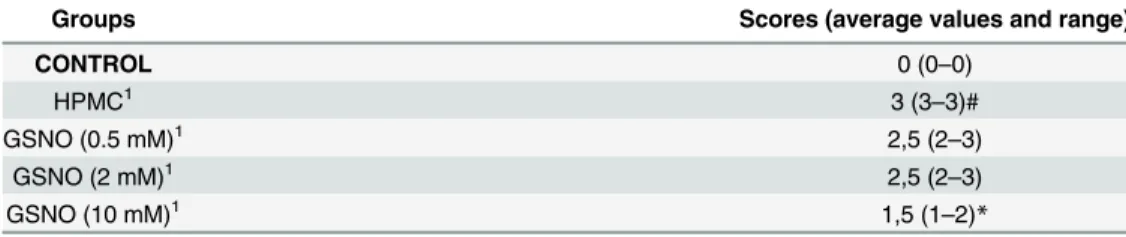
Histopathological analysis of the region between the first and second molars of the animal periodontium that received local applications of HPMC demonstrated accentuated inflammatory leukocyte infiltration, resorption of the alveolar bone and cementum and collagen fiber derange-ment within the periodontal ligaderange-ment, receiving a median score and range of 3(2–3) (Table 2;Fig 2D). The periodontium of rats treated with the HPMC/GSNO 10 mM solution showed preserva-tion of the alveolar process and cementum, reducpreserva-tion of the inflammatory cell infiltrapreserva-tion and partial preservation of the collagen fibers of the periodontal ligament, receiving a median score and range of 1(0–2) (Table 2;Fig 2F). The histological analysis of the region between the first and second molars of the control group that was not subjected to EPD (Normal), shows the structures of the normal periodontium, where fingiva (G), periodontal ligament (PDL), alveolar bone (AB), cementum (C) and dentin (D) can be observed [(0(0–0)] (Table 2;Fig 2B).
Plasma Bone Alkaline Phosphatase (BALP)
The treatment with GSNO/HPMC (2 and 10 mM) resulted in a significant increase in the BALP serum levels variation when compared with the control HPMC solution group (Fig 3).
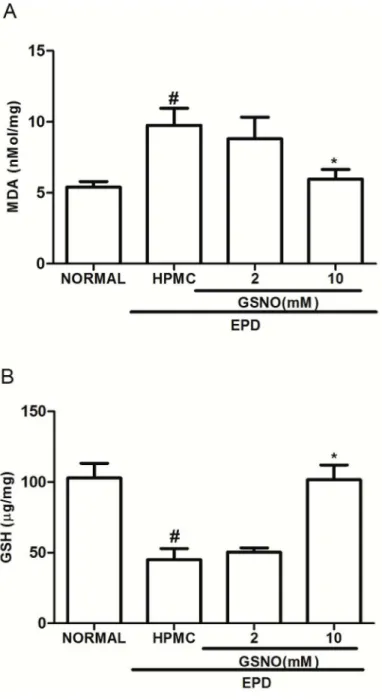
Malonaldehyde (MDA) and Glutathione (GSH) Gingival levels
At the 11thday the group subjected to EPD that received topical applications of the control HPMC solution showed a significant increase in the MDA level and a reduction in the GSH con-centration in the gingival tissue, when compared to the normal group. At the same time, treat-ment with GSNO/HPMC at 10 mM (but not 2.0 mM) significantly reduced the MDA gingival tissue levels (Fig 4A) and increased GSH levels (Fig 4B) when compared with HPMC group.
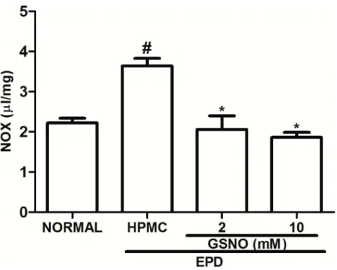
Nitrite/Nitrate (NOx) Gingival Levels
The group subjected to EPD that received topical applications of the control HPMC solution showed a significant increase in NOx levels when compared to the Normal group, on day 11. Treatment with the HPMC/GSNO solutions (2 and 10 mM) significantly reduced NOx con-centration when compared with the control HPMC solution group (Fig 5).
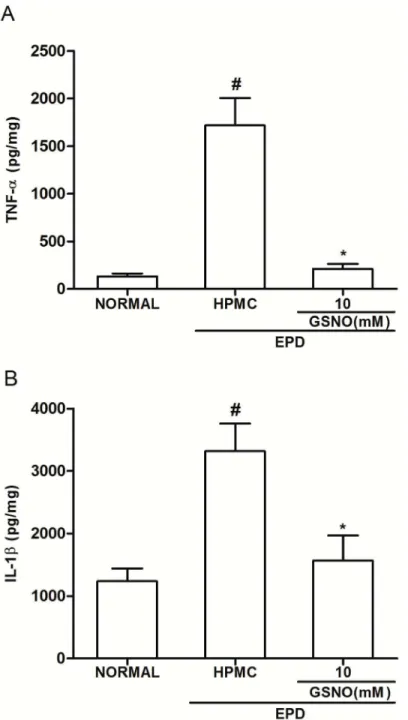
Gingival Tissue cytokine levels
It was observed a significant increase in the TNF-α(Fig 6A) and IL-1β(Fig 6B) levels in the gingival tissue of animals subjected to EPD that received topical applications of control HPMC solution, when compared with the Normal control group. Topical treatment with the HPMC/
Table 2. Effect of topical application of GSNO/HPMC formulation on histopathologic score of rat maxillae.
Groups Scores (average values and range)
CONTROL 0 (0–0)
HPMC1 3 (3–3)#
GSNO (0.5 mM)1 2,5 (2
–3)
GSNO (2 mM)1 2,5 (2
–3)
GSNO (10 mM)1 1,5 (1
–2)*
EPD: Experimental periodontal disease. 1Animals submitted to EDP;
#
p<0.05 versus the normal controls (CONTROL);
*p<0.05 versus HPMC (animals submitted to EDP and treated with vehicle).
GSNO 10 mM solution significantly reduced cytokines levels when compared with the control HPMC solution group (Fig 6A and 6B).
Immunohistochemical staining for iNOS, RANK and TRAP and
nitrotyrosine
Fig 7illustrates immunostaining for iNOS (Fig 7A), RANK (Fig 7B) and TRAP (Fig 7C) and nitrotyrosine (Fig 7D) immune expression in the periodontal tissue of animals subjected to EPD that received topical applications of control HPMC solution, when compared with the Normal control group. Topical treatment with the HPMC/GSNO 10 mM solution significantly reduced the immunostaining of iNOS, RANK, TRAP and nitrotyrosine, simultaneously, when compared with the control HPMC solution group. Accordingly,Fig 8illustrates immunostaining for iNOS, RANK, TRAP and nitrotyrosine in the periodontal tissue of animals subjected to EPD that received local applications of the control HPMC solution (Fig 8C, 8H, 8M and 8Rrespectively) on the 11thday compared with the weak staining observed in the Normal control group (Fig 8B, 8G, 8L and 8Qrespectively). Local treatment with the HPMC/GSNO 10 mM solution for 11 days resulted in a considerable reduction in iNOS, RANK and TRAP immunostaining (Fig 8D, 8I, 8N e 8Srespectively) when compared with the control HPMC solution group (Fig 8C, 8H, 8M and 8R, respectively). When the antibodies were replaced with PBS/BSA 5%, no immunostaining was detected (negative controls;Fig 8A, 8F and 8K and 8P).Fig 8E, 8J, 8O and 8Tillustrate the strong immunostaining observed in the osteoclasts in the control HPMC solution group.
Fig 3. Effect of topical application of HPMC/GSNO solutions on the plasma bone alkaline
phosphatase (BAP) in experimental periodontitis in rats.Data represent the mean±SEM of six animals for each group. #P<0.05 was considered significantly different compared to the normal control (NORMAL) and to the group of animals subjected to experimental periodontitis that received topical applications of the vehicle (HPMC). Analysis of variance (ANOVA); Bonferroni test.
Quantitative PCR analysis of RANK, TNF-
α
and RANKL/OPG mRNA
levels
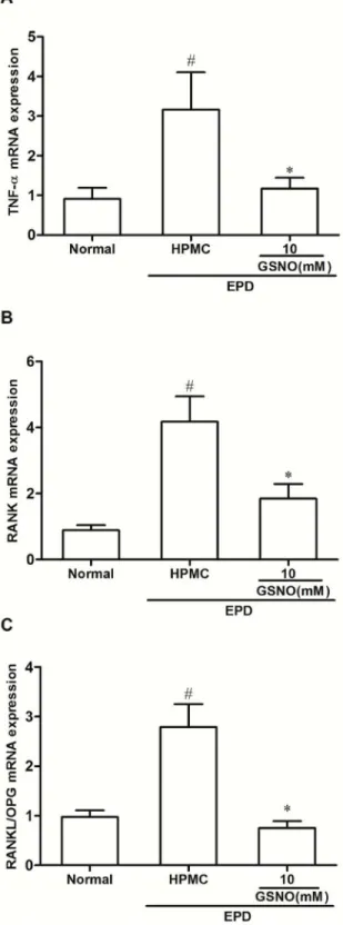
The group subjected to EPD that received local applications of the control HPMC solution showed a significant increase in the TNF-α(Fig 9A), RANK (Fig 9B) and in the ratio RANKL/ OPG (Fig 9C) mRNA expression when compared with the Normal control group. Topical
Fig 4. Effect of the topical application of HPMC/GSNO solutions on the oxidative stress markers malonaldehyde (MDA; A) and reduced glutathione (GSH; B) in experimental periodontitis in rats.Data represent the mean±SEM of six animals for each group. #P<0.05 was considered significantly different compared to the normal control (NORMAL);*P<0.05 was considered significantly different compared to the group of animals subjected to experimental periodontitis that received topical applications of the vehicle (HPMC solution). Analysis of variance (ANOVA); Bonferroni test.
treatment with the HPMC/GSNO 10 mM solution prevented the increase in all cases (Fig 9A, 9B and 9C).
Discussion
In the present study, we demonstrated that topical application of HPMC solution containing GSNO 10 mM reduces the alveolar bone loss associated with EPD when compared with the control group that received only the HPMC solution. This result is in accordance with previous studies demonstrating that both GSNO and S-nitroso-N-acetylpenicilamine (SNAP) reduce inflammation and bone loss in EPD [11,14]. The absence of anti-reabsorptive or anti-inflam-matory effects of topical applications of the HPMC/GSNO 2 mM solution suggests that this GSNO concentration is insufficient to provide a beneficial effect in the present EPD model. This result is consistent with the literature, which shows that the biological effect of exogenous NO administration is highly dependent on its local concentration [11,14,31]. HPMC/GSNO solutions were already shown to release free NO from the spontaneous thermal decomposition of GSNO according to the equation:
2 GSNO ! GSSGþ2 NO ð1Þ
where GS-SG is oxidized glutathione, formed after the homolytic S-N bond cleavage, followed by the formation of a covalent S-S bond [14,19,32]. Free NO locally released at the HPMC/ GSNO solution/gingiva interface is expected to diffuse through the gingiva and periodontal tis-sues and to be the chemical species responsible for the biological actions herein reported.
In the present HPMC/GSNO solutions the HPMC concentration is 1.25 wt%. At this con-centration, the HPMC chains are considered to be fully hydrated, with little polymer-polymer
Fig 5. Effect of HPMC/GSNO on the nitrite/nitrate (NOX) levels in experimental periodontitis in rats.
Data represent the mean±SEM of six animals for each group. # P<0.05 was considered significantly different compared to the normal control (NORMAL);*P<0.05 was considered significantly different compared to the group of animals subjected to experimental periodontitis that received topical applications of the vehicle (HPMC). Analysis of variance (ANOVA); Bonferroni test.
interactions, comprising thus a viscous polymeric solution and not a hydrogel. HPMC solu-tions can undergo gelation with HPMC concentrasolu-tions above 10 wt% and HPMC hydrogels are frequently used as vehicles for drug delivery [33,34]. The lower HPMC concentration used in the present study was chosen to facilitate the spreading of the formulation on the affected gingiva, avoiding thus any inflammation that could be caused by the topical of a hard hydrogel.
Fig 6. Effect of HPMC/GSNO on tumor necrosis factor alpha (TNF-α; A) and interleukin-1β(IL-1β; B) in experimental periodontitis in rats.Data represent the mean±SEM of six animals for each group. # P<0.05 was considered significantly different compared to the normal control (NORMAL);*P<0.05 was considered significantly different compared to the group of animals subjected to experimental periodontitis that received topical applications of the vehicle (HPMC). Analysis of variance (ANOVA); Bonferroni test.
The observed therapeutic effect of the HPMC/GSNO 10 mM solution on the bone resorp-tion was associated with an increase in the level of plasma bone alkaline phosphatase (BALP), a marker of osteoblastic activity, in the serum of animals and a considerable reduction in TRAP immunostaining (a histochemical marker of osteoclasts observed in multinuclear cells, mainly osteoclast). These changes were observed in the periodontal tissue of the animals treated with he HPMC/GSNO 10 mM solution for 11 days, when compared with the control HPMC solu-tion group, suggesting that the HPMC/GSNO 10 mM solusolu-tion prevents bone resorpsolu-tion and also induces bone formation. Accordingly, it has been described that NO donors have benefi-cial effects on the control of bone resorption (decreased osteoclast activity), and have a milder anabolic action on bone formation, enhancing osteoblast activity [12,13].
The present study suggests that the protective effect of GSNO on the alveolar bone resorp-tion is at least in part due to its anti-inflammatory, anti-oxidant and immunomodulatory prop-erties. We demonstrated that the HPMC/GSNO 10 mM solution significantly reduced the
Fig 7. Quantification of iNOS, RANK, TRAP and nitrotyrosine immunostainning in periodontal tissue of rats submitted to experimental periodontitis.Data represent the mean±SEM of four animals for each group. # P<0.05 was considered significantly different compared to the normal control (NORMAL);*P<0.05 was considered significantly different compared to the group of animals subjected to experimental periodontitis that received topical applications of the vehicle (HPMC). Analysis of variance (ANOVA); Bonferroni test.
Fig 8. Representative examples of iNOS (1strow), RANK (2ndrow) and TRAP (3rdrow)
immunostainning in experimental periodontitis in rats.Staining was performed using periodontal tissues from normal control animals (b, g, l, q), animals subjected to experimental periodontitis that received topical applications of HPMC (c, h, m, r) or 10 mM HPMC/GSNO (d, i, n, s). Negative controls were samples of periodontal tissue where the primary antibody was replaced with PBS-BSA (5%); no immunostaining was detected (a, f, k, p). Magnification x200. Arrows points to immunostaining osteoclasts in the periodontal tissue of the control HPMC solution group (Magnification x1000).
Fig 9. Effect of HPMC/GSNO on tumor necrosis factor alpha (TNF-α; A), RANK (B) and on the ratio RANKL/OPG (C) mRNA levels in experimental periodontitis in rats.Data represent the mean±SEM of six animals for each group. # P<0.05 was considered significantly different compared to the normal control (NORMAL);*P<0.05 was considered significantly different compared to the group of animals subjected to experimental periodontitis that received topical applications of the vehicle (HPMC). Analysis of variance (ANOVA); Bonferroni test.
TNF-αmRNA in gingival tissue of animals treated with this formulation as well as the gingival levels of TNF-αand IL-1β, when compared with the control HPMC solution group.
Cytokine participation in periodontal disease is well documented and these substances, par-ticularly TNF-αand IL-1β, may amplify the inflammatory response causing tissue destruction and bone loss [31]. It has been reported that IL-1 and TNF-αactivate the transcription of inducible nitric oxide synthase (iNOS) gene resulting in NO production [26]. The HPMC/ GSNO 10 mM solution markedly reduced both iNOS and nitrotyrosine expressions in the peri-odontal tissues and also reduced the NOx levels in the gingival tissues in comparison with the control HPMC solution. These findings are in accordance with those reported by Menezes et al. (2012) who showed that GSNO significantly reduces the gingival levels of IL-1, TNF-α and NOx, associated with the reduction of iNOS expression in a study conducted using the same experimental model of the present work [14]. The reduction of iNOS expression associ-ated with the administration of exogenous GSNO was also described in other studies, notably in the protective action against secondary injury in an animal model of traumatic brain injury [35]. This effect has been attributed to the S-nitrosation of the p65 subunit of nuclear factor kappa B (NFκB) by GSNO, via transnitrosation reaction in the endothelial cells, with conse-quent inhibition of NFκB activation and down regulation of iNOS. Alternatively, or concomi-tantly, the mechanism may involve the negative feedback modulation of iNOS by NO itself, including self-generated and exogenous NO [36]. In this case, the mechanism is considered to involve the coordination of NO to the iron center of the heme group of iNOS, and can be extended to other NOS isoforms [37]. The buildup of Heme-NO complex is expected to strongly inhibit the catalytic activity of the NOSs. In the present case, the source of NO for this negative feedback action on iNOS, could be attributed to the exogenous NO locally released by the HPMC/GSNO formulation, according toEq 1.
The release of large amounts of NO by iNOS has been shown to play a major role in immune reactions and inflammatory processes, including rheumatoid arthritis and periodontal disease [23]. NO reacts with oxygen-derived free radicals such as superoxide anion to form highly reactive molecules such as peroxynitrite anion and the hydroxyl radical and production of such species may contribute to tissue damage by inducing lipid peroxidation [38]. In accor-dance, we demonstrated that EPD leads to an oxidative stress in periodontal tissue with conse-quent generation of reactive oxygen species, indicated by the significant increase in MDA levels associated with an increase in the nitrotyrosine expression in the periodontal tissue and a decrease in GSH levels in the gingival tissue. Topical application of HMPC/GSNO 10 mM solu-tion attenuated the gingival oxidative stress, reestablishing the levels of GSH and decreasing nitrotyrosine expression and MDA concentration. Together, these results suggest that the pro-tective effect of the HPMC/GSNO solution is at least partially related to its inhibitory effect on oxidative stress. Moreover, other studies have indicated that GSNO decreases the formation of nitrotyrosine and lipid peroxidation in blood, increasing the reduced GSH/oxidized GSH (GSH/GS-SG) ratio in the brain [39–41].
results revealed an enhanced expression of RANK in the periodontal tissues of rats submitted to EPD, mainly in osteoclasts, associated with a decrease in the OPG expression and a signifi-cant increase in the RANK mRNA levels, as well as in the ratio RANKL/OPG mRNA levels, in the gingival tissue of animals submitted to EPD (HPMC solution group). The topical applica-tion of HPMC/GSNO 10 mM soluapplica-tion was able to reduce the RANK expression and increase the OPG expression in the periodontal tissue of the animals. In addition, the topical application of the HPMC/GSNO 10 mM solution led to a significant decrease in the RANK and RANKL/ OPG mRNA levels in the gingival tissue, compared with the control HPMC solution group. In summary, our results provide evidence that the topical application of HPMC/GSNO 10 mM solution is capable of reducing alveolar bone loss, pro-inflammatory cytokines production and oxidative stress in an experimental model of periodontal disease, representing a new potential treatment for periodontal diseases.
Author Contributions
Conceived and designed the experiments: CSM RFCL MGO GACB. Performed the experi-ments: CSM DVSC IMM GSS VB DVTW LEB. Analyzed the data: CSM RFCL DVSC MGO GACB. Contributed reagents/materials/analysis tools: VL VB DVTW CBM MGO. Wrote the paper: RFCL VL MGO.
References
1. Raitapuro-Murray T, Molleson TI, Hughes FJ. The prevalence of periodontal disease in a Romano-Brit-ish population c.200-400 AD. Brit Den J. 2014; 217:459–466.
2. Kinney JS, Ramseier CA, Giannobile WV. Oral fluid-based biomarkers of alveolar bone loss in peri-odontitis. Ann NY Acad Sci. 2007; 1098:230–251. PMID:17435132
3. Pihlstrom BL, Michalowicz BS, Johnson NW. Periodontal diseases. Lancet. 2005; 366:1809–1820. PMID:16298220
4. Dahiya P, Kamal R, Gupta R, Bhardwaj R, Chaudhary K, Kaur S. Reactive oxygen species in periodon-titis. J Indian Soc Periodontol. 2013; 17(4):411–416. doi:10.4103/0972-124X.118306PMID:
24174716
5. Joshi MS, Ponthier JL, Lancaster JR Jr. Cellular antioxidant and pro-oxidant actions of nitric oxide. Free Radiacal Biol Med. 1999; 27:1357–1366.
6. Zhang M, Zhao Zm, HE L, Wan CL. A meta-analysis of oxidative stress markers in schizophrenia. Sci China Life Sci. 2010; 53:112–124. doi:10.1007/s11427-010-0013-8PMID:20596963
7. Wimalawansa SJ, Chapa MT, Yallampalli C, Zhang R, Simmons DJ. Prevention of corticosteroid-induced bone loss with nitric oxide donor nitroglycerin in male rats. Bone 1997; 21:275–80. PMID:
9276093
8. Wimalawansa SM, Shankar VS, Simmins DJ, Wimalawansa SJ. The mechanism of bone resorption by cyclosphorin: involvement of the NO-Cgmp pathway. J Musculoskelet Neuronal Interact. 2001; 1 (2):141–143.
9. Wimalawansa SJ. Nitric oxide and bone. Ann N Y Acad Sci. 2010; 1192:391–403. doi: 10.1111/j.1749-6632.2009.05230.xPMID:20392265
10. Chae HJ, Park RK, Chung HT, Kang JS, Kim MS, Choi DY, et al. Nitric oxide is a regulator of bone remodeling. J Pharm Pharmacol. 1997; 49:897–902. PMID:9306258
11. Leitão RFC, Rocha FAC, Chaves HV, Lima V, Cunha FQ, Ribeiro RA, et al. Locally applied isosorbide decreases bone resorption in experimental periodontitis in rats. J Periodontol. 2004; 75:1227–1232. PMID:15515338
12. Wimalawansa SJ. Rationale for using nitric oxide donor therapy for prevention of bone loss and treat-ment of osteoporosis in humans. Ann NY Acad Sci. 2007; 1117: 283–297. PMID:18056048
13. Wimalawansa SJ. Nitric oxide: Evidence for novel therapeutic indications. Exp Opin Pharmacol. 2008; 9:1935–1954.
15. Fini M, Torricelli P. Giavaresi G, Carpi A, Nicolini A, Giardino R. Effect of L-lysine and L-arginine on pri-mary osteoblast cultures from normal and osteopenic rats. Biomed Pharmacother. 2001; 55:213–220. PMID:11393808
16. de Souza GFP, Yokoyama-Yasunaka JK, Seabra AB, de Oliveira MG, Uliana SR. Leismanicidal activity of primary S-nitrosothiols against Leismania major and Leismania amazonensis: implications for the treatment of cutaneous leismaniasis. Nitric Oxide 2006; 15:209–216. PMID:16527502
17. Cariello AJ, de Souza GFP, Foronda AS, Yu MCZ, Hofling-Lima AL, de Oliveira MG. In vitro amoebici-dal activity of S-nitrosoglutathione and S-nitroso-N-acetylcysteine against trophozoites of Acantha-moeba castellani. J Antimicrob Chemother. 2010; 65:588–591. doi:10.1093/jac/dkp485PMID:
20085996
18. Skeff MA, Brito GA, de Oliveira MG, Braga CM, Cavalcante MM, Baldim V, et al. S-nitrosoglutathione accelerates recovery from 5-fluorouracil induced oral mucositis. Plos One. Dec 5; 9(12):e113378. doi:
10.1371/journal.pone.0113378PMID:25478918
19. Schanuel FS, Santos KSR, Monte-Alto-Costa A, de Oliveira MG. Combined nitric oxide-releasing poly (vinyl alcohol) film/F127 solution for accelerating wound healing. Colloids & Surfaces B-Biointerfaces 2015; 130:182–191.
20. Sallay K, Sanavi F, Ring I, Pham P, Behling UH, Nowotny A. Alveolar bone destruction in the immunos-suppressed rat. J Periodont Res. 1982; 17:263–274. PMID:6213757
21. Lima V, Bezerra MM, Alencar VBM, Vidal ýP, Da Rocha FAC, Brito GAC, et al. Effects of chlorproma-zine on alveolar bone loss in experimental periodontal disease in rats. Eur J Oral Sci. 2000; 108 (2):123–129. PMID:10768725
22. Kuhr A, Popa-Wagner A, Schmoll H, Schwahn C, Kocher T. Observations on experimental marginal periodontitis in rats. J Periodontal Res. 2004; 39:101–106. PMID:15009517
23. Leitão RF, Ribeiro RA, Chaves HV, Rocha FA, Lima V, Brito GA. Nitric oxide synthase inhibition pre-vents alveolar bone resorption in experimental periodontitis in rats. J Periodontol. 2005; 76:947–954.
24. Whitby LG, Moss DW. Analysis of heat inactivation curves of alkaline phosphatase isoenzymes in serum. Clin Chim Acta. 1975; 59:361–367. PMID:1126023
25. Mihara M, Uchiyama M. Determination of malonaldehyde precursor in tissues by thiobarbituricacid test. Anal Biochem. 1978; 86:271–278. PMID:655387
26. Ding AH, Nathan CF, Stuehr DJ. Release of reactive nitrogen intermediates and reactive oxygen inter-mediates from mouse peritoneal macrophages. Comparison of activating cytokines and evidence for independent production. J Immunol. 1988; 141:2407–2412. PMID:3139757
27. Bayer EA, Wilchek M. The use of the avidin-biotin complex as a tool in molecular biology. Methods Bio-chem Anal. 1980; 26:1–45. PMID:7392958
28. Hsu SM, Raine L. Protein A, avidin, and biotin in immunohistochemistry. J Histochem Cytochem. 1981; 29:1349–1353. PMID:6172466
29. Brey EM, Lalani Z, Johnston C, Wong M, McIntire LV, Duke PJ, Patrick CW Jr. Automated selection of DAB-labeled tissue for immunohistochemical quantification. J Histochem Cytochem. 2003; 51 (5):575–584. PMID:12704205
30. Livak KJ, Schmittgen TD. Analyses of relative gene expression data using real-time quantitative PCR and the 2-ΔΔCTmethod. Methods 2001; 25 (4):402
–408. PMID:11846609
31. Van’t Hof RJ, Ralston H. Nitric oxide and bone. Immunology 2001; 103: 255–261. PMID:11454054 32. Simões MMSG, de Oliveira MG. Poly(vinyl alcohol) films for topical delivery of S-nitrosoglutathione: effect of freezing-hawing on the diffusion properties. J Biomed Mat Res–Part B–App Biomat 2010; 93B:416–424.
33. Fyfe CA, Blazek AI. Investigation of hydrogel formation from hydroxypropylmethylcellulose (HPMC) by NMR spectroscopy and NMR imaging techniques. Macromol 1997: 30:6230–6237.
34. Silva SMC, Pinto FV, Antunes FE, Miguel MG, Sousa JJS, Pais AACC. Aggregation and gelation in hydroxypropylmethyl cellulose aqueous solutions. J Coll Interf Sci 2008; 327:333–340.
35. Khan M, Im YB, Shunmugavel A, Gilg AG, Dhindsa RK, Singh AK, et al. Administration of S-nitrosoglu-tathione after traumatic brain injury protects the neurovascular unit and reduces secondary injury in a rat model of controlled cortical impact. J Neuroinflammation 2009; 6:1–12.
36. Colasanti M, Persichini T, Menegazzi M, Mariotto S, Giordano E, Caldarera CM, et al. Induction of nitric oxide synthase mRNA expression. Suppression by exogenous nitric oxide 1995; J Biol Chem 270: 26731–26733. PMID:7592903
38. Lipton SA, Choi YB, Pan ZH, Lei SZ, Chen HS, Sucher NJ et al. A redox-based mechanism for the neu-roprotective and neurodestructive effects of nitric oxide and related nitroso-compounds. Nature 1993; 364(6438):626–632. PMID:8394509
39. Chiueh CC, Rauhala P. The redox pathway of S- nitrosoglutathione, glutathione and nitric oxide in cell to neuron communications. Free Radic Res. 1999; 31:641–650. PMID:10630687
40. Prasad R, Giri S, Nath N, Singh I, Singh AK. GSNO attenuates EAE disease by S-nitrosylation-medi-ated modulation of endothelial-monocyte interactions. Glia 2007; 55:65–77. PMID:17019693 41. Khan M, Jatana M, Elango C, Paintlia AS, Singh AK, Singh I. Cerebrovascular protection by various
nitric oxide donors in rats after experimental stroke. Nitric Oxide 2006; 5:114–124.
42. Assuma R, Oates T, Cochram D, Amar S, Graves DT. IL-1 and TNF antagonists inhibit the inflamma-tory response and bone loss in experimental periodontitis. J Immunol. 1998; 160(1):403–409. PMID:
9551997
43. Wasa T, Nakashima T, Hiroshi N, Penninger JM. RANKL-RANK signaling in osteoclastogenesis and bone disease. Trends Mol Med. 2006; 12:17–25. PMID:16356770
44. Simonet WS, Lacey DL, Dunstan CR, Kelley M, Chang MS, Lüthy R, et al. Osteoprotegerin: a novel secreted protein involved in the regulation of bone density. Cell 1997; 89:309–319. PMID:9108485 45. Tay JY, Bay BH, Yeo JF, Harris M, Meghji S, Dheen ST. Identification of RANK-L inosteolytic lesions of