RESEARCH
Immunoexpression of Metalloproteinases 2 and 14 and TIMP-2
Inhibitor in Main Types of Primary Gastric Carcinomas
and Lymph Node Metastasis
Daniel Cordeiro Gurgel&José Telmo Valença-Junior&Conceição Aparecida Dornelas& Renato Braga Vieira&João Tarcisio Alves Maia-Filho&Roberto Cesar Pereira Lima-Junior& Ronaldo Albuquerque Ribeiro&Paulo Roberto Carvalho Almeida
Received: 20 December 2013 / Accepted: 21 April 2014 #Arányi Lajos Foundation 2014
Abstract Metalloproteinase-2 (MMP-2) and−14 (MMP-14) and the tissue inhibitor of metalloproteinases type 2 (TIMP-2) participate in epithelial-mesenchymal transition and tumor pro-gression in many cancers. However, the correlation between these enzymes in gastric cancer and the metastatic potential to their respective lymph node needs to be determined. Here, we evaluated the expression of these enzymes in gastric carcinoma and lymph node metastases and their possible involvement in tumor progression. Histological samples from 83 patients with gastric cancer and their respective lymph nodes were used. MMP-2, MMP-14 and TIMP-2 immunoexpression was scored. TIMP-2 expression in tumor-associated macrophages occurred more frequently than in normal mucosa (P=0.0128). Female tumor samples presented higher MMP-2 expression (P= 0.0248), while TIMP-2 occurred mainly in patients over
50 years old (P=0.0034). MMP-2 was higher expressed in primary tumor macrophages than in neoplastic cells (P= 0.0118), and was also seen in macrophages from metastatic-affected lymph nodes of intestinal and diffuse
histotypes (P=0.0006). MMP-2, MMP-14 and TIMP-2 ex-pression in mononuclear cells might be correlated with pro-gression of gastric cancer. MMP-14 production by macro-phages appears to be more involved indiffusegastric cancer progression.
Keywords Gastric cancer . Immunohistochemistry . Macrophage . Metalloproteinase . Metastasis
Introduction
Gastric cancer is the world’s fourth most common neoplasia in developing countries, affecting more men than women [1]. It is the second cause of cancer-related mortality with approxi-mately one million deaths per year [2]. In Brazil, gastric cancer is still considered an important cause of morbidity and mortality and represents a public health problem [2,3].
The genetic status of neoplastic cells, such as epithelial-mesenchymal transition (EMT) and tumor microenvironment, are key mutually related factors for cancer development [4,5]. In this context, paracrine activation of stromal cells, such as macrophages and fibroblasts, by mesenchymal phenotype neoplastic cells leads to metalloproteinase activation. In turn, these enzymes that act on the extracellular matrix (ECM) and inhibit E-cadherin synthesis promote a mesenchymal pheno-type in cancer cells [6,7].
Matrix metalloproteinases (MMPs) are a group of 25 zinc-dependent endopeptidases that play a major role in ECM ho-meostasis [8]. MMP-2 is secreted in an inactive pro-enzymatic form, and unlike other MMPs, its activity is modulated by the
D. C. Gurgel
:
J. T. Valença-Junior:
C. A. Dornelas:
R. B. Vieira:
J. T. A. Maia-Filho:
P. R. C. AlmeidaDepartment of Pathology and Forensic Medicine, Faculty of Medicine, Federal University of Ceará, Fortaleza, Ceará, Brazil
D. C. Gurgel
e-mail: [email protected]
R. C. P. Lima-Junior
:
R. A. RibeiroDepartment of Physiology and Pharmacology, Faculty of Medicine, Federal University of Ceará, Fortaleza, Ceará, Brazil
R. A. Ribeiro
Department of Clinical Oncology, Haroldo Juaçaba Hospital/Cancer Institute of Ceará, Fortaleza, Ceará, Brazil
P. R. C. Almeida (*)
Departamento de Patologia e Medicina Legal/FAMED/UFC, Rua Monsenhor Furtado S/N, Rodolfo Teófilo, 60430-350 Fortaleza, Ceará, Brazil
e-mail: [email protected]
P. R. C. Almeida
tissue inhibitor of metalloproteinases (TIMP)-2 and the mem-brane type 1 MMP (MMP-14) [9,10]. Both TIMPs and MMPs are produced by the same cells and are important modulators of ECM remodeling [11]. A growing body of evidence shows that increased expression and production of MMPs by tumor and stromal cells are essential for tumor invasion and metastasis in several types of cancer, such as gastric, pancreatic, lung, ovar-ian and breast carcinomas [12,13].
In gastric cancer, the enzymes MMP-2 [14], MMP-14 [15], and TIMP-2 [16] have been correlated with the invasion process. However, the exact role of the potential interaction of these three enzymes in gastric cancer and their respective lymph node metastases need to be determined.
Therefore, this study aimed to investigate the expression of MMP-2, MMP-14 and TIMP-2 in theintestinalanddiffuse
gastric cancer histotypes and possible correlations between this expression and lymph node metastasis.
Material and Methods
Case Selection
Our study was carried out using 83 cases of male and female subjects who had undergone gastric cancer gastrectomy. Tumor samples were obtained from the files of the Department of Pathology and Forensic Medicine at Federal University of Ceará. The collected material was fixed in 10 % formalin, embedded in paraffin, sectioned 3μm thick and then stained with hematoxylin-eosin.
Data were analyzed according to gender, age, anatomic location of the tumor in the stomach, pathological staging (degree of local invasion and lymph node involvement) [17] and tumor macroscopic dimensions. Cases were classified as
intestinaland diffuse tumors according to Lauren’s criteria [18]. Exclusion criteria were poorly fixed samples with exten-sive necrotic areas and samples with less than 100 cells. This study was approved by the Research Ethics Committee (Protocol number 010.03.10).
Tissue Microarray
A tissue microarray assay was performed according to GURGEL et al., 2012 [19]. Briefly, tissue samples were obtained from the donor paraffin block with a manual puncher and arrayed in a matrix of four rows and six columns, with up to 24 samples per recipient block. Liquid paraffin was gently added to melt the wax cylinders at 60 °C. Then, typical H&E staining and an immunohistochemistry assay were performed. The analysis was conducted by two independent observers who were unaware of the clinicopathological data. Slides were observed under a microscope (Olympus BX41™).
Immunohistochemistry
Paraffin sections were dewaxed and rehydrated. Treatment with a 3 % H2O2 solution in methanol for 10 min was used to block endogenous peroxidase. A 1 % retrieval solution (Vector Laboratories, Burlingame, California, USA) in 98 °C warm water was applied for 20 min to unmask the antigens. Ultra V block (TA-125-UB; LabVision) was utilized for 10 min to inhibit unspecified ground reactions. The slides were incubated with mouse monoclonal antibodies anti-MMP-2 (SCBT® sc-13595; placenta as positive control) [20], anti-MMP-14 (SCBT® sc-80210; kidney as positive control) [21] and anti-TIMP-2 (SCBT®, sc-21735; lung as positive control) [22] at a 1:50 dilution. A mouse monoclonal anti-human CD68 (KP1 clone, DakoTM) was also utilized at 1:800 for 1.5 h. Negative controls, in which no primary antibody was applied, were included. Following primary an-tibody incubation, a secondary biotinylated anan-tibody donkey anti-mouse IgG (SCBT® sc-2098) was applied at 1:100. Then, the streptavidin-coupled peroxidase complex (TS-125-HR; LabVision) was used for 15 min. An automated immunostainer (Ventana Benchmark XT / Roche™) was uti-lized to process the reactions, and diaminobenzidine was used as the chromogen. The sections were counterstained with haematoxylin, dehydrated, diaphanized, mounted and analyzed.
Score Analysis
The following score criteria for tumor and stromal cells, adapted from the previous publication of Buskens et al., 2003 [23], were employed: 0 = absence of immunoreac-tivity or rare labeled cells (<5 %); 1 = discrete staining in more than 50 % of tumor/mononuclear inflammatory cells or less than 50 % of cells moderately stained; 2 = mod-erate staining in most (>50 %) tumor/mononuclear inflam-matory cells or less than 50 % of cells strongly stained; and 3 = strong staining in more than 50 % of tumor or inflammatory mononuclear cells (confirmed by CD68 ex-pression). Expression was analyzed and always considered negative when score 0 was observed and positive when scores 1, 2 and 3 were seen.
Statistical Analysis
The scores were compared using Fisher’s exact test. A value of
P<0.05 was considered statistically significant.
Results
including sex, age, site of neoplasia in the stomach, tumor size, angiolymphatic and perineural invasion, local tumoral invasion (T1 & T2-T4) and lymph node stage (N0 & N1-N3), were studied. In most cases, no correlation was found (P>0.05). Positive findings are expressed in Table1.
Table1shows that MMP-2 was markedly more expressed in female subjects compared with male subjects, both in neoplastic (P= 0.0248) and mononuclear cells (P= 0.0028). A significantly higher positivity for TIMP-2 immunoexpression was detected in neoplastic cells of subjects who were 50 or more years old (P= 0.0034), and we also found a more frequent expression of this inhibitor in mononuclear cells (P= 0.044) of individuals with larger tumors (≥5 cm).
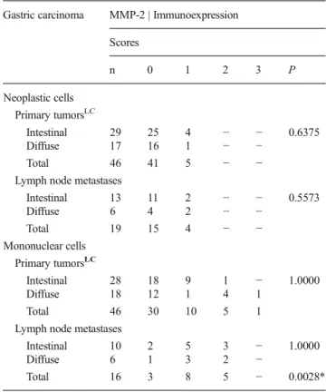
In Table2, MMP-2 expression in neoplastic and mononu-clear cells ofintestinalanddiffusehistotypes of gastric carci-nomas was compared both in primary tumors and in the respective lymph node metastasis. There was no difference in MMP-2 expression in neoplastic or mononuclear cells based on tumor histotype (intestinaland diffuse). The same result was observed in neoplastic cells of both sites (no dif-ference in primary tumors and respective lymph node metas-tasis). However, mononuclear cells of lymph node metastases presented a higher positivity for MMP-2 compared with pri-mary tumors (13/16=81 % in lymph nodes vs 16/46=35 % in primary gastric tumor,P=0.0028).
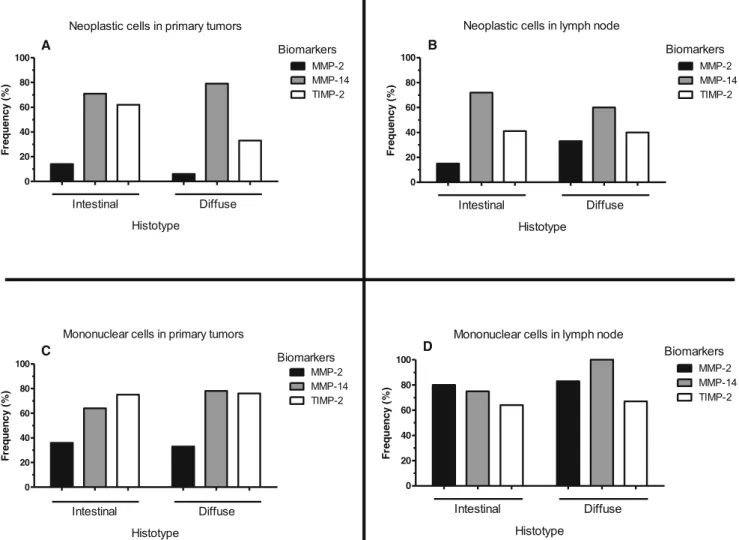
MMP-2 expression in mononuclear cells was higher in metastatic than in primary tumors of both histotypes, with a significant difference in intestinal carcinomas (metastatic site: 8/10 = 80 % vs primary site: 10/28 = 36 %; P= 0.0265; Fig. 1) and a marginal difference in
diffuse carcinomas (5/6 = 83 % vs 6/18 = 33 %;
P=0.0608).
MMP-14
In contrast to MMP-2, MMP-14 expression was predominant-ly positive in neoplastic cells of carcinomas present in the
Table 1 Association of MMP-2 and TIMP-2 immunoexpression with clinicopathological parameters
*P<0.05; **P<0.005; Fisher’s exact test
Immunoexpression–scores
Clinicopathologic parameters n 0 1 2 3 P Biomarker Cell type
Sex
Males 32 31 1 − − 0.0248* MMP-2 Neoplastic Cell
Females 14 10 4 − −
Males 31 25 5 1 0 0.0028** MMP-2 Mononuclear Cell
Females 15 5 5 4 1
Age (yrs.)
<50 7 7 − − − 0.0034** TIMP-2 Neoplastic Cell
≥50 50 20 29 1 −
<50 6 2 1 1 2 0.6274 TIMP-2 Mononuclear Cell
≥50 47 11 21 10 5
Tumor size
<5 cm 16 5 11 − − 0.2357 TIMP-2 Neoplastic Cell
≥5 cm 40 21 18 1 −
<5 cm 16 1 8 6 1 0.0440* TIMP-2 Mononuclear Cell
≥5 cm 36 12 13 5 6
Table 2 MMP-2 immunoexpression in neoplastic and mononuclear cells of primary gastric carcinomas ofintestinalanddiffusehistotypes and in the respective lymph node metastases
Gastric carcinoma MMP-2 | Immunoexpression
Scores
n 0 1 2 3 P
Neoplastic cells Primary tumorsLC
Intestinal 29 25 4 − − 0.6375
Diffuse 17 16 1 − −
Total 46 41 5 − −
Lymph node metastases
Intestinal 13 11 2 − − 0.5573
Diffuse 6 4 2 − −
Total 19 15 4 − −
Mononuclear cells Primary tumorsLC
Intestinal 28 18 9 1 − 1.0000
Diffuse 18 12 1 4 1
Total 46 30 10 5 1
Lymph node metastases
Intestinal 10 2 5 3 − 1.0000
Diffuse 6 1 3 2 −
Total 16 3 8 5 − 0.0028*
LCHistological type (Lauren classification)
stomach (51/69=74 %) and in the respective lymph node (19/ 28=68 %), without a significant difference between these sites (Table3; Fig.2).
In addition, we found no significant difference while eval-uating MMP-14 expression in neoplastic cells of the same histotype in primary and metastatic lesions or between stomach and lymph node gastric carcinomas (intestinal
histotype: 32/45 vs 13/18,P=1.0000;diffusetumors: 19/24 vs 6/10,P=0.3951).
MMP-14 was also frequently expressed in mononuclear cells of both sites (stomach: 47/68=69 %; lymph node: 11/ 13=85 %). Score 1 predominated in gastric samples of both histotypes, but without a statistical difference between these anatomical sites (Table3).
TIMP-2
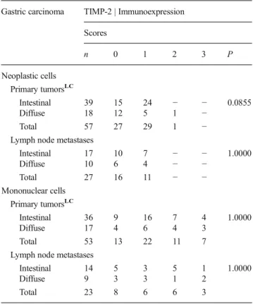
TIMP-2 positive immunoexpression predominated in neo-plastic cells of primary carcinomas (30/57 = 53 %)
compared with the lymph node metastasis (11/27=41 %), but no significant difference was found (P= 0.5118, Table 4). In the stomach, TIMP-2 was marginally more expressed in the intestinal histotype (24/39=62 %) than in
diffuse tumors (6/18=34 %; P=0.0855). In addition, the expression of this marker in primary and metastatic lesions of each histotype was found to be similar (intestinal:P=0.2425;
diffuse:P=1.0000).
Taking into consideration mononuclear cells, TIMP-2 positivity was more frequent in both sites (compared to negative immunoexpression), without a difference between histotypes in each site (Table 4). Similarly, there was no difference in the whole group (stomach: 40/53=75 %; lymph node: 15/23=65 %, P=0.4083) or in isolated histotypes of gastric compared to lymph node lesions (intestinal: 27/36 vs 9/14, respectively,P=0.4955; diffuse: 13/17 vs 6/9, respec-tively,P=0.6613).
Figure 1 shows a synoptic view of the most important findings concerning the immunoexpression of MMP-2,
Fig. 1 MMP-2, MMP-14 and TIMP-2 immunoexpression in neoplastic and mononuclear cells in primary and metastatic gastric carcinomas of intestinaland diffuse histotypes. Note the generally predominant
MMP-14 and TIMP-2 in our samples. Figure2demonstrates the immunostaining patterns of these biomarkers in primary and metastatic sites in both histotypes, and Fig.3shows that most mononuclear cells correspond to macrophages, with conspicuous CD68 positivity.
Discussion
In this study, we found a much more frequent positivity for MMP-2 immunostaining in tumors of female subjects, as demonstrated in neoplastic cells and mainly in mononuclear cells. This finding contrasts with that of other authors who found higher MMP-2 expression in males [12,24]. The results are controversial, and in other reports, there was no difference in expression between men and women [25, 26]. Possible explanation for these discrepancies could be regional poly-morphisms of the MMP-2 gene in gastric cancer. Some reports have shown differences in MMP-2 genotypes according to intensity of expression [27,28] or gender [28], but extensive comparative regional studies are lacking.
We found few reports comparing TIMP-2 expression and clinicopathological features. Our data showed a more frequent TIMP-2 positivity in the neoplastic cells of older patients
(≥50) than in those of younger patients. We do not know the
reason for this association. Joo et al. found no significant differences in their cases considering the same age limit cut off [29]. We have also found that cases with positive expres-sion of TIMP-2, especially in mononuclear cells, occurred mainly in association with smaller tumors (<5 cm), in accor-dance with the modulating action of this enzyme [9]. Zhang et al. did not find any difference in TIMP-2 expression be-tween gastric tumors smaller or larger than 5 cm in diameter [30].
Out of these cited correlations, our data showed no associ-ations between the expression of the three biomarkers studied, MMP-2, MMP-14 and TIMP-2 and other clinicopathological variables, such as site of the tumor in the stomach, local angiolymphatic infiltration, perineural invasion and lymph node staging. Previous reports have widely divergent results, ranging from complete absence of correlations between the clinical parameters analyzed and each specific biomarker [29] to full association between protein expression and all clinico-pathological variables [31]. However, most investigators have found a link between a few variables and a specific biomarker expression [12,26,30,32,33], resulting in a very large range of heterogeneity and controversies about which correlations are most compelling.
In our study, we found that MMP-2 positive expression was low in primary gastric carcinomas (11 %). According to other reports, this immunomarker expression largely varies in the range of 25 to 74 % [12,34]. These differences could be partially explained by environmental or genotypic features, which are influenced by geographical aspects or population characteristics and are related to other variables. Furthermore, ulcers, erosions and intense inflammatory processes in select-ed tumor samples can significantly alter positivity of staining [35]. In addition, methodological and technical issues are likely to occur, including fixation time of samples, variable sensitivity and specificity of monoclonal or polyclonal anti-bodies according to different manufacturers and the different criteria used to evaluate the immunohistochemistry [34–36]. Several studies have demonstrated the role of MMP-2 in the lysis or breakdown of the extracellular matrix, which favors local tumor invasion and dissemination of cancer cells [8,11,
14,25]. However, the only report we found that evaluated the expression of this enzyme in the microenvironment of the lymph node with metastases of gastric carcinoma was that of Wang et al. [37].
MMP-2 was expressed preferentially in neoplastic cells of metastatic samples (21 %) compared to primary tumors, but the difference was not significant. Wang et al. have found a larger difference in favor of a more frequent expression in lymph nodes metastases of gastric cancer [37]. In the present study, positive MMP-2 immunoexpression predominated in mononuclear cells compared to epithelial malignant cells, mainly in the lymph node metastases. These findings
Table 3 MMP-14 immunoexpression in neoplastic and mononuclear cells of primary gastric carcinomas ofintestinalanddiffusehistotypes and in the respective lymph node metastases
Gastric carcinoma MMP-14 | Immunoexpression
Scores
n 0 1 2 3 P
Neoplastic cells Primary tumorsLC
Intestinal 45 13 17 12 3 0.5715
Diffuse 24 5 15 4 −
Total 69 18 32 16 3
Lymph node metastases
Intestinal 18 5 5 7 1 0.6775
Diffuse 10 4 4 2 −
Total 28 9 9 9 1
Mononuclear cells Primary tumorsLC
Intestinal 45 16 17 9 3 0.2806
Diffuse 23 5 14 4 −
Total 68 21 31 13 3
Lymph node metastases
Intestinal 8 2 2 3 1 0.4872
Diffuse 5 − 5 − −
Total 13 2 7 3 1
LCHistological type (Lauren classification)
emphasize the role of MMP-2 in favoring metastatic potential and agree with other publications related to gastric carcinomas [14,38].
The prominent MMP-2 expression in mononuclear cells of the lymph node also reinforces the great importance of these cells (mainly macrophages) in the process of tumor progres-sion. Our data also showed that higher MMP-2 expression in these macrophages in the lymph node was evident in both histotypes, significantly inintestinaltumors and marginally significant indiffusetumors (Table2). Mrena et al. have found a higher frequency of MMP-2 expression inintestinaltumors in the stomach [12], and Lee et al. have reported that MMP-2 expression was significantly higher inintestinal-type than in
diffuse-type gastric cancer; however, they only evaluated the expression in the cancer cells and in the primary gastric
tumors [39]. In accordance with the last authors, our findings in gastric primary lesions also showed a higher frequency in
intestinal than in diffuse carcinomas, but without statistical significance. We did not find any difference in MMP-2 ex-pression among both histotypes in metastases to lymph nodes. To the best of our knowledge, there are no previous reports concerning MMP-2 expression in both sites of gastric carci-nomas (stomach and lymph nodes) in both types of cells (neoplastic and stroma mononuclear cells) and in the two main histotypes of Lauren classification (diffuse and intestinal
tumors).
Our data showed that MMP-14 presented the highest pos-itivity in both neoplastic and mononuclear stromal cells, whether in primary or lymph node metastases, without signif-icant differences between these anatomical sites or gastric
Fig. 2 a MMP-2: Positive control, placenta. Decidual cells slightly stained (400×).b Stomach (MMP-2)Diffuse carcinoma: Neoplastic cells, score = 0; Mononuclear cells, score = 2; Case 75 (400×).c Lymph node (MMP-2)Intestinalcarcinoma: Neoplastic cells, score = 1; Mono-nuclear cells, score = 2; Case 96 (400×).d MMP-14: Positive control, kidney. Tubular cells strongly stained (100×).e Stomach (MMP-14)
Intestinalcarcinoma: Neoplastic cells, score = 3; Mononuclear cells, score = 3; Case 65 (400×).f Lymph node (MMP-14) Intestinal
carcinoma: Neoplastic cells, score = 3; Mononuclear cells, score = 3; Case 46 (400×).g TIMP-2: Positive control, lung. Alveolar macrophages strongly stained (400×).h Stomach (TIMP-2)Intestinalcarcinoma: Neoplastic cells, score = 1; Mononuclear cells, score = 2; Case 83 (400×).
carcinoma histotypes. The substantial presence of MMP-14 is in agreement with the known role of this membrane-bound metalloproteinase, such as the activation of other metallopro-teinases (for example, MMP-2) [40], with a final effect favor-ing local tumor invasion and metastasis [41]. According to some reports, mononuclear stromal cells in the tumor micro-environment are mainly macrophages associated with tumor or peritumoral fibroblasts that are associated with carcinomas [42–45]. There are scarce publications about the expression of
MMP-14 using immunohistochemistry in cancer cells and/or stromal adjacent cells. Ishigaki et al. [46] and Mylona and colleagues [47] have detected important expression of the pro-tein in both types of cells in breast primary carcinomas. Karadag et al. have described the presence of this membrane-type MMP in all of their cases of gallbladder carcinomas in tumor and stromal cells (including muscle fibers, vascular endothelium, fibroblasts and lymphoid cells) [48], and Markovic et al. have shown that MMP-14 is over-regulated in microglia associated with glioma but not in glioma cells [49].
The only report we found for gastric cancer regarding the theme of the present paper was written by Mori and col-leagues, which described positive staining in carcinoma cells and negative expression in stromal cells (except in some positive cells adjacent to the margin of cancer invasive cells) [50]. Unlike this report, our findings showed high frequency of positivity in both cell types, equivalently in the two sites, but with a predominance in the mononuclear cells of stroma. In the present study, the CD68 positivity in most (>50 %) of the mononuclear cells, which were also frequently positive for MMP-14 in gastric and, mainly, lymph node lesions, is a compelling suggestion for the participation of macrophages in tumor microenvironment and metastasis in gastric carcino-mas. We did not find any report that evaluated MMP-14 expression in the two cell types, in both sites (primary and metastatic lesions) and in the two gastric carcinoma histotypes.
The expression of TIMP-2 in neoplastic cells of primary tumors in our samples was predominantly positive (>50 %), with a frequency similar to that reported by some authors [51]. A higher positivity has been described in other reports [29,32]. We found a greater frequency of immunostaining in mononu-clear cells compared to neoplastic cells in the two sites, stom-ach and lymph nodes, and no differences were observed be-tweenintestinal anddiffuse histotypes. In the lymph nodes, TIMP-2 positivity was lower than in gastric lesions in both mononuclear and neoplastic cells. This difference is compati-ble with a possicompati-ble diminished inhibitory effect of this enzyme on MMP-2 and MMP-14 activities in the lymph nodes, which
Table 4 TIMP-2 immunoexpression in neoplastic and mononuclear cells of primary gastric carcinomas ofintestinalanddiffusehistotypes and in the respective lymph node metastases
Gastric carcinoma TIMP-2 | Immunoexpression
Scores
n 0 1 2 3 P
Neoplastic cells Primary tumorsLC
Intestinal 39 15 24 − − 0.0855
Diffuse 18 12 5 1 −
Total 57 27 29 1 −
Lymph node metastases
Intestinal 17 10 7 − − 1.0000
Diffuse 10 6 4 − −
Total 27 16 11 − −
Mononuclear cells Primary tumorsLC
Intestinal 36 9 16 7 4 1.0000
Diffuse 17 4 6 4 3
Total 53 13 22 11 7
Lymph node metastases
Intestinal 14 5 3 5 1 1.0000
Diffuse 9 3 3 1 2
Total 23 8 6 6 3
LC
Histological type (Lauren classification)
*P= Fisher’s exact test (positivity: lymph node and stomach)
consequently allows an elevated expression of these metallo-proteinases. This effect is in accordance with our findings, as shown previously, and in agreement with the TIMP-2 classic inhibitory action described in the literature [9,51].
Some studies have shown a close relationship between MMP-2, MMP-14 and TIMP-2. MMP-14 is classically an activator of MMP-2, while TIMP-2 has a controversial role. By definition, TIMP-2 inhibits MMP-2 [11,51]. As we have shown, our data are consistent with this role of TIMP-2. However, other reports have demonstrated its ability to act together with MMP-14 for binding to MMP-2 and to then form a trimolecular proteolytic complex capable of degrading the extracellular matrix [52–54].
Some authors have shown a relationship between the three biomarkers studied here in some types of tumors, such as breast, prostate and colorectal primary cancers [55–57]. To the best of our knowledge, the immunohistochemical assess-ment of MMP-2, MMP-14 and TIMP-2 in the same cases of primary gastric carcinomas and lymph node metastases in neoplastic and mononuclear stromal cells of both histotypes of stomach cancer is lacking in the previous reports.
Apart from the biological traditional roles of the matrix metalloproteinases associated with the degradation and turn-over of many components of the extracellular matrix, studies ranging from transgenic models to recent proteomic screens are changing the dogma about MMP functions. New sub-strates have been described, including cytokines and chemokines, growth factors, angiogenic factors, cell adhesion molecules, receptors, metabolic enzymes and components of the complement system, among others [58]. Therefore, many of the mechanisms involved in the ancient and newly discov-ered actions of these proteases are unknown, which stimulates further studies.
Conclusions
The immunoexpression of MMP-2 was found predominantly in women, whereas TIMP-2 was expressed preferentially after 50 years of age, regardless of sex, and in larger tumors. The importance of such findings has yet to be demonstrated. The higher expression of MMP-2, MMP-14 and TIMP-2 in mono-nuclear stromal cells compared to neoplastic cells in both anatomical sites and in the two gastric cancer histotypes suggests the possible role of these biomarkers for the progres-sion of gastric cancer in the tumor microenvironment. The increased lymph nodal expression of MMP-2 and MMP-14 might be correlated with cancer progression. In addition, the increased expression of TIMP-2 in primary cancer is in accor-dance with its inhibitory effect on MMP-2 and MMP-14, and the reduced immunostaining of this inhibitor in the lymph node might be correlated with metastatic potential. Furthermore, we verified a higher MMP-14 expression in
mononuclear cells in both sites of the diffuse tumors, which suggests the possible participation of this biomarker in the tumor progression of this particular gastric cancer histotype.
Acknowledgments We are grateful to the American Journal Experts for the English edition. This work was supported by CNPq (Conselho Nacional de Desenvolvimento Científico e Tecnológico), CAPES (Fundação Coordenação de Aperfeiçoamento de Pessoal de Nível Supe-rior) and FUNCAP (Fundação Cearense de Apoio ao Desenvolvimento Científico). RAR is recipient of a CNPq fellowship.
Conflict of Interest The authors indicate that they have no potential conflicts of interest.
References
1. Fitzgerald R, Hardwick R, Huntsman D et al (2010) Hereditary diffuse gastric cancer: update consensus guidelines for clinical man-agement and directions for future research. J Med Genet 47:436–444 2. Instituto Nacional de Câncer (Brasil) (2012) Estimativa 2012: incidência de câncer no Brasil. INCA, Rio de Janeiro, http:// www.inca.gov.br/estimativa/2012/
3. Rodrigues M, Queiroz D, Rodrigues R et al (2005) Prevalence of Helicobacter pylori infection in Fortaleza, Northeastern Brazil. Rev Saude Publica 39:847–849
4. Gao D, Vahdat L, Wong S et al (2012) Microenvironmental regula-tion of epithelial-mesenchymal transiregula-tions in cancer. Cancer Res 72(19):4883–4889
5. Talbot L, Bhattacharya S, Kuo P (2012) Epithelial-mesenchymal transition, the tumor microenvironment, and metastatic behavior of epithelial malignancies. Int J Biochem Mol Biol 3(2):117–136 6. Munshi H, Stack M (2006) Reciprocal interactions between adhesion
receptor signaling and MMP regulation. Cancer Metastasis Rev 25: 45–56
7. Bourboulia D, Stetler-Stevenson W (2010) Matrix MetalloProteinases and Tissue Inhibitors of Metalloproteinases: positive and negative reg-ulators in tumor cell adhesion. Semin Cancer Biol 20(3):161–168 8. Al-Dasooqi N, Gibson R, Bowen J, Keefe D (2009) Matrix
metallo-proteinases: key regulators in the pathogenesis of chemoterapy-induced mucositis? Cancer Chemother Pharmacol 64:1–9
9. Ries C, Egea V, Karow M et al (2007) MMP-2, MT1-MMP, and TIMP-2 are essential for the invasive capacity of human mesenchy-mal stem cells: differential regulation by inflammatory cytokines. Blood 109(9):4055–4063
10. Azar D, Casanova F, Mimura T et al (2010) Corneal epithelial MMP-14 inhibits vascular endothelial cell proliferation and migration. Cornea 29:321–330
11. Brew K, Nagase H (2010) The tissue inhibitors of metalloproteinases (TIMPs): an ancient family with structural and functional diversity. Biochim Biophys Acta 1803:55–71
12. Mrena J, Wiksten J, Nordling S et al (2006) MMP-2 but not MMP-9 associated with COX-2 and survival in gastric cancer. J Clin Pathol 59:618–623
13. Ra H, Parks W (2007) Control of matrix metalloproteinase catalytic activity. Matrix Biol 26:587–596
14. Kubben F, Sier C, Van Duijn W et al (2006) Matrix metalloproteinase-2 is a consistent prognostic factor in gastric cancer. Brit J Cancer 95:1035– 1040
16. Hong S, Park I, Hong W et al (1996) Overexpression of tissue inhibitors of metalloproteinase−1 and−2 in the stroma of gastric cancer. J Korean Med Sci 11:474–479
17. Sobin L, Gospodarowicz M, Wittekind C (2010) TNM classification of malignant tumours. International union against cancer, 7th edn. Wiley-Blackwell, New York
18. Lauren P (1965) The two histological main types of gastric carcino-ma: diffuse and so-called intestinal-type carcinoma. An attempt at a histo-clinical classification. Acta Pathol Microbiol Scand 64:31–49 19. Gurgel D, Dornelas C, Lima-Junior R et al (2012) An adapted tissue
microarray for the development of a matrix arrangement of tissue samples. Pathol Res Pract 208:167–168
20. Rojiani M, Alidina J, Esposito N, Rojiani A (2010) Expression of MMP-2 correlates with increased angiogenesis in CNS metastasis of lung carcinoma. Int J Clin Exp Pathol 3(8):775–781
21. Bonfil R, Dong Z, Trindade-Filho J et al (2007) Prostate cancer-associated membrane type 1–matrix metalloproteinase. A pivotal role in bone response and intraosseous tumor growth. Am J Pathol 170:2100–2111
22. Dong B, Sato M, Sakurada A et al (2005) Computed tomographic images reflect the biologic behavior of small lung adenocarcinoma: they correlate with cell proliferation, microvascularization, cell adhe-sion, degradation of extracellular matrix, and K-ras mutation. J Thorac Cardiovasc Surg 130:733–739
23. Buskens C, Sivula A, Van Rees B et al (2003) Comparison of cyclooxygenase 2 expression in adenocarcinomas of the gastric cardia and distal oesophagus. Gut 52:1678–1683
24. Hwang TL, Lee LY, Wang CC et al (2010) Claudin-4 expression is associated with tumor invasion, MMP-2 and MMP-9 expression in gastric cancer. Exp Ther Med 1(5):789–797
25. Zhou Y, Li G, Wu J et al (2010) Clinicopathological significance of E-cadherin, VEGF, and MMPs in gastric cancer. Tumour Biol 31(6):549– 558
26. Wu ZY, Li JH, Zhan WH, He YL (2006) Lymph node micrometastasis and its correlation with MMP-2 expression in gas-tric carcinoma. World J Gastroenterol 12(18):2941–2944
27. Kubben FJ, Sier CF, Meijer MJ et al (2006) Clinical impact of MMP and TIMP gene polymorphisms in gastric cancer. Br J Cancer 95(6): 744–751
28. Alakus H, Afriani N, Warnecke-Eberz U et al (2010) Clinical impact of MMP and TIMP gene polymorphisms in gastric cancer. World J Surg 34(12):2853–2859
29. Joo YE, Seo KS, Kim HS et al (2000) Expression of tissue inhibitors of metalloproteinases (TIMPs) in gastric cancer. Dig Dis Sci 45(1): 114–121
30. Zhang JF, Zhang YP, Hao FY et al (2005) DNA ploidy analysis and expression of MMP-9, TIMP-2, and E-cadherin in gastric carcinoma. World J Gastroenterol 11(36):5592–5600
31. Zheng H, Takahashi H, Murai Y et al (2006) Expressions of MMP-2, MMP-9 and VEGF are closely linked to growth, invasion, metastasis and angiogenesis of gastric carcinoma. Anticancer Res 26(5A):3579– 3583
32. Alakus H, Grass G, Hennecken JK et al (2008) Clinicopathological significance of MMP-2 and its specific inhibitor TIMP-2 in gastric cancer. Histol Histopathol 23(8):917–923
33. He L, Chu D, Li X et al (2013) Matrix metalloproteinase-14 is a negative prognostic marker for patients with gastric cancer. Dig Dis Sci 58(5):1264–1270
34. Dicken B, Graham K, Hamiltom S et al (2006) Lymphovascular inva-sion is associated with poor survival in gastric cancer: an application of gene-expression and tissue array techniques. Ann Surg 243:64–73 35. Gu Q, Wang D, Gao Y et al (2002) Expression of MMP1 in surgical
and radiation-impaired wound healing and its effects on the healing process. J Environ Pathol Toxicol Oncol 21(1):71–78
36. Murnane M, Cai J, Shuja S et al (2009) Active MMP-2 effectively identifies the presence of colorectal cancer. Int J Cancer 125:2893–2902
37. Wang LB, Jiang ZN, Fan MY et al (2008) Changes of histology and expression of MMP-2 and nm23-H1 in primary and metastatic gastric cancer. World J Gastroenterol 14(10):1612–1616
38. Mönig S, Baldus S, Hennecken J et al (2001) Expression of MMP-2 is associated with progression and lymph node metastasis of gastric carcinoma. Histopathology 39:597–602
39. Lee LY, Wu CM, Wang CC et al (2008) Expression of matrix metalloproteinases MMP-2 and MMP-9 in gastric cancer and their relation to claudin-4 expression. Histol Histopathol 23(5):515–521 40. Freudenberg J, Chen W (2007) Induction of Smad1 by MMP-14
contributes to tumor growth. Int J Cancer 121:966–977
41. Seiki M, Yana I (2003) Roles of pericellular proteolysis by membrane type-1 matrix metalloproteinase in cancer invasion and angiogenesis. Cancer Sci 94(7):569–574
42. Allavena P, Sica A, Solinas G et al (2008) The inflammatory micro-environment in tumor progression: the role of tumor-associated mac-rophages. Crit Rev Oncol Hematol 66:1–9
43. Kang J, Chen J, Lee C et al (2010) Intratumoral macrophage counts correlate with tumor progression in colorectal cancer. J Surg Oncol 102:242–248
44. Roa M, Asbun J, Ruiz A et al (2008) Expression of MMP-1 and MMP-11 in squamous cell carcinoma of the nasal cavity and paranasal sinuses. Patol Rev Latinoam 46:209–214
45. Ribeiro B, Iglesias D, Nascimento G et al (2009) Immunoexpression of MMPs-1, −2 and −9 in ameloblastoma and odontogenic adenomatoid tumor. Oral Dis 15:472–477
46. Ishigaki S, Toi M, Ueno T et al (1999) Significance of membrane type 1 matrix metalloproteinase expression in breast cancer. Jpn J Cancer Res 90:516–522
47. Mylona E, Nomikos A, Magkou C et al (2007) The clinicopatholog-ical and prognostic significance of membrane type 1 matrix metallo-proteinase (MT1-MMP) and MMP-9 according to their localization in invasive breast carcinoma. Histopathology 50(3):338–347 48. Karadag N, Kirimlioglu H, Isik B et al (2008) Expression of matrix
metalloproteinases in gallbladder carcinoma and their significance in carcinogenesis. Appl Immunohistochem Mol Morphol 16(2):148–152 49. Markovic D, Vinnakota K, Chirasani S et al (2009) Gliomas induce
and exploit microglial MMP-14 expression for tumor expansion. PNAS 106:12530–12535
50. Mori M, Mimori K, Shiraishi T et al (1997) Analysis of MT1-MMP and MMP2 expression in human gastric cancers. Int J Cancer 74(3):316–321 51. Murray GI, Duncan ME, Arbuckle E et al (1998) Matrix
metallopro-teinases and their inhibitors in gastric cancer. Gut 43(6):791–797 52. Sternlicht M, Werb Z (2001) How matrix metalloproteinases regulate
cell behavior. Annu Rev Cell Dev Biol 17:463–516
53. Munshi H, Wu Y, Mukhopadhyay S et al (2004) Differential regula-tion of membrane type 1-matrix metalloproteinase activity by ERK 1/2- and p38 MAPK-modulated tissue inhibitor of metalloproteinases 2 expression controls transforming growth factor-β1-induced pericelular collagenolysis. J Biol Chem 279:39042–39050 54. Sounni N, Rozanov D, Remacle A et al (2010) TIMP-2 binding with
cellular MMP-14 stimulates invasion-promoting MEK/ERK signal-ing in cancer cells. Int J Cancer 126:1067–1078
55. Têtu B, Brisson J, Wang CS et al (2006) The influence of MMP-14, TIMP-2 and MMP-2 expression on breast cancer prognosis. Breast Cancer Res 8(3), 1-9-R28
56. Reis ST, Antunes AA, Pontes J Jr et al (2012) Underexpression of MMP-2 and its regulators, TIMP2, MT1-MMP and IL-8, is Associated with Prostate Cancer. Int Braz J Urol 38:167–174 57. Kikuchi R, Noguchi T, Takeno S et al (2000) Immunohistochemical
detection of membrane-type-1-matrix metalloproteinase in colorectal carcinoma. Br J Cancer 83(2):215–218