Introduction
Squamous cell carcinoma (SCC) of the head and neck may be classified by anatomic sites such as the maxilla, oropharynx, larynx and hypopharynx. Although the recommendations for treatment had been developed by tumor site, tumor-node-metastasis (TNM) staging and histological diagnosis, they do not always predict clinical results precisely (1). Despite the slow decline in its incidence (about 4% since 1980), head and neck SCC remains a clinical challenge. In the Brazilian population, the floor of the mouth represents the second most prevalent anatomic site in oral SCC after the tongue (2).
Tumor invasion is a dynamic, complex process with several stages, which involves the displacement of malignant cells from their point of origin, traversing the extracellular matrix and basal membrane and invasion of blood and lymphatic vessels. Proteolysis of the components of the basement membrane and extracellular matrix are essential steps in tissue invasion, therefore specific proteases are required for the degradation of collagen, laminin and fibronectin. For this reason, matrix metalloproteinases (MMPs), which are zinc-dependent proteases, have been studied extensively (3).
The aim of this study was to evaluate the immunoexpression of MMP-2, MMP-9 and CD31/microvascular density in squamous cell carcinomas of the floor of the mouth and to correlate the results with demographic, survival, clinical (TNM staging) and histopathological variables (tumor grade, perineural invasion, embolization and bone invasion). Data from medical records and diagnoses of 41 patients were reviewed. Histological sections were subjected to immunostaining using primary antibodies for human MMP-2, MMP-9 and CD31 and streptavidin-biotin-immunoperoxidase system. Histomorphometric analyses quantified positivity for MMPs (20 fields per slide, 100 points grade, ×200) and for CD31 (microvessels <50 μm in the area of the highest vascularization, 5 fields per slide, 100 points grade, ×400). Statistical design was composed by non-parametric Mann-Whitney U test (investigating the association between numerical variables and immunostainings), chi-square frequency test (in contingency tables), Fisher’s exact test (when at least one expected frequency was less than 5 in 2x2 tables), Kaplan-Meier method (estimated probabilities of overall survival) and Iogrank test (comparison of survival curves), all with a significance level of 5%. There was a statistically significant correlation between immunostaining for MMP-2 and lymph node metastasis. Factors associated negatively with survival were N stage, histopathological grade, perineural invasion and immunostaining for MMP-9. There was no significant association between immunoexpression of CD31 and the other variables. The intensity of immunostaining for MMP-2 can be indicative of metastasis in lymph nodes and for MMP-9 of a lower probability of survival.
A general concept is that increased angiogenesis leads to a greater ability to invade and produce metastases. The precise role of microvascular density (MVD)/CD31+ cells in the prognosis of SCC is yet to be fully established. Some authors believe that a greater MVD leads to a poorer prognosis (4) whereas others believe the opposite (5). In addition, the majority of studies in this field do not make a distinction between different head and neck tumor sites that can display heterogeneous vascular networks (4,6).
In this way, besides showing their importance in the knowledge of tumor pathogenesis, MMPs and angiogenesis could contribute as potential biomarkers in diagnosis and prognosis of oral cancer. For this reason, this study aimed to evaluate the MMP-2, MMP-9 and CD31/MVD immunoexpression in SCC of the floor of the mouth, correlating microscopic parameters with tumor aggressiveness or patient survival.
Material and Methods
PatientsThis study was approved by the institutional Ethics Committee (registration protocol #632/04 on January 28th 2004 at A.C. Camargo Cancer Hospital and #177/2004 on
Immunohistochemical Evaluation
o f M M P - 2 , M M P - 9 a n d C D 31 /
Microvascular Density in Squamous Cell
Carcinomas of the Floor of the Mouth
Flávio Monteiro-Amado1, Igor Iuco Castro-Silva2, Cristina Jardelino de Lima2,
Fernando Augusto Soares3, Luiz Paulo Kowalski3, José Mauro Granjeiro2,4
1Hospital for Rehabilitation of Craniofacial Anomalies (HRAC), USP - University of São Paulo, Bauru, SP, Brazil 2Dental School, UFF - Fluminense Federal University, Niterói, RJ, Brazil 3AC Camargo Cancer Hospital, São Paulo, SP, Brazil
4INMETRO - National Institute of Metrology, Quality and Technology, Duque de Caxias, RJ, Brazil
Correspondence: Prof.Dr. José Mauro Granjeiro, Avenida Nossa Senhora das Graças, 50, prédio 6, 1o andar, DIPRO/Programa de Bioengenharia, Xerém, Duque de Caxias, RJ, Brasil. Tel: +55-21-2145-3320. e-mail: [email protected]
4
F
. Monteiro-Amado et al.
March 8th 2005 at Bauru Dental School, University of São Paulo).
The eligibility requirements for the 41 individuals participating in this study carried out at the A.C. Camargo Cancer Hospital - Antônio Prudente Foundation (São Paulo, SP, Brazil) were as follows: i) medical records registered with histological diagnosis of primitive malignant neoplasm in floor of the mouth between 1970 and 1993; ii) patient adherence to the treatment with curative purpose (excluding isolated antineoplastic chemotherapy) until its termination; and iii) availability of formalin-fixed and paraffin-embedded human carcinoma samples for immunohistochemical analysis.
Demographic, survival, clinical (TNM staging) and histopathological variables were also reviewed.
Demographic and Survival Variables
Medical records provided the data for demographic analysis (gender, race, habits, age of diagnosis), as well as for the evaluation of overall survival of the patients (lifetime after diagnosis/treatment and duration of follow-up). The last evaluation of the medical records was performed on November 2007.
TNM Staging
Neoplasm staging described in medical records followed the TNM system according to International Union Against Cancer (U.I.C.C.), where: T (Tx - primary tumor could not be identified, T0 - no evidence of primary tumor, Tis - carcinoma in situ; T1 - tumor extension <2 cm; T2 - tumor extension >2 cm and <4 cm; T3 - tumor extension >4 cm and T4 - tumor invading adjacent structures), N (Nx - lymph node could not be identified; N0 - no evidence of lymph node involvement; N1 - single ipsilateral lymph node involvement <3 cm; N2a - single ipsilateral lymph node involvement >3 cm and <6 cm; N2b - multiple ipsilateral lymph nodes involvement <6 cm; N2c - bilateral lymph nodes involvement <6 cm; N3 - lymph nodes involvement >6 cm) and M (Mx - distant metastasis could not be identified; M0 - no evidence of distant metastasis; M1 - presence of distant metastasis).
The clustering of cases in clinical stages obeyed the following organization: Stage 0 (Tis N0 M0), Stage I (T1 N0 M0), Stage II (T2 N0 M0), Stage III (T3 N0 M0 or T1-T2-T3 N1 M0), Stage IV (T4 N2-N3 M1).
Histopathological Variables
From the corresponding paraffin blocks, 5-μm-thick sections were prepared, stained with hematoxylin and eosin and examined by a pathologist experienced in this field. Detailed description of the samples included evaluation of perineural invasion, presence of vascular tumor emboli and
histological tumor grading (well differentiated, moderately differentiated and poorly differentiated carcinomas).
Immunohistochemical Procedures
Histological sections were mounted on silanized slides with poly-D-lysine, heated in oven at 56oC for 1
h, deparaffinized using 3 baths of xylol and rehydrated with 2 baths of ethanol 100% and 1 bath of ethanol 95% (5 min in each step). Next, they were submitted to 2 baths of phosphate buffered saline (PBS, pH=7.2-7.4) for 5 min. Endogenous peroxidases and serum proteins were inactivated for 15 min with 3% hydrogen peroxide and serum-free protein block, respectively, and washed in PBS. The recovery of the epitopes of protein antigens was accomplished by heating the sections at 95ºC in a 10 mM sodium citrate buffer for 30 min and a new wash in PBS. Primary antibodies against human MMP-2 (AF902; R&D Systems, Minneapolis, MN, USA), MMP-9 (AF911; R&D Systems) and CD31 (SC-1506; Santa Cruz Biotechnology, Santa Cruz, CA, USA) were diluted 1:250 in PBS containing 1% bovine serum albumin (BSA) and dropped upon the slides which remained in wet chamber at room temperature for 1 h. Then the sections were washed in PBS, aspirated and exposed to the biotinylated secondary antibody rabbit anti-goat IgG-HRP (E066; Dako Cytomation Inc., Mississauga, ON, Canada) at room temperature for 15 min. All sections were washed three times in PBS and incubated in streptavidin-peroxidase for 30 min. The negative control was obtained by substituting the primary antibody with PBS/BSA in each batch. The slides were washed tree times with PBS, and after aspiration of the excess, they were exposed to a chromogenic substrate 3,3’-diaminobenzidine (DAB; Dako Cytomation Inc.) for 5 min. The slides were washed with distilled water, counter-stained with Harris Hematoxylin for 1 min and the excess removed with rinse in distilled water. Additionally, 3 shorter baths of ethanol 100%, 3 baths of xylol and bonding of coverslips with syntethic resin Entellan (Merck, Darmstadt, Germany) completed the procedure.
Histological and Histomorphometric Analyses of Immunostainings
M
M
P
-2,
M
M
P
-9 and
C
D3
1/
M
V
D i
n o
ral
c
arc
ino
m
as
MMP-2 and MMP-9 expressions were evaluated in photomicrographs of SCC with ×200 magnification in 20 fields per slide. The number of points that coincided with positive marking for MMPs was counted in each area. The representative average percentage of the immunostained fields in each lesion was obtained. Values above 10% were considered positive in the classification of MMP status.
The CD31/MVD expression was determined in photomicrographs of SCC with total ×400 magnification in 5 fields from area of highest vascular density in the tumor per slide. Vessels up to 50 μm in diameter were considered for counting. Isolated cells or groups of vascular cells without a visible lumen were not included.
Statistical Design
The numeric variables and the frequency distribution of the category variables were evaluated using descriptive statistics of measurements with central tendencies and variability. The non-parametric Mann-Whitney U test was used to evaluate the association between the numeric variables and the markers, and the chi-square test of frequencies was applied to the contingency tables. Fisher’s exact test was applied when at least one value was less than 5 in 2x2 tables. The probabilities of overall survival were estimated by the Kaplan-Meier method and the comparison of survival curves was carried out using the Iogrank test. The follow-up period was defined as the interval between the date of surgery and the date of either death or the last status information. A significance level of 5% was applied to all statistical tests.
Results
In the obtained sample, there was a predominance of men and poor survival rate. Thirty-six subjects were males and 5 were females and their ages ranged from 39 to 95 years (mean age: 54.8 years). At the last check of the records, only 4 subjects were alive, 24 had died of cancer, 8 had died from other causes, and 5 subjects were lost to follow-up. The shortest follow-up was 12 days and the longest was 8,171 days, with a mean follow-up period of 1,897 days. More than one half (51.2%) of the tumors were well differentiated, 36.6% were moderately differentiated and 12.2% were poorly differentiated. Approximately 80% of the subjects were in an advanced clinical stage (III and IV) and 56.1% had lymph node metastases. The number of vessels found per slide ranged from 20 to 46, with an average of 21.3 ± 7.8 used as an analysis parameter of MVD.
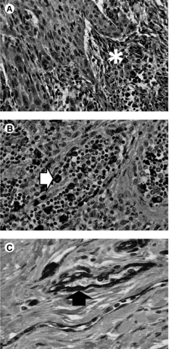
All three biomarkers were positive for SCC of floor of the mouth. MMP-2 had stronger staining for fibroblasts in the tumor stroma and weaker for epithelial tumor cells and inflammatory cells. MMP-9 expression was concentrated in the macrophage-rich inflammatory infiltrate but had
also weak staining for fibroblast-like cells and tumor cells in the basal layer of epithelium. CD31 staining vascular endothelium aided identification of blood microvessels and obtaining the value of MVD (Fig. 1).
The distribution of demographic and clinical variables according to MMP-2 (Table 1) and MMP-9 expressions (Table 2) showed labeling with MMP-2 correlated to N positive stage, indicating lymph node metastasis at the time of initial diagnosis while the positivity for MMP-9 is not related to the demographic data (p>0.05). The analysis of demographic and clinical variables in function of MVD showed a lower degree of embolization (Table 3). The presence of only one case of bone invasion this category inappropriate for the assessment of correlation with molecular variables and the prognosis of patients.
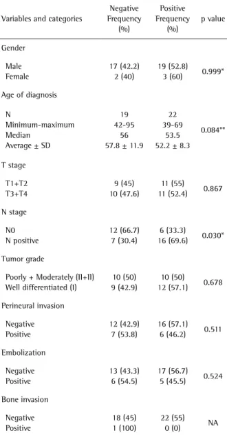
Table 1. Distribution of demographic variables, TNM staging and histopathological variables for MMP-2 immunostaining
Variables and categories
Negative Frequency
(%)
Positive Frequency
(%)
p value
Gender
Male Female
17 (42.2) 2 (40)
19 (52.8)
3 (60) 0.999*
Age of diagnosis
N
Minimum-maximum Median
Average ± SD
19 42-95
56 57.8 ± 11.9
22 39-69
53.5 52.2 ± 8.3
0.084**
T stage
T1+T2 T3+T4
9 (45) 10 (47.6)
11 (55)
11 (52.4) 0.867
N stage
N0 N positive
12 (66.7) 7 (30.4)
6 (33.3)
16 (69.6) 0.030*
Tumor grade
Poorly + Moderately (II+II) Well differentiated (I)
10 (50) 9 (42.9)
10 (50)
12 (57.1) 0.678
Perineural invasion
Negative Positive
12 (42.9) 7 (53.8)
16 (57.1) 6 (46.2) 0.511
Embolization
Negative Positive
13 (43.3) 6 (54.5)
17 (56.7)
5 (45.5) 0.524
Bone invasion
Negative Positive
18 (45) 1 (100)
22 (55)
0 (0) NA
6
F
. Monteiro-Amado et al.
Analysis of distribution in negative and positive cases among molecular variables showed no correlation of MMPs expression with MVD. However, there was negative correlation between MMP-9 and MMP-2 (p=0.018, Mann-Whitney U test, data not shown).
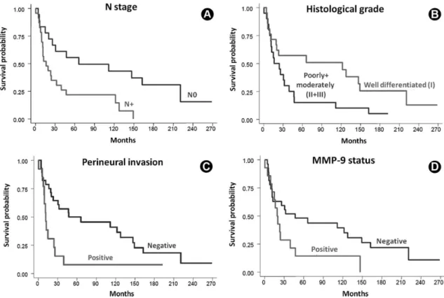
The relationship between the analyzed variables and patient survival is shown in Figure 2. The 5-year survival rate was 36.6% and the 10-year rate was 30.6%. The Iogrank test revealed that positive N stage, a poorly differentiated histological grade, the perineural invasion and MMP-9 expression were associated with poorer survival rates.
Discussion
Tumorigenesis and tumor progression are based on at least seven fundamental changes in cell physiology: 1)
production of signs of autocrine growth; 2) insensitivity to signs of growth inhibition; 3) escape from apoptosis; 4) loss of senescence; 5) maintenance of angiogenesis; 6) tissue invasion; 7) metastases (7). The ability to invade tissues and establish colonies in remote sites certainly are characteristics that define malignant neoplasms. Since metastases are the main cause of death in cancer patients, a better understanding of the processes of tumor invasion and metastasis is indispensable and it can lead to the identification of new forms of treatment (3).
There is no consensus about the cell types most directly involved in the production of MMP-2 and MMP-9 in the tumor progression. Some authors state that tumor cells produce higher levels of proteolytic enzymes than normal cells (8) or indirectly stimulate the adjacent stromal cells
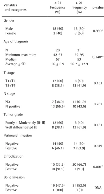
Table 3. Distribution of demographic variables, TNM staging and histopathological variables for CD31/MVD immunostaining
Variables and categories
≤ 21 Frequency (%) > 21 Frequency (%) p-value Gender Male Female 18 (50) 2 (40) 18 (50)
3 (60) 0.999*
Age of diagnosis
N
Minimum-maximum Median
Average ± SD
20 42-67
57 56 ± 6.9
21 39-95
53 56.7 ± 12.9
0.140** T stage T1+T2 T3+T4 12 (60) 8 (38.1) 8 (40)
13 (61.9) 0.161
N stage N0 N positive 7 (38.9) 13 (56.5) 11 (61.9)
10 (43.5) 0.262
Tumor grade
Poorly + Moderately (II+II) Well differentiated (I)
12 (60) 8 (38.1)
8 (40)
13 (61.9) 0.161
Perineural invasion Negative Positive 14 (50) 6 (46.1) 14 (50)
7 (53.9) 0.819
Embolization Negative Positive 10 (33.3) 10 (91.9) 20 (66.7)
1 (9.1) 0.001*
Bone invasion Negative Positive 19 (47.5) 1 (100) 21 (52.5)
0 (0) DNA
SD: standard deviation; NA: not applicable; p: Chi square frequency test; *p: Fisher’s exact test; **p: Mann-Whitney U test.
Table 2. Distribution of demographic variables, TNM staging and histopathological variables for MMP-9 immunostaining
Variables and categories Negative Frequency (%) Positive Frequency (%) p value Gender Male Female 25 (69.4) 2 (40) 11 (30.4)
3 (60) 0.317*
Age of diagnosis
N
Minimum-maximum Median
Average ± SD
27 40-95
54 55.48 ± 11.5
14 39-67
55 53.5 ± 8.1
0.783** T stage T1+T2 T3+T4 15 (75) 12 (57.1) 5 (25)
9 (42.9) 0.228
N stage N0 N positive 14 (77.8) 13 (56.5) 4 (22.2)
10 (43.5) 0.154
Tumor grade
Poorly + Moderately (II+II) Well differentiated (I)
12 (60) 15 (71.4)
8 (40)
6 (28.6) 0.440
Perineural invasion Negative Positive 20 (71.4) 7 (53.8) 8 (28.6)
6 (46.2) 0.307*
Embolization Negative Positive 19 (63.3) 8 (72.7) 11 (36.7)
3 (27.7) 0.709*
Bone invasion Negative Positive 27 (67.5) 0 (0) 13 (32.5) 1 (100) NA
MMP-2, MMP-9 and CD3
1/MVD in oral carcinomas
to secrete them, opening a tissue space for tumor invasion (3,9). The production of MMP by the stroma can also be interpreted as a feature of epithelial tumors that have lost their ability to produce MMP due to the dedifferentiation process (3). The most accepted hypothesis is the main production of MMP-2 and MMP-9 by stromal tumor cells including fibroblasts, inflammatory cells and endothelial cells, playing an active role in carcinogenesis and tumor invasion (9) similar to the outcomes of this study, with marked expression of MMP-2 and MMP-9 in the stroma. It is important to highlight the participation of the
inflammatory component of a neoplasm, which includes a diverse cell population (neutrophils, dendritic cells, macrophages, eosinophils, mast cells and lymphocytes) able to producing cytokines, mediators, proteases and MMPs, keeping close correlation with modulation of extracellular matrix degradation and tumor invasion (2,10).
MMP-2 was more strongly immunostained in fibroblast-like cells of tumor stroma. Fibroblasts are important in cancer progression due to release of MMP-2 on a larger scale and facilitation for the tumor invasion and metastasis (11). MMP-2 in this work was a rare condition in inflammatory cells. This suggests that the invasion afforded by MMP-2 is not influenced by local inflammation close to the tumor. The significant relationship between MMP-2 and lymph node metastasis corroborates other findings (3). A less expressive staining for MMP-2 in the basal membrane could suggest that tumor cells stimulate the MMPs expression in their environment to promote catabolism of extracellular matrix (type IV collagen) by the phagocytic host cells, paving the way for the tumor expansion (11).
Predominant immunostaining for MMP-9 in inflammatory cells does indicate that an expressive inflammatory reaction around the tumor increases the MMP-9 concentration and the risk of tumor invasion resulting from the action of gelatinase. A greater dissolution of extracellular matrix creates space for the tumor mass, facilitates intratumoral angiogenesis, decreases cellular adhesion and sustains the metastasis (12). However, one might consider that chronic inflammation as the body’s normal reaction can promote the release of compounds that prevent tumor progression (10). MMPs also influence the angiogenesis promoting release of pro-angiogenic molecules (VEGF and angiopoetin) and anti-angiogenic molecules (angiostatin) (11). In particular, MMP-9 action also generates tumstatin and endostatin, cleavage fragments of the alpha-3 chain of type IV collagen and alpha-1 chain of type XVIII collagen with anti-angiogenic properties (12). For this dual influence in the mechanism of angiogenesis, it was considered plausible the lack of correlation between MMPs and MVD studied in this work, corroborating other similar results (13). On the other hand, the relationship found between MMP-9 and poor prognosis refutes other findings (14) and deserves further study.
Tumor vascularization is an essential factor for tumor progression. Without angiogenesis, the tumor mass cannot exceed a critical size and is restricted to a tissue diffusion distance of about 0.2 mm (15). In the present study, no relationship was found between MVD and clinical or histological parameters such as T or N stage, histological grade, perineural invasion or patient survival. Other authors have equally failed to find such associations in SCC of head and neck, which may indicate that the tumor Figure 1. Immunohistochemical evaluation of SCC of the floor of the
8
F
. Monteiro-Amado et al.
physiopathology is more complex than the mere presence or absence of vessels, and the interaction of various factors that influence tumor progression makes it difficult to objectively evaluate the effect of each individual factor (4,16). This behavior could be explained in the use of hot spot method, highly variable for counting of MVD in low-vascularization regions between SCC islets leading to misinterpretation (4), or due to unspecific immunostaining of newly formed blood vessels and pre-existing ones, disabling its prognostic importance (16). Furthermore, the fact that different sites have different degrees of vascularization may hinder the evaluation of recurrence and metastasis rates (4,6,17). The present study comprises only SCCs of the floor of the mouth and yet no such correlations were found, as opposite to other studies with correlation between MVD and tumor aggressiveness (13), tumor size (18) or tumor grade (19). There was a negative correlation between MVD and embolization, which may be attributed to a deficiency of the newly formed vessels in performing their usual functions, such as gas exchange and supplying nutrients to the neoplasm (15). Furthermore, the presence of endothelial cells may favor tumor invasion through cytokine production, thus creating a chemically attractive environment for tumor cells (20). On the other hand, if one considers that new vessels formed in an uncontrolled way in a tumor mass are not always functional, their capabilities of chemoattraction and of supplying nutrients to the tumor
may be impaired, thus hindering the penetration of tumor cells and explaining the lack of correlation between the MVD and the other evaluated parameters.
The literature attempts to establish an association between some histological variables and SCC prognosis. Factors such as lymph node metastases and histological grade have a clear correlation with cancer prognosis (2,21,22). The present study found a significant correlation between survival, lymph node metastases, more aggressive histological grades (II and III) and perineural invasion. The latter has been reported as a negative prognostic factor in the development of SCC (2,5). Tumor propagation could occur through the perineural connective tissue that envelops the peripheral nerves because of its low resistance (23). Other authors reported that malignant tumors of the floor of mouth with perineural space invasion are more prone to metastasize to cervical lymph nodes, suggesting higher aggressiveness and more recurrences (24). The overexpression of neural growth factor in salivary gland carcinomas may be a reason for their affinity for nerves, perhaps being a marker for perineural invasion in such lesions in humans (25). Although the present study has shown a significant correlation between perineural invasion and poor survival, data that could aid in the selection of a more effective therapy, more studies are needed in order to determine the molecular mechanisms involved in this process.
MMP-2, MMP-9 and CD3
1/MVD in oral carcinomas
Based on the results obtained in SCC of the floor of the mouth, it is possible to conclude that MMP-2 expression could be indicative of lymph node metastasis and MMP-9 is associated with lower probability of survival. There was no significant association between immunoexpression of CD31/MVD and the other variables.
Resumo
O objetivo deste estudo foi avaliar a imunoexpressão de 2, MMP-9 e CD31/densidade microvascular em carcinomas espinocelulares de soalho bucal e correlacionar os resultados com variáveis demográficas, de sobrevida, clínicas (estadiamento TNM) e histopatológicas (grau de diferenciação tumoral, invasão perineural, embolização e invasão óssea). Dados de prontuários e de diagnósticos de 41 pacientes foram revisados. Cortes histológicos foram submetidos à imunomarcação usando anticorpos primários para MMP-2, MMP-9 e CD31 humanos e sistema streptoavidina-biotina-imunoperoxidase. Análise histomorfométrica quantificou a positividade para MMPs (20 campos, grade de 100 pontos por lâmina, ×200) e para CD31 (microvasos <50 μm na área de maior vascularização, 5 campos, grade de 100 pontos por lâmina, ×400). O planejamento estatístico foi composto pelo teste não paramétrico U de Mann-Whitney (verificação da associação entre variáveis numéricas e imunomarcações), teste de frequências do qui-quadrado (em tabelas de contingência), teste exato de Fisher (quando pelo menos uma frequência esperada foi menor do que 5 em tabelas 2x2), método de Kaplan-Meier (estimativa de probabilidades de sobrevida global) e teste de Iogrank (comparação das curvas de sobrevida), todos com nível de significância de 5%. Houve correlação estatisticamente significante entre imunomarcação para MMP-2 e metástase em linfonodo. Os fatores relacionados negativamente com a sobrevida foram estadiamento N, gradação histopatológica, invasão perineural e imunomarcação de MMP-9. Não houve associação significativa entre imunoexpressão de CD31 e as demais variáveis. A intensidade de imunomarcação para MMP-2 pode ser indicativa de metástase em linfonodo e para MMP-9 de uma menor probabilidade de sobrevida.
Acknowledgements
The following Brazilian Funding Agencies supported this research: CAPES, CNPq (Grant #503209/2004-0), FAPESP (Grant #2001/10707-7) and FINEP-MCT (Grant #01.05.0972.03).
References
1. Ludwig JA, Weinstein JN. Biomarkers in cancer staging, prognosis and treatment selection. Nat Rev Cancer 2005;5:845-856.
2. Lindenblatt RC, Camisasca DR, Martinez GL, Lourenço SQC, Faria PAS. WHO classification and HRA association with clinicopathological features of oral cancer prognostic factors. Rev Bras Odontol 2009;66:65-70.
3. Görögh T, Beier UH, Bäumken J, Meyer JE, Hoffmann M, Gottschlich S, et al.. Metalloproteinases and their inhibitors: influence on tumor invasiveness and metastasis formation in head and neck squamous cell carcinomas. Head Neck 2006;28:31-39.
4. Tse GM, Chan AW, Yu KH, King AD, Wong KT, Chen GG, et al.. Strong immunohistochemical expression of vascular endothelial growth factor predicts overall survival in head and neck squamous cell carcinoma. Ann Surg Oncol 2007;14:3558-3565.
5. Kurtz KA, Hoffman HT, Zimmerman MB, Robinson RA. Perineural and vascular invasion in oral cavity squamous carcinoma: increased incidence on re-review of slides and by using immunohistochemical enhancement. Arch Pathol Lab Med 2005;129:354-359.
6. Franchi A, Santucci M, Masini E, Sardi I, Paglierani M, Gallo O.
Expression of matrix metalloproteinase 1, matrix metalloproteinase 2, and matrix metalloproteinase 9 in carcinoma of the head and neck. Cancer 2002;95:1902-1910.
7. Hanahan D, Weinberg RA. Hallmarks of cancer: the next generation. Cell 2011;144:646-674.
8. Patel BP, Shah PM, Rawal UM, Desai AA, Shah SV, Rawal RM, et al.. Activation of MMP-2 and MMP-9 in patients with oral squamous cell carcinoma. J Surg Oncol 2005;90:81-88.
9. DeClerck YA. Interactions between tumor cells and stromal cells and proteolytic modification of the extracellular matrix by metalloproteinases in cancer. Eur J Cancer 2000;36:1258-1268. 10. Coussens LM, Werb Z. Inflammation and cancer. Nature
2002;420:860-867.
11. Saad S, Gottlieb DJ, Bradstock KF, Overall CM, Bendall LJ. Cancer cell-associated fibronectin induces release of matrix metalloproteinase-2 from normal fibroblasts. Cancer Res 2002;62:283-289.
12. Björklund M, Koivunen E. Gelatinase-mediated migration and invasion of cancer cells. Biochim Biophys Acta 2005;1755:37-69.
13. Guttman D, Stern Y, Shpitzer T, Ulanovski D, Druzd T, Feinmesser R. Expression of MMP-9, TIMP-1, CD-34 and factor-8 as prognostic markers for squamous cell carcinoma of the tongue. Oral Oncol 2004;40:798-803.
14. Kim SH, Cho NH, Kim K, Lee JS, Koo BS, Kim JH, et al.. Correlations of oral tongue cancer invasion with matrix metalloproteinases (MMPs) and vascular endothelial growth factor (VEGF) expression. J Surg Oncol 2006;93:330-337.
15. Carmeliet P, Jain RK. Angiogenesis in cancer and other diseases. Nature 2000;407:249-257.
16. Gleich LL, Biddinger PW, Duperier FD, Gluckman JL. Tumor angiogenesis as a prognostic indicator in T2-T4 oral cavity squamous cell carcinoma: a clinical-pathologic correlation. Head Neck 1997;19:276-280. 17. Dantas DDL, Ramos CCF, Costa ALL, Souza LB, Pereira Pinto L.
Clinical-pathological parameters in squamous cell carcinoma of the tongue. Braz Dent J 2003;14:22-25.
18. Shang ZJ, Li ZB, Li JR. VEGF is up-regulated by hypoxic stimulation and related to tumour angiogenesis and severity of disease in oral squamous cell carcinoma: in vitro and in vivo studies. Int J Oral Maxillofac Surg 2006;35:533-538.
19. Li C, Shintani S, Terakado N, Klosek SK, Ishikawa T, Nakashiro K, et al.. Microvessel density and expression of vascular endothelial growth factor, basic fibroblast growth factor, and platelet-derived endothelial growth factor in oral squamous cell carcinomas. Int J Oral Maxillofac Surg 2005;34:559-565.
20. Warner KA, Miyazawa M, Cordeiro MM, Love WJ, Pinsky MS, Neiva KG, et al.. Endothelial cells enhance tumor cell invasion through a crosstalk mediated by CXC chemokine signaling. Neoplasia 2008;10:131-139. 21. Neves AC, Mesquita RA, Novelli MD, Toddai E, Sousa SOM. Comparison
between immunohistochemical expression of cyclin D1 and p21 and histological malignancy graduation of oral squamous cell carcinomas. Braz Dent J 2004;15:93-98.
22. Siriwardena BS, Tilakaratne A, Amaratunga EA, Tilakartne WM. Demographic, aetiological and survival differences of oral squamous cell carcinoma in the young and the old in Sri Lanka. Oral Oncol 2006;42:831-836.
23. Selander D, Sjöstrand J. Longitudinal spread of intraneurally injected local anesthetics. An experimental study of the initial neural distribution following intraneural injections. Acta Anaesthesiol Scand 1978;22:622-634.
24. Brown B, Barnes L, Mazariegos J, Taylor F, Johnson J, Wagner RL. Prognostic factors in mobile tongue and floor of mouth carcinoma. Cancer 1989;64:1195-1202.
25. Wang L, Sun M, Jiang Y, Yang L, Lei D, Lu C, et al.. Nerve growth factor and tyrosine kinase A in human salivary adenoid cystic carcinoma: expression patterns and effects on in vitro invasive behavior. J Oral Maxillofac Surg 2006;64:636-641.