Rectal dose assessment in patients submitted to
high-dose-rate brachytherapy for uterine cervix cancer*
Avaliação da dose no reto em pacientes submetidas a braquiterapia de alta taxa de dose para o tratamento do câncer do colo uterino
Jetro Pereira de Oliveira1, Luiz Antonio Ribeiro da Rosa2, Delano Valdivino Santos Batista3, Lúcia Helena Bardella4, Arnaldo Rangel Carvalho5
OBJECTIVE: The present study was aimed at developing a thermoluminescent dosimetric system capable of assessing the doses delivered to the rectum of patients submitted to high-dose-rate brachytherapy for uterine cervix cancer. MATERIALS AND METHODS: LiF:Mg,Ti,Na powder was the thermoluminescent material utilized for evaluating the rectal dose. The powder was divided into small portions (34 mg) which were accommodated in a capillary tube. This tube was placed into a rectal probe that was introduced into the patient’s rectum. RESULTS: The doses delivered to the rectum of six patients submitted to high-dose-rate brachytherapy for uterine cervix cancer evaluated by means of thermoluminescent dosimeters presented a good agreement with the planned values based on two orthogonal (anteroposterior and lateral) radiographic images of the patients. CONCLUSION: The thermoluminescent dosimetric system developed in the present study is simple and easy to be utilized as compared to other rectal dosimetry methods. The system has shown to be effective in the evaluation of rectal doses in patients submitted to high-dose-rate brachytherapy for uterine cervix cancer.
Keywords: Rectum; Brachytherapy; HDR; Thermoluminescent dosimetry; Uterine cervix cancer.
OBJETIVO: O objetivo deste trabalho foi desenvolver um sistema dosimétrico termoluminescente capaz de avaliar as doses administradas ao reto de pacientes submetidas a braquiterapia de alta taxa de dose para o tratamento do câncer do colo uterino. MATERIAIS E MÉTODOS: O material termoluminescente utilizado para a avaliação da dose no reto foi o LiF:Mg,Ti,Na na forma de pó. O pó foi separado em pequenas porções de 34 mg, que foram acomodadas em um tubo capilar. Este tubo foi colocado em uma sonda retal, que era introduzida no reto da paciente. RESULTADOS: As doses administradas ao reto de seis pacientes submeti-das a braquiterapia de alta taxa de dose para o tratamento do câncer do colo uterino foram avaliasubmeti-das com dosímetros termoluminescentes e apresentaram boa concordância com os valores planejados, com base em duas radiografias ortogonais da paciente, imagens ântero-posterior e lateral. CONCLUSÃO: O sistema de dosimetria termoluminescente utilizado no presente trabalho é simples e de fácil utilização quando comparado a outros métodos de dosimetria do reto. Ele mostrou-se eficiente na avaliação da dose no reto de pacientes submetidas a braquiterapia de alta taxa de dose para o tratamento do câncer do colo uterino.
Unitermos: Reto; Braquiterapia; HDR; Dosímetro termoluminescente; Câncer do colo uterino. Abstract
Resumo
* Study developed at Instituto Nacional de Câncer (INCA), Rio de Janeiro, RJ, Brazil.
1. Master, Fellow PhD degree, Faculdade de Medicina da Uni-versidade Federal do Rio de Janeiro (UFRJ), Rio de Janeiro, RJ, Brazil.
2. PhD, Researcher at Instituto de Radioproteção e Dosime-tria/Comissão Nacional de Energia Nuclear (IRD/CNEN) in the area of Medical Physics, Rio de Janeiro, RJ, Brazil.
3. Bachelor of Science, Head for the Unit of Medical Physics at Instituto Nacional de Câncer (INCA), Rio de Janeiro, RJ, Brazil. 4. Master, Physicist, Unit of Medical Physics of Instituto Nacio-nal de Câncer (INCA), Rio de Janeiro, RJ, Brazil.
5. Bachelor of Electronic Engineering, Operator at Laboratory of Thermoluminescent Dosimetry, Unit of Medical Physics in Radiotherapy and Nuclear Medicine of Instituto de Radioprote-ção e Dosimetria (SEFME/IRD), Rio de Janeiro, RJ, Brazil.
Mailing address: Dr. Luiz Antonio Ribeiro da Rosa. Instituto de Radioproteção e Dosimetria. Avenida Salvador Allende, s/nº, Recreio dos Bandeirantes. Rio de Janeiro, RJ, Brazil, 22780-160. E-mail: lrosa@ird.gov.br
Received September 14, 2007. Accepted after revision De-cember 12, 2008.
INTRODUCTION
The uterine cervix is a cylindrical and narrow portion of the uterus, measuring 2–4 cm in length, connecting to the ante-rior wall of the vagina, in most of cases lying perpendicularly to this structure. It joins the lower segment of the uterus at the isthmus level, where a subtle narrowing of the lumen is observed.
Uterine cervix cancer represents a con-tinuing challenge to the clinical practice. According to the Instituto Nacional de Câncer (INCA), in 2008 19,000 new cases of the disease are expected to be detected
Oliveira JP, da Rosa LAR, Batista DVS, Bardella LH, Carvalho AR. Rectal dose assessment in patients submitted to high-dose-rate brachytherapy for uterine cervix cancer. Radiol Bras. 2009;42(2):83–88.
in Brazil(1). Among Brazilian women,
uter-ine cervix cancer is the third most frequent type of cancer. Not considering non-mela-noma skin tumors, uterine cervix cancer is the most common one in the Northern re-gion of the country. In the Southern, West-ern and NortheastWest-ern regions this form of cancer is the second most common and in the Southeastern region it is the fourth most common(2).
this disease in less developed countries is twice as high as that in more developed countries(3).
One of the methods used in the treat-ment of uterine cervix cancer with ioniz-ing radiation is radiotherapy. Radiotherapy is divided into teletherapy and brachy-therapy. In brachytherapy, the sources placed next to the tumor are radioisotopes whose radiation penetrates the tissues of interest, releasing energy locally and lead-ing to the death of neoplastic(4) cells.
Brachytherapy is typically utilized as a complementary treatment after the patient has been submitted to teletherapy. In Janu-ary of 2001, the Brazilian radiotherapy completed ten years of experience with high-dose-rate (HDR) brachytherapy, de-fined as the treatment whose doses are higher than 0.2 Gy/min(5).
Undoubtedly, radiotherapy is an effec-tive option for the treatment of uterine cer-vix cancer, but also it poses a risk to radi-osensitive organs adjacent to the uterus, such as the rectum and the bladder. In the case of the rectum, a high dose can cause complications, with possibilities ranging from episodic diarrhea, rectal spasmus and occasional bleeding, up to local ulceration, partial stenosis, reoccurring abundant hem-orrhage, with necrosis and obstruction and, finally, development of rectovaginal fis-tula. An appropriate treatment plan can assure with a high degree of accuracy, a high radiation dose to the tumor, with si-multaneous protection against high dose exposure for the rectum and bladder.
The planning systems utilized in brachytherapy for treatment of uterine cer-vix cancers are based on mathematical models which are naturally limited. When-ever possible, procedures for evaluation of doses to organs at risk are recommended so that it becomes possible to compare these dose values with those established in the treatment plan. Thus, not only the plan correctness is evaluated, but also the irra-diation procedure utilized in the patient is validated. This can be done by adopting in vivo procedures that is, assessing the doses
during treatment by means of thermolumi-nescent dosimeters (TLDs), radiophoto-luminescent glass dosimeters (RPLGDs), metal oxide semiconductor field effect transistors (MOSFETs) or diodes(6–9).
In the present study, the authors pro-posed the utilization of TLD LiF:Mg,Ti,Na detector in the form of a powder, for in vivo
measurement of doses to the rectum of patients during the treatment for uterine cervix cancer utilizing a 192Ir source in
HDR brachytherapy.
Criterion for limitation of doses to the rectum
In brachytherapy, because of the non-homogeneous dose distribution, the dose prescription is more complex than in tele-therapy. Different systems have been devel-oped with different prescription items. The Report 38 of International Commission of Radiation Units and Measurement (ICRU)(10) suggests the standardization of
the manner in which dose and treatment dose are reported. Such report includes a number of items that should be included in the prescription of intracavitary therapy. The technique description, including dose and fractioning, and the temporal ratio with the implant, the type of intracavitary sys-tem applied, the isotope utilized, the ab-sorbed dose in a number of relevant points to the tumor management, the dose to the rectum and to the bladder are some of the items that should be included in the report of intracavitary therapy.
The points of relevance in the manage-ment of the tumor are located in the pelvic wall and in the lymphatic Fletcher trap-ezoid. The critical reference points for the bladder and the rectum are defined in the vesical trigone (on the surface of the Foley catheter balloon filled with 7 ml of contrast agent) and on the anterior rectal wall (0.5 cm beyond the posterior vaginal wall, on a line passing by the medium point of the colpostat source). The vesical trigone is a geometric figure formed by an inverted tri-angle, whose vertices correspond to the points where the urethers connect to the bladder and to the urethral orifice in the bladder(10).
One of the first essential steps to deter-mine the dose distribution in brachytherapy is based on the determination of the source positioning in relation to the target volume and other anatomic characteristics of inter-est. The point A prescription, in spite of being criticized by many authors and by the ICRU Report 38(10), is still the most
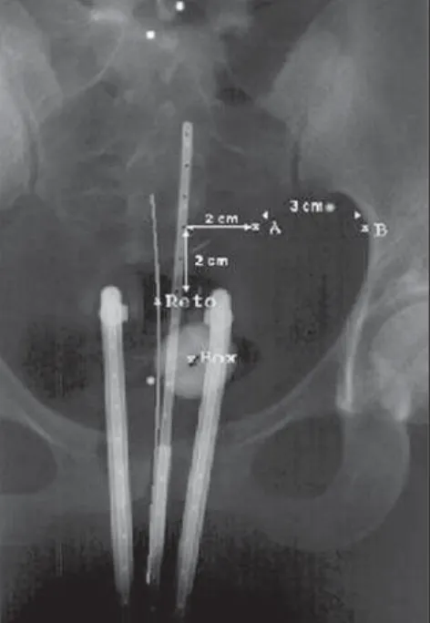
fre-quently utilized method of dose prescrip-tion. Point A was originally described as being located 2.0 cm above the vaginal fornix and 2.0 cm laterally in relation to the uterine canal, having been defined to re-flect an average dose inside the paracer-vical triangle containing critical vascular structures (tolerance pyramid). Ideally, point A represents the intersection of the urether with the uterine artery. The point B, 2.0 cm above and 5.0 cm laterally, follow-ing the same origin system utilized in the localization of point A, represents the pel-vic wall and, particularly, the lymph nodes, and can be seen on Figure 1. The dose to point B typically corresponds to one third of the dose applied to point A.
MATERIALS AND METHODS
The thermoluminescent powder utilized was lithium fluoride doped with magne-sium, titanium and sodium (LiF:Mg,Ti,Na). This compound is produced by the French company Philitech (Philitech; Paris, France) under the code DTL937. This prod-uct is enriched with 7Li (99.994%).
The pre-irradiation thermal therapy uti-lized for material regeneration was 450 °C for three hours. An ETT oven (Fimel; Paris, France) was utilized for this thermal
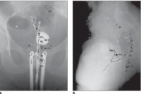
therapy. The thermoluminescent powder, once irradiated, is evaluated in a PCL3 automatic reader. The post-irradiation ther-mal therapy is carried out in the thermolu-minescent reader itself at a temperature of 125 °C for five seconds. This temperature is lower than the temperature utilized in the evaluation of the TLDs which is 440 °C. The equipment is produced by the French company Fimel, and can read about 34 mg of the material placed in small stainless steel containers, as seen on Figure 2.
The dose limitation criterion adopted by INCA was four applications at one-week intervals, each one of 700 cGy (prescribed dose on point A), with a maximum of 65% of this dose reaching the rectum. For the bladder and the sigmoid colon, the dose is limited to 70% and 55% respectively, to the dose on point A.
Before assessing the doses in the rec-tum, the reproducibility and linearity of the dosimetric system was evaluated. The ex-periment to evaluate the linearity was con-ducted in a cubic acrylic phantom measur-ing 38.0 × 38.0 × 30.5 cm3, filled with
water. Approximately 34 mg of the dosim-etric powder were placed in 20.0 cm long capillaries with 3.0 mm in diameter. The powder was placed in the posterior part of the capillaries. These were positioned at 3.0 cm from the 192Ir source and were irradiated
three times at different dose levels, from 4.9 to 902.3 cGy.
Blocks of “solid water” were utilized for TLDs calibration as a function of the absorbed in water. This procedure was cho-sen in order to minimize the uncertainty level in the dosimeter positions relative to the source, considering that because of the high dose gradient, the accuracy in such positioning is very important.
The use of solid water in the current study, instead of normal liquid water, was based on the results of Meli et al.(11), who,
by using Monte Carlo simulation, demon-strated the appropriateness of the simula-tor material for measurements with 192Ir
sources.
The thermoluminescent powder samples were calibrated at a 6.0 cm-distance from the 192Ir source. The absorbed dose values
in water in the points where the samples of thermoluminescent material were placed, was calculated using the mathematical
for-mulas developed in the Report 51 of the American Association of Physicists in Medicine (AAPM)(12), based on
measure-ment of air kerma rates at 10.0 cm from the source using the Farmer chamber with the calibration factor determined by Maré-chal(13–15). After TLDs calibration for the
energy spectrum of gamma radiation from
192Ir, the method was tried with patients
undergoing treatment at INCA.
In the routine gynecological procedure, before the treatment is actually initiated a probe is introduced into the patient’s
rec-tum. This probe is a disposable tube made of non-toxic siliconized polyvinyl chloride and sterilized by gamma radiation. Radio-paque wires are placed within this probe in the gynecologic applicators. Before expo-sure to therapeutic 192Ir radiation, the
pa-tient is exposed to x-rays, and anterior-pos-terior and lateral images are obtained for therapy planning, as seen on Figure 3. Therefore, the doses delivered along the rectum, more precisely along the radio-paque wire within the rectal probe, can be planned. By replacing the wire by the
cap-Figure 2.A: Automatic PCL3 reader. B: Stainless steel containers with millimetric dimensions for con-tainment of thermoluminescent material during its evaluation on the PCL3 reader.
A B
Figure 3. Anteroposterior and lateral radiographic images of a patient to be submitted to HDR brachytherapy or uterine cervix cancer, showing the gynecologic applicator and the rectal probe containing radiopaque wires.
Figure 4. Representation of a capillary with ten measurement positions (small compartments containing thermoluminescent material) inserted in the rectal probe.
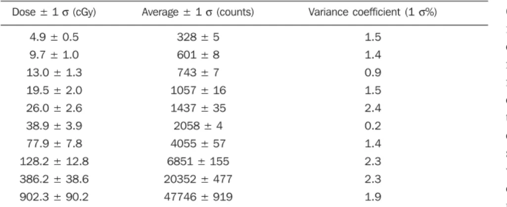
Table 1 Study of linearity andreproducibility of DTL937 response to gamma radiation from 192Ir.
Dose ± 1 σ (cGy)
4.9 ± 0.5 9.7 ± 1.0 13.0 ± 1.3 19.5 ± 2.0 26.0 ± 2.6 38.9 ± 3.9 77.9 ± 7.8 128.2 ± 12.8 386.2 ± 38.6 902.3 ± 90.2
Average ± 1 σ (counts)
328 ± 5 601 ± 8 743 ± 7 1057 ± 16 1437 ± 35 2058 ± 4 4055 ± 57 6851 ± 155 20352 ± 477 47746 ± 919
Variance coefficient (1 σ%)
1.5 1.4 0.9 1.5 2.4 0.2 1.4 2.3 2.3 1.9
ted to HDR brachytherapy for uterine cer-vix cancer. The limit dose determined for the rectum according to the criterion adopted by INCA is shown, as well as the maximum dose obtained by the planning system for the rectum, the maximum dose measured in the rectum with TLDs, the average dose obtained by means of the planning system for the rectum, and the average dose in the rectum measured by the TLDs.
Although the TLDs response presents a good reproducibility, (1.6 ± 0.7)%, on av-erage, the uncertainty in experimental mea-surements and in the dose values obtained by the planning system achieves about 10%. The TLDs calibration was based on mathematical formulas developed in the AAPM Report 51(12). The planning system
utilized for the calculation of delivered dose to the target volume and to the rectum in brachytherapy is based the same formu-las. In the AAPM(12) document, with basis
on the quadratic propagation of uncertainty involved in the formulas, estimated uncer-tainty level is 10% for the calculated dose.
DISCUSSION
Considering the uncertainty associated with measured doses and the planned ones (Table 2), one observes that in the six cases included in the present study the doses delivered to the patients rectums were at a maximum, equal to the limit doses deter-mined for the organ, following the adopted criterion and taking the estimated uncer-tainties into account. As far as the average dose in the rectum is concerned, the mea-surements presented a good agreement with the planned values. The same can be observed in relation to maximum dose val-ues. In the cases of patients 1 and 2, there
Table 2 Results from rectal dosimetry obtained from patients submitted to HDR brachytherapy for uterine cervix cancer. The rectal limit dose determined according to criterion adopted by INCA, the maximum dose obtained by the planning system for the rectum, the maximum measured dose in the rectum with the aid of TLDs, the average dose obtained by the planning system for the rectum, and the average measured rectal dose via thermoluminescent dosimetry are shown.
Patient
1 2 3 4 5 6
Limit dose (cGy)
455 455 455 455 455 455
Maximum planned dose (cGy)
382.7 ± 38 437.2 ± 44 317.0 ± 31 295.5 ± 29 382.0 ± 38 457.0 ± 45
Maximum measured dose(cGy)
415.1 ± 42 406.0 ± 40 308.0 ± 30 288.4 ± 29 390.1 ± 39 465.9 ± 46
Deviation (%)
7.8 –7.6 –2.8 –2.5 2.1 1.9
Maximum planned dose (cGy)
216.0 ± 22 280.3 ± 28 226.5 ± 23 170.9 ± 17 275.3 ± 27 368.3 ± 37
Maximum measured dose(cGy)
228.7 ± 23 263.8 ± 26 222.7 ± 22 165.2 ± 16 276.4 ± 28 373.6 ± 37
Deviation (%)
5.5 –6.3 –1.7 –3.5 0.4 2.2
illary containing thermoluminescent pow-der, before the patient is exposed to 192Ir, it
is possible to evaluate the dose along this organ during treatment and comparing the experimental values with those determined by the planning system, allowing a new planning for the following therapy applica-tion, in cases where the measured doses were higher than the limit value for the rectum. The capillary utilized for measur-ing the rectal dose is 20.0 cm long and 3.0 mm in diameter. It is divided in ten small compartments, each filled with 34 mg of the dosimetric powder. The capillary is very carefully introduced into the rectal probe, so that such probe is kept in the same po-sition it held when the radiopaque wire was in place. Figure 4 shows a drawing of this capillary.
Before collaborate with the investiga-tion, all patients were explained about the type of experiment to be developed, and
agreed in participating. In total, six patients participated in the investigation.
RESULTS
Table 1 presents the study results on the reproducibility of the DTL937 response to gamma radiation energy from 192Ir. Three
irradiations were made for each value of absorbed dose. The thermoluminescent response of the dosimetric material pre-sents good reproducibility, lower than 2.4% for the considered dose interval, between 4.9 and 902.3 cGy (mean and standard deviation: 1.6 ± 0.7%).
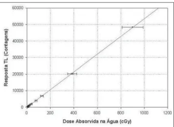
The TLD response presents a linear be-havior, making it unnecessary to apply lin-earity correction factors to the results as it can be seen on Figure 5. The linear regres-sion adjustment factor (r) is 0.9999.
submit-are bigger differences between planned values and the measured ones. Possibly, because internal movements of the organs, the positioning of the thermoluminescent dosimeters may not have been as accurate as in the other patients. It is extremely im-portant that the dosimeters are positioned in the same point utilized by the planning system for dose calculation.
The uncertainty of experimental mea-surements and of dose values obtained by the planning system achieves about 10%. This is due to the inherent uncertainty of dose calculation based on the AAPM docu-ment TG 43(12) which is approximately 10%.
Lambert et al.(7) have studied different
dosimeters for in vivo dosimetry applica-tions during treatment with HDR brachy-therapy. Thermoluminescent, MOSFETs, diamonds and scintillation detectors were evaluated. The diamond detectors pre-sented the most accurate results; however they are too large for dose measurements in HDR brachytherapy, in which the dose gradients are very high. The MOSFETs pre-sented errors rates between 30% and 40% in measurements at more than 5.0 cm from the source, although for measurements at a distance between 2.0 and 5.0 cm, the er-rors rate were at the order of 5%. The scin-tillation detector presented a dosimetric accuracy of 3% for measurements made between 1.0 and 10.0 cm from the brachy-therapy source. The authors’ criticism in relation to TLDs was due to their incapac-ity of carrying out real-time measurements, although their dosimetric performance was better than the MOSFETs performance. It is important note that diamond and
scintil-lation detectors are not commonly utilized in radiotherapy centers, and that TLDs, di-odes and MOSFETs are most widely uti-lized in medical physics for in vivo mea-surements(16).
Sakata et al.(6) have measured doses in
the rectum utilizing semiconductor dosim-eters during intracavitary brachytherapy procedures for treatment of gynecologic cancer in 105 patients. The results indicate differences of up to 5% between planned and measured doses for 30.8% of the pa-tients. This difference may reach 10% for 56% of the patients and up to 20% for 85% of the patients. For 15% of the patients, the differences between planned and measured doses were higher than 20%.
Waldhäusl et al.(9), using diodes, have
measured the dose in the rectum in 55 HDR brachytherapy procedures in patients with gynecologic cancer. Differences from – 31% to +90% were found between mea-sured values and calculated values for de-livered doses to the organ during treatment for uterine cancer. The main reason for such high differences was due to uncer-tainty in the detectors positioning.
Contrary to results reported by Wald-häusl et al.(9) and Sakata et al.(6), in the
present study the differences between planned dose values and measured dose values were always bellow 8%, with sev-eral values lower than 3.5%. Forty four percent of the differences between calcu-lated and measured dose values in the rec-tum by Sakata et al.(6) were greater than
10%, and 15% of these were greater than 20%. Waldhäusl et al.(9) have found
differ-ences ranging from –31% to +90%
be-tween calculated and measured doses in the rectum of patients submitted to HDR brachytherapy. It is important to note that Sakata et al.(6) have studied 105 patients,
and the results reported by Waldhäusl et al.(9) were based on 55 applications of HDR
brachytherapy. In the present study, only six patients were studied and, therefore, its sta-tistical significance is very poor when com-pared to that of the mentioned bibliogra-phy(6,9). Thus, based on the present results
one cannot say that the procedure herein described is superior to the procedures uti-lized by the mentioned authors(6,9),
how-ever, the method has shown to be appropri-ate for the evaluation of rectal doses, and can indicate a need for reevaluation of the therapy plan in the case of detection of rec-tal doses considerably higher (> 10%) than planned.
Sakata et al.(6) and Waldhäusl et al.(9)
have utilized diodes in their studies. It is important to mention that according to Lambert et al.(7), the diodes are less
accu-rate than TLDs in in vivo dose evaluations. In the present study, thermoluminescent dosimetry was the technique utilized.
TLDs have the inconvenience of not allowing a real-time dose evaluation, con-trary to diodes or MOSFETs. However, due to the fact that they do not require wires and electrometers during measurement, they are more suitable to in vivo measurements, causing less trouble to patients. They are also smaller than the diodes, which is also an advantage for a dosimeter for in vivo dose evaluations. The MOSFETs can also be very small, such as the micro-MOS-FETs. TLDs are also less subject to patient temperature variations when compared to diodes(17).
The dosimetric procedure described in the present study allows the evaluation of the dose delivered to the rectum as a con-sequence of the HDR brachytherapy for uterine cervix cancer. Such evaluation makes it possible for the medical team to make decisions on the best approaches for the continuation of the treatment adminis-tered to the patients, when the measured doses are higher than the planned ones, in order to protect the organ, which contrib-utes to minimizing complications that may occur when the rectum is exposed to high radiation doses.
CONCLUSION
The thermoluminescent dosimetry sys-tem utilized in the present study, is simple and easy to use when compared to other possible dosimetric techniques applicable to in vivo dosimetry in the rectum, and has shown to be efficient in the evaluation of rectal dose in patients submitted to HDR brachytherapy for uterine cervix cancer, allowing the reevaluation of the therapy plan between the first and second weekly applications, when doses above the limit (> 10%) are measured in the organ to be protected.
Acknowledgements
To Comissão Nacional de Energia Nu-clear (CNEN) for the financial support, to Dr. Andrés Reinaldo Rodrigues Papa, first Coordinator of the Comissão de Pós-Gra-duação do Instituto de Radioproteção e Dosimetria (CPG/IRD), for the support given to the first author during his Master’s course, to the brachytherapy team at INCA, for the constant support during the mea-surements phase, as well as to the persons who extended their help in the conception of this study.
REFERENCES
1. Instituto Nacional de Câncer. Estimativa 2008: incidência de câncer no Brasil. Rio de Janeiro: INCA; 2007. p. 24.
2. Instituto Nacional de Câncer. Estimativa 2008: incidência de câncer no Brasil. Rio de Janeiro: INCA; 2007. p. 32.
3. Salvajoli JV, Souhami L, Faria LS. Radioterapia em oncologia. 1ªed. Rio de Janeiro: Editora Mé-dica e Científica; 1999.
4. Calcina CSG, Almeida A, Rocha JRO. Análises de protocolos de braquiterapia, por alta taxa de dose, do controle de qualidade de alguns servi-ços locais, baseados no TG40, TG56 e ARCAL XXX. Radiol Bras. 2001;34:225–32. 5. Esteves SCB, Oliveira ACZ, Feijó LFA.
Braqui-terapia de alta taxa de dose no Brasil. Radiol Bras. 2004;37:337–41.
6. Sakata K, Nagakura H, Oouchi A, et al. High-dose-rate intracavitary brachytherapy: results of analyses of late rectal complications. Int J Radiat Oncol Biol Phys. 2002;54:1369–76.
7. Lambert J, Nakano T, Law S, et al. In vivo dosim-eters for HDR brachytherapy: a comparison of a diamond detector, MOSFET, TLD, and scintilla-tion detector. Med Phys. 2007;34:1759–65. 8. Nose T, Koizumi M, Yoshida K, et al. In vivo
do-simetry of high-dose-rate interstitial brachytherapy in the pelvic region: use of radiophotolumi-nescence glass dosimeter for measurement of 1004 points in 66 patients with pelvic malignancy. Int J Radiat Oncol Biol Phys. 2008;70:626–33. 9. Waldhäusl C, Wambersie A, Pötter R, et al.
In-vivo dosimetry for gynaecological brachytherapy: physical and clinical considerations. Radiother Oncol. 2005;77:310–7.
10. International Commission on Radiation Units and Measurements. Dose and volume specification for reporting intracavitary therapy in gynaecology. ICRU Report 38. Bethesda: ICRU; 1985. p. 1–16. 11. Meli J, Meigooni A, Nath R. On the choice of phan-tom material for the dosimetry of 192Ir sources. Int J Radiat Oncol Biol Phys. 1988;14:587–94.
12. American Association of Physicists in Medicine. Dosimetry of interstitial brachytherapy sources: recommendations of the AAPM Radiation Therapy Committee Task Group No. 43. AAPM Report No. 51. Woodbury: American Institute of Physics; 1995. [Reprinted from Med Phys. 1995; 22:209–34].
13. Maréchal MH. Recomendações para calibração de fontes de 192Ir de alta taxa de dose [tese de dou-torado]. Rio de Janeiro: Universidade do Estado do Rio de Janeiro; 2000.
14. Ferreira IH, Almeida CE, Marre D, et al. Monte Carlo calculations of the ionization chamber wall correction factors for 192Ir and 60Co gamma rays and 250 kV x-rays for use in calibration of 192Ir HDR brachytherapy sources. Phys Med Biol. 1999;44:1897–904.
15. Maréchal MH, Almeida CE, Ferreira IH, et al. Experimental derivation of wall correction factors for ionization chambers used in high dose rate 192Ir source calibration. Med Phys. 2002;29:1–5.
16. Kron T. Applications of thermoluminescence do-simetry in medicine. Radiat Prot Dodo-simetry. 1999;85:333–40.