167
Radiol Bras. 2008 Mai/Jun;41(3):167–172
Features of cystic breast lesions at ultrasound
elastography*
Apresentação das lesões mamárias císticas à ultra-sonografia utilizando a elastografia
Eduardo de Faria Castro Fleury1, José Francisco Rinaldi2, Sebastião Piato3, José Carlos Fleury4, Décio Roveda Jr.5
OBJECTIVE: To demonstrate the most frequent features of cystic breast lesions at ultrasound elastography, discussing the applicability of this method. MATERIALS AND METHODS: The present casuistic included 150 patients referred for percutaneous breast biopsy of 175 lesions. Histologically diagnosed solid lesions (153 lesions) were excluded; lesions histologically diagnosed as cystic (22 lesions), including complicated cysts, papillary lesions, inflammatory lesions, typical columnar cell hyperplasia and duct ectasia were retrospectively classified by means of elastography, according to a scoring system developed by the authors, with categories ranging between 1 and 4. RESULTS: Thirteen (59%) of the 22 lesions evaluated corresponded to cysts, one (4.6%) to duct ectasia, two (9.2%) to inflammatory lesions, five (22.6%) to papillary lesions, and one (4.6%) to columnar cell hyperplasia. The scoring system was applied with the following results: 17 category 2 lesions, four category 3 lesions, one category 4 lesion, and none category 1 lesion, with a 95% specificity. CONCLUSION: Different features of cystic breast lesions are demonstrated by elastography according to histological results, representing a useful and easily applicable method for differentiating benign from malignant breast lesions. Keywords: Breast cyst; Ultrasonography; Breast.
OBJETIVO: Demonstrar a apresentação mais freqüente das lesões mamárias císticas utilizando a elastogra-fia e discutir a sua aplicabilidade. MATERIAIS E MÉTODOS: A casuística compôs-se de 150 pacientes enca-minhadas para realização de biópsia mamária percutânea com 175 lesões. Foram excluídas as lesões com diagnóstico histológico de lesões sólidas (153 lesões) e incluídas as lesões com características císticas à histologia (22 lesões), incluindo cistos complicados, lesões papilíferas, lesões inflamatórias, hiperplasia de células colunares típica e ectasia ductal. Estas lesões foram classificadas de forma retrospectiva por meio da elastografia, conforme escores criados pelos autores, variando de 1 a 4. RESULTADOS: Das 22 lesões en-caminhadas, 13 (59%) correspondiam a cistos, uma (4,6%) a ectasia ductal, duas (9,2%) a lesões inflama-tórias, cinco (22,6%) a lesões papilíferas e uma (4,6%) a hiperplasia de células colunares. Foram encontra-dos 17 escores 2, quatro escores 3, um escore 4 e nenhum escore 1, com especificidade de 95%. CONCLU-SÃO: As lesões císticas mamárias têm diferentes apresentações à elastografia, conforme o resultado histo-lógico, sendo este um método útil para a sua diferenciação e de fácil aplicabilidade na clínica diária. Unitermos: Cisto mamário; Ultra-sonografia; Mama.
Abstract
Resumo
* Study developed at Faculdade de Ciências Médicas da Santa Casa de São Paulo, São Paulo, SP, Brazil.
1. PhD degree, MD, Second Assistant at Santa Casa de São Paulo, São Paulo, SP, Brazil.
2. Assistant Professor, Head for Clinic of Mastology, Depart-ment of Obstetrics and Gynecology at Irmandade da Santa Casa de Misericórdia de São Paulo, São Paulo, SP, Brazil.
3. Full Professor, Head for Department of Obstetrics and Gynecology at Irmandade da Santa Casa de Misericórdia de São Paulo, São Paulo, SP, Brazil.
4. Specialist in Imaging Diagnosis, Coordinator for the Unit of Breast Intervention at Centro de Tomografia Computadorizada – CTC Gênese, São Paulo, SP, Brazil.
5. Professor/Instructor, Director for the Unit of Imaging Diag-nosis at Faculdade de Ciências Médicas da Santa Casa de São Paulo, São Paulo, SP, Brazil.
Mailing address: Dr Eduardo de Faria Castro Fleury. Alameda Ministro Rocha Azevedo, 1368, ap. 52, Jardins. São Paulo, SP, Brazil, 01410-002. E-mail: [email protected]
Received August 23, 2007. Accepted after revision October 16, 2007.
screening for breast cancer in young women who have presented dense breasts at mammography (BI-RADS® categories 3 and 4)(2–4).
One of the problems resulting from the widespread utilization of ultrasonography as a method for screening in these patients was the visualization of new alterations in the breast tissue generally not related to malignancy. Frequently, nodules usually associated with benignity which typically could not be visualized started being de-tected, with the presence of cysts with a thick content (complicated cysts). These cysts can hardly be differentiated from true nodules by the conventional method, and
Fleury EFC, Rinaldi JF, Piato S, Fleury JC, Roveda Jr D. Features of cystic breast lesions at ultrasound elastography. Radiol Bras. 2008;41(3):167–172.
INTRODUCTION
The utilization of breast ultrasonogra-phy was disseminated in the eighties as an ancillary method in the differentiation be-tween solid and cystic lesions of the breast, aiding in the diagnosis of nodules detected by mammography(1). Since the decade of
1990, with the introduction of higher fre-quency transducers, ultrasonography has allowed not only the differentiation be-tween solid and cystic lesions, but also a clear-sighted analysis of the lesions, con-solidating its role as an adjuvant diagnos-tic method up to nowadays when it is pro-posed by some authors as a method of
168 Radiol Bras. 2008 Mai/Jun;41(3):167–172 generally are classified as indeterminate
nodules, causing anxiety in the patients who end up opting for diagnostic breast biopsy(5,6).
One of the greatest challenges for ultra-sonography is to allow the differentiation between these two entities without increas-ing costs or necessity of interventional pro-cedures. Studies aiming at increasing the ultrasonography accuracy have been devel-oped about methods supplementary to ul-trasonography to increase its accuracy, with the development of Doppler fluxmetry, ultrasound harmonic imaging, ultrasound elastography and the streaming detection technique in breast ultrasonography(7-10).
The present study approaches the fea-tures of cystic breast lesions at ultrasound elastography in correlation with a scoring system developed by the authors, in pa-tients referred to the Institution for diagnos-tic biopsy. Also, the clinical applicability of the method is discussed.
MATERIALS AND METHODS
Retrospective study approved by the Institutional Committee for Ethics in Re-search, developed at the Unit of Imaging Diagnosis of Santa Casa de Misericórdia de São Paulo, evaluating histological results of 150 patients in the age range between 24 and 70 years (mean, 45 years) who pre-sented 170 lesions at conventional ultra-sonographic studies and were referred to the Center of Computed Tomography for percutaneous breast biopsy in the period between May 1st and June 30, 2007. The mean diameter of lesions was 1.4 cm (me-dian, 1.2 cm; range, 0.5–3.2 cm). One hun-dred and thirty patients with 148 exclu-sively solid lesions at histology were ex-cluded. The remaining 20 patients with 22 lesions histologically diagnosed as purely cystic (complicated cysts), inflammatory lesions and ductal ectasia, or cystic lesions associated with solid components, such as papillary lesions and typical columnar cell hyperplasia, based on 17 (91.9%) fragment biopsies and 5 (8.1%) preoperative needle localization.
Pathological diagnosis
Specimens were sent for histological study and were analyzed by a specialized
pathologist with 17-year experience in breast lesions. The lesions were classified into cysts, papillary lesions, inflammatory lesions, typical columnar cells hyperplasia and ductal ectasia(11,12).
Equipment
Both the conventional study and the elastography were performed by a same radiologist with six-year experience in breast imaging, utilizing a Sonix SP (Ultrasonix Medical Corporation; Vancouver, Canada) ultrasonography system with a 5–14 MHz multifrequency linear transducer. A special software specifically designed for Ultra-sonix equipment (version 3.0.2 [Beta 1]), whose license for experimental utilization in research had been granted to the main author, was utilized. No adverse reaction was reported during the development of the present study.
Technique
Firstly, a conventional imaging of the breast was performed, with the patients positioned in horizontal dorsal decubitus with the hands under their heads. Mode B and color Doppler images were obtained to evaluate the nodules vascularization, ac-cording to the BI-RADS® criteria. Mea-surements were performed by B mode on the longitudinal and antero-posterior axes, the highest measurement being considered for analysis. Subsequently, ultrasound elastography was performed, also with the patients positioned in horizontal dorsal decubitus, and with the transducer perpen-dicular to the chest wall. Previously to the scan, compression was exerted on the le-sion to assure that it was not laterally dis-placed. Once the elastography mode was activated, serial compressions and decom-pressions were performed on the area of interest, with compressions not > 1% of the total breast thickness, allowing the inves-tigator a real time monitoring of the behav-ior of the breast tissue under compression. The area selected for investigation in-cluded from the subcutaneous cellular tis-sue to the pectoral musculature and tistis-sues adjacent to the nodule up to 0.5 cm. After the images acquisition, a reevaluation was undertaken by means of cinememory. The examination time did not exceeded five minutes.
Ultrasonographic analysis
The sonographic analysis followed the BI-RADS® Atlas criteria, where anechoic, circumscribed masses with imperceptible walls, with accoustic shadowing are clas-sified as simple cysts(13); complicated cysts,
lesions with a homogeneous internal con-tent, slightly thickened walls, fine tissue débris in suspension or intermingled fine septa, and posterior accoustic shadowing; indeterminate lesions, lesions with homo-geneous content intermingled with fine echoes, with no evident posterior accoustic shadowing and imperceptible walls; com-plex cystic lesions, with gross septa > 0.5 mm or with a mural nodule occupying less than 50% of the cyst; nodules with a solid component of more than 50% of the cyst. No simple cyst was considered for the pur-poses of the present study, because of the BI-RADS® classification criteria including it in category 2(14,15).
Classification of elastography findings
Elastography reflects a variation in a color spectrum corresponding to the elas-ticity of the different tissues present in a sonographic sample, where red corre-sponds to softest components like fat, yel-low and green to intermediate components, and blue to the hardest components like hypercellular lesions or those with an in-tense fibrosis (Figure 1)(16).
169
Radiol Bras. 2008 Mai/Jun;41(3):167–172
Figure 1. Normal breast tissue specimen at conventional imaging (left image), utilizing compression (central image), and post-decompression elastography (right image). The arrowheads demonstrate fibroadipous tissue, the arrow indicates normal glandular tissue, and the curved arrow demonstrates the pectoral musculature.
both images. Scores 1 and 2 corresponded to benign lesions; score 3, to low likelihood of malignancy; and score 4, to high likeli-hood of malignancy (Table 1).
RESULTS
Pathological diagnosis
Thirteen (59%) of the 22 lesions corre-sponded to apocrine cysts, one (4.6%) to ductal ectasia, two (9.2%) to inflammatory lesions, five (22.6%) to papillary lesions, one (4.6%) to columnar cell hyperplasia.
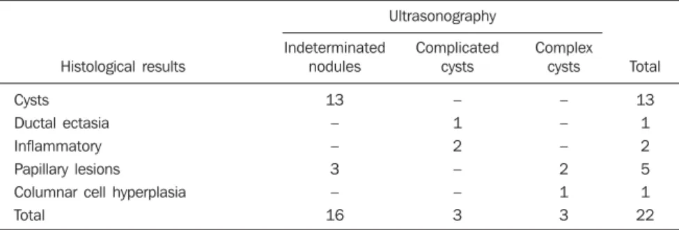
Sonographic presentation
All the apocrine cysts presented like in-determinate nodules. Ductal ectasia and two inflammatory lesions presented like complicated cysts. Three (60%) of the five papillary lesions presented like indetermi-nate nodules, and two (40%) like complex cysts; and the columnar cell hyperplasia like complex cyst (Table 2).
Elasticity scoring
All of the 13 apocrine cysts were as-signed score 2 (Figure 2). The ductal
ecta-sia and inflammatory lesions were also as-signed score 2. One of the five papillary lesions was assigned score 2 and the other four, score 3 (Figure 3). The columnar cell hyperplasia was assigned score 4 (Figure 4; Table 3).
DISCUSSION
In the last decade, ultrasound elasto-graphy has attracted a lot of attention to the assessment of soft tissues with the clinical prospect of allowing the early detection of
lesions which determine pathological alter-ations, and providing appropriate manage-ment of these lesions with the consequen-tial improvement in the prognosis for the patients(17). Information provided by this
method are similar, but more sensitive and less subjective than to the ones obtained with manual palpation(18).
The pioneering study developed in 1991 by Ophir et al.(10), proposed a classification
according to the elasticity variation, based Table 1 Elasticity scoring according to color spectrum during the sample compression and after decompression.
Score
1
2
3
4
During compression
Color similar to the normal tissue
Variation from yellow to greenish blue
Variation from greenish blue to dark blue
Generally blue to dark blue
Decompression
Color similar to the normal tissue
Color variation to softer in more than 50% of the nodule
Color variation to softer ranging between 10% and 50% of the nodule
With no significant variation
Result
Benign
Benign
Low likelihood of malignancy
High likelihood of malignancy
Table 2 Sonographic cystic lesions presentation according to histological results.
Histological results
Cysts
Ductal ectasia Inflammatory
Papillary lesions Columnar cell hyperplasia
Total
Indeterminated nodules
13
– –
3 –
16
Complicated cysts
–
1 2
– –
3
Complex cysts
–
– –
2 1
3
Total
13
1 2
5 1
170 Radiol Bras. 2008 Mai/Jun;41(3):167–172 Table 3 Cystic lesions presentation at elastography according to histological results.
Histological results
Cysts
Ductal ectasia
Inflammatory
Papillary lesions
Columnar cell hyperplasia
Total
1
–
–
–
–
–
–
2
13
1
2
1
–
17
3
–
–
–
4
–
4
4
–
–
–
–
1
1
Total
13
1
2
5
1
22 Elastography scores
on the principle that benign lesions were softer, whereas most of the malignant ones were harder. Elastographic images were obtained by means of comparison between pre- and post-breast tissue compression images. Since then, several studies have been published, although with no standard-ization of the technique or classification, most of them approaching only a compari-son between pre- and post-compression images.
Figure 2. Example of score 2 elastogram. Indeterminate nodule at conventional mode (left image), utilizing compression (central image), and post-decom-pression elastrography (right image). During compost-decom-pression, the greenish blue color in the center of the nodule is observed; after decompost-decom-pression, almost the whole nodule presents a greenish color.
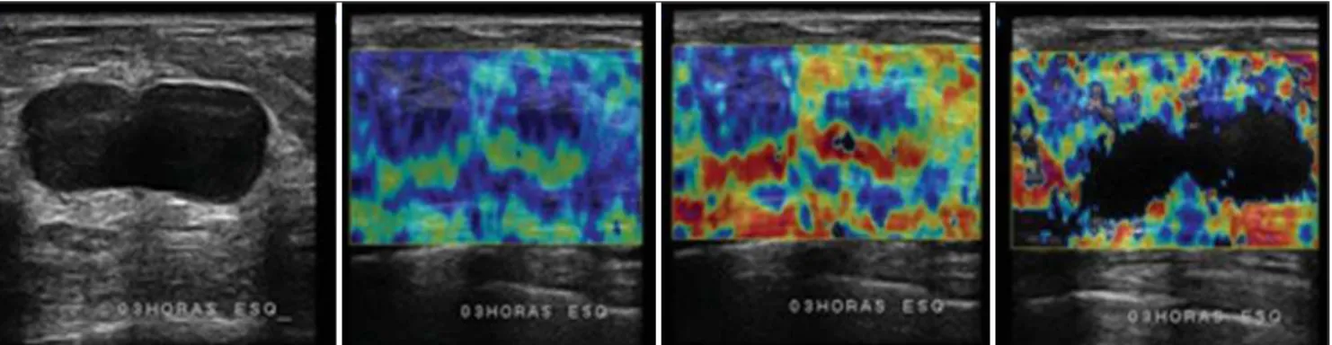
Figure 3. Example of score 3 elastogram. A complex cyst at the conventional mode (left image) utilizing compression (central image), and post-decompression elastography (right image). During compression a greenish color is observed in the peripheral cystic area, and a bluish color is observed in the mural nodule; after decompression, the cystic area changes to red, while the mural nodule presents a change of about 10–50% to greenish blue.
171
Radiol Bras. 2008 Mai/Jun;41(3):167–172 No study describes the findings in cys-tic alterations of the breast that many times may generate anguish in women similar to the one caused by malignant nodules, al-though they generally are associated with benign lesions or lesion with low malig-nancy potential. Frequently, these lesions are interpreted as indeterminate nodules by the conventional approach, requiring an short-term follow-up and invariably lead-ing to unnecessary diagnostic biopsies(19).
Simple cystic lesions present a typical aspect at US, as a circumscribed nodule with imperceptible walls, with an anechoic content and posterior accoustic shadowing, classified as BI-RADS® 2, whereas solid lesions typically present like circum-scribed, ovoid nodules parallel to the skin, classified as BI-RADS® 3. However not all the cystic lesions present with the same features; some of them are hardly differen-tiated from solid nodules, particularly those with a thick fluid content, sometimes with fine debris in suspension, being classified as indeterminate nodules. Considering their low malignancy potential (about 2%), a short-term follow-up is recommended, de-spite the controversy and lack of a consen-sus about this matter. Most frequently, the short-term follow-up protocol adopted is a new study performed in a ten-month term, evaluating the area partially darkened at the mammogram and, in cases of lesions un-detected by mammography, only ultra-sonography was performed. The subse-quent follow-up study is performed 12 months after the first one, but with bilateral mammography and ultrasonography, con-sidering that one year is the term for breast cancer screening, followed by another study one year afterwards. Provided the lesion has remained stable for this two-year period, the final BI-RADS® category is altered to 2. Cases where there is an alter-ation in the borders of the lesion or an ap-proximate increase of 10% in its initial di-ameter, demand percutaneous biopsy(4,5,7).
However, according to some authors, a solid nodule characterized only by ultra-sonography also requires diagnostic biopsy, considering that US does not allow differ-entiating benign from malignant lesions(20).
It is believed that these incidental US find-ings result in an increase in the number of negative biopsies(21,22).
On the other hand, a consensus has been achieved as regards the necessity of surgi-cal excisional biopsy - the golden standard in cases of complex cysts. With the intro-duction of vacuum-assisted biopsy for breast lesions diagnosis, this method started being adopted because it can be easily performed on an outpatient basis and with low complication rates. In these cases, the B-mode ultrasonographic image is a determining factor in the approach to be adopted, as far as BI-RADS descriptive criteria are utilized. Little controversy re-mains about the approach to be adopted in these cases(5).
An attempt of a conventional approach with harmonic imaging and supplementary Doppler fluxmetry has been undertaken aiming at minimizing this limitation of ul-trasonography; however, no significant in-crement was achieved in relation to the conventional method. In recent studies uti-lizing the so called “streaming detection” - where the response of the cystic internal content to the accoustic energy generated by the US transducer detected by Doppler is evaluated -, cystic lesions would have presented a response to Doppler, and the solid lesions would not. Experiments are still in development, with few studies pub-lished in the literature, but this method may be useful in the differentiation of indeter-minate nodules(7).
Elastography, originally introduced to ultrasonography for differentiating benign from malignant breast lesions, can also be utilized for differentiating solid from cys-tic lesions, considering that the cyst elas-ticity is higher than the one of the adjacent parenchyma. Additionally, this method can be useful as an adjuvant in the evaluation of complex cysts, especially in the presence of mural nodules, whose hardness can be determined. In the present study, all of the cysts histologically diagnosed were sonographically characterized as indeter-minate nodules, and assigned score 2, be-nign by elastography. Cysts with inflamma-tory content and ductal ectasia were sono-graphically characterized as complicated cysts, also with score 2 by elastography. These lesions presented a low malignancy potential and biopsies could be avoided if the features at ultrasound elastography had been taken into consideration.
Three (60%) of the papillary lesions presented as indeterminate nodules, and two (40%), as complex cysts at the conven-tional method; at elastography, one (20%) was assigned score 2, and the other four (80%), score 3. The score 2 lesion was sonographically classified as indeterminate nodule and measured 0.5 mm in its largest axis. This lesion would be considered as benign by elastography, and the patient would return for breast cancer screening in one year. However, this lesion was surgi-cally excised and confirmed as benign. Two (50%) of the other four (80%) lesions with low suspicion for malignancy at elasto-graphy (score 3) were submitted to surgi-cal excision and had their benignity con-firmed; and for the other two lesions (50%) follow-up was recommended, considering their two-year stability.
On the other hand, the typical columnar cell hyperplasia was sonographically char-acterized as a complex cyst, with score 4 at elastography, indicating a malignancy potential. This lesion was also submitted to surgical excision, with histologically con-firmed benignity; i.e., this was the false-positive result found in the present study. One of the limitations for cystic lesions evaluation by elastography is the serial compressions intensity; The higher the in-tensity, the more the superficial tension of the internal fluid content is increased, de-termining the characterization as solid le-sion according to the color spectrum (Fig-ure 5). The advantage is that, as a real-time method, the investigator is allowed to mea-sure the compression intensity during the examination according to the different types of breast and lesion to be evaluated. This increase in the superficial tension of the intracystic fluid also can be observed in secretory lesions, like in the case of the columnar cell hyperplasia and also in some inflammatory cysts.
Currently, the authors are developing studies based on their own criteria aiming at demonstrating the elastography sensitiv-ity, specificity and diagnostic accuracy. Preliminary data of 170 nodules have dem-onstrated 80% positive predictive value, 97.5% specificity, and 97.7% diagnostic accuracy for the proposed classification.
diagno-172 Radiol Bras. 2008 Mai/Jun;41(3):167–172 sis of cystic breast lesions, confirming their
etiology, and that the introduction of this method in the clinical routine could reduce the number of unnecessary biopsies and conventional follow-up. This method could also be utilized as a supplementary study in cases of complex cysts for evaluating their internal content, but diagnostic biopsy should not be contra-indicated, considering the significance of B-mode findings.
Acknowledgement
The authors thank the Núcleo de Apoio à Publicação (Center for Support to Publi-cation), Faculdade de Ciências Médicas da Santa Casa de São Paulo (NAP-SC), for the technical-scientific support for publication of this manuscript.
REFERENCES
1. Kasumi F. The diagnostic criteria for breast le-sions of the Japan Society of Ultrasonics in Medi-cine and topical issues in the field of breast trasonography in Japan. In: Topics in breast ul-trasound: Seventh International Congress on the Ultrasonic Examination of the Breast. Tokyo: Shinohara; 1991. p. 19–26.
2. Stavros AT, Thickman D, Rapp CL, et al. Solid breast nodules: use of sonography to distinguish between benign and malignant lesions. Radiol-ogy. 1995;196:123–34.
3. Kolb TM, Lichy J, Newhouse JH. Comparison of the performance of screening mammography,
physical examination, and breast US and evalu-ation of factors that influence them: an analysis of 27,825 patient evaluations. Radiology. 2002; 225:165–75.
4. Kaplan SS. Clinical utility of bilateral whole-breast US in the evaluation of women with dense breast tissue. Radiology. 2001;221:641–9.
5. Berg WA, Campassi CI, Ioffe OB. Cystic lesions of the breast: sonographic-pathologic correlation. Radiology. 2003;227:183–91.
6. Buchberger W, Niehoff A, Obrist P, et al. Clini-cally and mammographiClini-cally occult breast le-sions: detection and classification with high-reso-lution sonography. Semin Ultrasound CT MR. 2000;21:325–36.
7. Soo MS, Ghate SV, Baker JA, et al. Streaming
de-tection for evaluation of indeterminate sono-graphic breast masses: a pilot study. AJR Am J Roentgenol. 2006;186:1335–41.
8. Cha JH, Moon WK, Cho N, et al.
Characteriza-tion of benign and malignant solid breast masses: comparison of conventional US and tissue har-monic imaging. Radiology. 2007;242:63–9.
9. Chang RF, Huang SF, Moon WK, et al. Solid breast masses: neural network analysis of vascular fea-tures at three-dimensional power Doppler US for benign or malignant classification. Radiology. 2007;243:56–62.
10. Ophir I, Céspedes I, Ponnekanti H, et al. Elas-tography: a quantitative method for imaging the elasticity of biological tissues. Ultrason Imaging. 1991;13:111–34.
11. Ellis IO, Humphreys S, Michell M, et al. Best Practice No. 179. Guidelines for breast needle core biopsy handling and reporting in breast screening assessment. J Clin Pathol. 2004;57: 897–902.
12. Courtillot C, Plu-Bureau G, Binart N, et al. Benign
Figure 5. A complicated cyst at the conventional mode (left image) utilizing compression (second image), post-decompression elastography (third image), and after inappropriate compression (right image). Typically, the cyst presents a score 2 elastogram, compatible with benignity. Observe that, inappropriate com-pression results in a blackish cystic area suggesting a solid lesion, probably because of the superficial tension of the fluid.
breast diseases. J Mammary Gland Biol Neoplasia. 2005;10:325–35.
13. American College of Radiology. Breast Imaging Reporting and Data System® (BI-RADS®) – ul-trasound. 4th ed. Reston: American College of Radiology; 2003.
14. Kopans DB, Monsees B, Feig SA. Screening for cancer: when is it valid? Lessons from the mam-mography experience. Radiology. 2003;229:319– 27.
15. Mendelson EB, Tobin CE. Critical pathways in using breast US. Radiographics. 1995;15:935–45. 16. Itoh A, Ueno E, Tohno E, et al. Breast disease:
clinical application of US elastography for
diag-nosis.Radiology. 2006;239:341–50.
17. Hoyt K, Forsberg F, Ophir J. Analysis of a hy-brid spectral strain estimation technique in elastography. Phys Med Biol. 2006;51:197–209. 18. Hall TJ. AAPM/RSNA physics tutorial for resi-dents: topics in US: beyond the basics: elasticity imaging with US. Radiographics. 2003;23:1657– 71.
19. Graf O, Helbich TH, Hopf G, et al. Probably be-nign breast masses at US: is follow-up an accept-able alternative to biopsy? Radiology. 2007;244: 87–93.
20. Moy L, Slanetz PJ, Moore R, et al. Specificity of
mammography and US in the evaluation of a palpable abnormality: retrospective review. Radiology. 2002;225:176–81.
21. Kopans DB. Sonography should not be used for breast cancer screening until its efficacy has been proven scientifically. AJR Am J Roentgenol. 2004;182:489–91.