Radiation dose optimization in routine computed
tomography: a study of feasibility in a University
Hospital*
Otimização da dose em exames de rotina em tomografia computadorizada: estudo de viabilidade em um hospital universitário
Juciléia Dalmazo1, Jorge Elias Júnior2, Marco Aurélio Corte Brocchi3, Paulo Roberto Costa4, Paulo Mazzoncini de Azevedo-Marques5
OBJECTIVE: To study the feasibility of reducing radiation dose in protocols for acquisition of helical computed tomography images in a University Hospital. MATERIALS AND METHODS: A survey of radiation doses in computed tomography protocols was performed with phantoms and ionization chamber. Changes in kVp and mAs were proposed, determining the average noise. Protocols with noise values ≤≤≤≤≤ 1% were submitted to qualitative assessment of contrast and spatial resolution by three observers. RESULTS: Tests of variations were performed with 22 protocols for pediatric skulls, 26 for adult skulls, 28 for abdomen, and 18 for chest. The reduction in dose achieved ranged between 7.4% and13% for pediatric skull, 3.8% and 25% for adult skull, 9.6% and 34.3% for abdomen, 6.4% and 12% for chest. It was also noted that the use of windowing and zoom tools supported the acceptance of images by the observers. CONCLUSION: Radiation dose levels can be reduced by up to 34.4% in comparison with routine protocols, keeping the noise at acceptable levels. The use of digital manipulation tools allowed the acceptance of images with higher noise levels, thus resulting in radiation dose reduction.
Keywords: Computed tomography; Radiation dose reduction; ALARA; Signal-to-noise ratio; CTDI; Optimization.
OBJETIVO: Estudar a viabilidade de redução da dose de radiação em protocolos de aquisição de imagens de tomografia helicoidal em um hospital universitário. MATERIAIS E MÉTODOS: Foi realizado levantamento de dose de radiação de protocolos de tomografia com objetos simuladores e câmara de ionização. Foram pro-postas variações de kVp e mAs, determinando-se a média de ruído. Protocolos com valores de ruído meno-res ou iguais a 1% foram submetidos à avaliação qualitativa de contraste e meno-resolução espacial por três ob-servadores. RESULTADOS: Foram realizados 22 testes de variações para o protocolo de crânio pediátrico, 26 para crânio adulto, 28 para abdome e 18 para tórax. A redução da dose conseguida variou entre 7,4–13% para protocolo de crânio pediátrico, 3,8–25% para crânio adulto, 9,6–34,3% para abdome e 6,4–12% para tórax. Notou-se também que a utilização de ferramentas de janelamento e zoom favoreceu o aceite das imagens
pelos observadores. CONCLUSÃO: É possível reduzir os níveis de dose de radiação em até 34,4%, compa-rativamente aos protocolos utilizados na rotina, mantendo-se o ruído em níveis aceitáveis. O uso de ferra-mentas de manipulação digital das imagens possibilitou a aceitação de imagens com níveis maiores de ruído, favorecendo o processo de redução de dose de radiação.
Unitermos: Tomografia computadorizada; Redução da dose de radiação; ALARA; Relação sinal-ruído; CTDI; Otimização.
Abstract
Resumo
* Study developed at Centro de Ciências das Imagens e Física Médica (CCIFM) – Hospital das Clínicas da Faculdade de Medi-cina de Ribeirão Preto da Universidade de São Paulo (HCFMRP-USP), Ribeirão Preto, SP, Brazil.
1. Physicist, Fellow PhD degree, Faculdade de Medicina de Ribeirão Preto da Universidade de São Paulo (FMRP-USP), Ri-beirão Preto, SP, Brazil.
2. MD, PhD, Professor and Coordinator at Centro de Ciências das Imagens e Física Médica (CCIFM) – Faculdade de Medicina de Ribeirão Preto da Universidade de São Paulo (FMRP-USP), Ribeirão Preto, SP, Brazil.
3. Master, Physicist in Radiodiagnosis at Hospital das Clíni-cas da Faculdade de Medicina de Ribeirão Preto da Universidade de São Paulo (HCFMRP-USP), Ribeirão Preto, SP, Brazil.
4. PhD, Professor at Department of Nuclear Physics, Instituto de Física da Universidade de São Paulo (IFUSP), São Paulo, SP, Brazil.
creased since its introduction in the clini-cal practice(1,2). Various factors contribute to this growth, including technological hardware improvements, leading to faster data acquisition with significant reduction in the images acquisition time, as well as the increase in the number of clinical indi-cations for CT, associated to a greater avail-ability of CT installed units and a relative tendency to costs reduction(3,4).
The increasing utilization of diagnostic imaging methods employing ionizing
ra-Dalmazo J, Elias Jr J, Brocchi MAC, Costa PR, Azevedo-Marques PM. Radiation dose optimization in routine computed tomography: a study of feasibility in a University Hospital. Radiol Bras. 2010;43(4):241–248.
INTRODUCTION
The annual number of computed to-mography studies (CT) has constantly
in-5. PhD, Associate Professor at Centro de Ciências das Ima-gens e Física Médica (CCIFM), Department of Medical Practice, Faculdade de Medicina de Ribeirão Preto da Universidade de São Paulo (FMRP-USP), Ribeirão Preto, SP, Brazil.
Mailing address: Dr. Jorge Elias Júnior. Centro de Ciências das Imagens e Física Médica, HCFMRP-USP. Avenida Bandeirantes, 3900, Monte Alegre. Ribeirão Preto, SP, Brazil, 14048-090. E-mail: [email protected]
242
diation, particularly CT, is the main cause for the marked increase in the mean medi-cal radiation dose per capita per year(2). Currently, the annual per capita radiation dose considered as secondary to medical care, particularly for diagnosis purposes, has overcome the dose received from en-vironmental factors (food, radon gas and others)(1). As a result, there is an increas-ing concern of the medical community, equipment manufactures and even patients with the control of doses determined by the different diagnostic methods that rely on ionizing radiation(5,6). Besides occupational radiation protection, the clinical practice adopts the ALARA (As Low As Reason-ably Achievable) principle as a guideline for the rational use of these imaging meth-ods(3,4,7,8).
Specifically considering the pediatric population, it is important to highlight that children present a considerably higher risk for development of radiation related neo-plasia as compared with the adult popula-tion(1,9,10). Such higher risk is explained by the presence of a greater cell population undergoing division in the different organs and tissues in development, and also for the greater life expectancy both in absolute and relative terms. As an example, a one-year-old child presents a 10 to 15 times higher risk than a 50-year-old adult for de-veloping a malignant neoplasm, for the same radiation dose(1). For these reasons, there is an increasing concern with the ra-diation dose utilized in pediatric radiologi-cal studies, and particularly in the case of CT scans. Several published studies report strategies and actions to reduce radiation dose(3,5,9,11–13).
There are several strategies in develop-ment or already in use in more modern equipment, such as, for example, tube cur-rent modulation according to the variation in the slice thickness for the evaluated ana-tomic region(6,14). However, in Brazil there are many relatively old CT systems in use with no software or parameter manipula-tion capabilities, which ultimately influ-ence the dose delivered at each exam.
Therefore, the present study is aimed to evaluate the feasibility of an optimization strategy to reduce radiation dose in single-slice helical CT protocols in a University Hospital.
MATERIALS AND METHODS
Strategy description
Initially, data on the absorbed radiation dose in routine protocols for both adult and pediatric skull, chest and abdomen CT studies were collected. With such data, an evaluation of the impact of variations in the voltage parameters (kVp) and current vs. time (mAs) was undertaken considering the radiation dose and image quality, the latter studied by the noise measurement and sub-jective analysis of images obtained with specific phantoms.
Equipment
The tests were performed with a Soma-tom Emotion single-slice helical computed tomography unit (Siemens Medical Solu-tions; Erlangen, Germany).
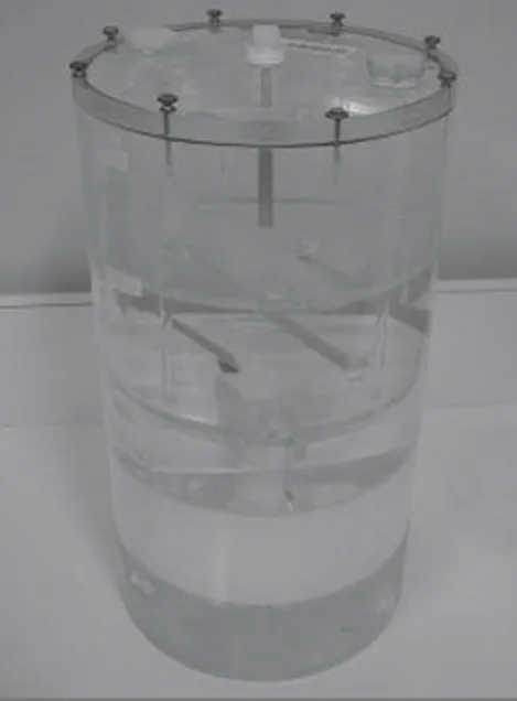
For the radiation dose measurements, cylindrical 15 cm long polymethylmeta-crylate phantoms were utilized, one with 32 cm in diameter, representing the torso, and another with 16 cm in diameter, represent-ing the head (Figure 1), accordrepresent-ing to the American Association of Physicists in Medicine (AAPM) specifications. Such phantoms presented absorption and scatter-ing characteristics that are similar to those of the anatomic structures of the skull and of the torso with holes in the center and at
predetermined locations, at 1 cm from the periphery at the 12, 3, 6 and 9 o’clock po-sitions, allowing the insertion of ionization chambers.
The radiation dose measurements were obtained by means of a pencil type ioniza-tion chamber model 10X5-3CT (Radcal Corporation; Monrovia, CA, USA), coupled with a Radcal electrometer model 9015. Figure 1 shows the assembly for absorbed dose measurement for the standard body phantom.
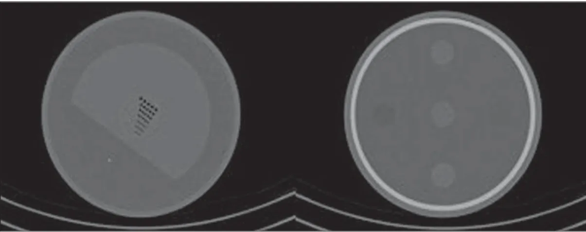
The images for the quality study with the proposed parameter variations were obtained by utilizing the specific AAPM standard phantom, model 76-410 which was freely borrowed by Instituto de Eletrotécnica e Energia da Universidade de São Paulo (IEE-USP) (Figure 2).
Studied variables
The kVp and mAs values were duly noted for each protocol, as well as their suggested variations for protocols that were developed for radiation dose reduction.
The initial evaluation of radiation dose in routine protocols as well as the CT equipment calibration were performed by means of the correlation between the CTDIvol obtained with the ionization chamber positioned within the phantoms, considering the acquisition of a section at
Figure 1. Electrometer, ionization chamber and body phantom exposed to the tomographic beam for
the central position of the chamber, and the CTDIvol provided by the equipment. The CTDIvol is obtained from the ratio between the weighted computed tomography dose index (CTDIw)(15,16)and the pitch, which is defined as the distance travelled by the table and one rotation of the X-ray source, divided by the total collimated beam width. The CTDIvol is referenced on the equip-ment by the DICOM tag (0018,9345). Con-sidering that the index of correlation be-tween the values obtained in the measure-ments performed with the ionization cham-ber and those provided by the equipment was practically 100% (r = 0.99; p < 0.0001), the equipment CTDIvol measurements were selected for results presentation of and discussion.
Strategy for the proposal of changes in routine CT studies protocols
Based on the initial standard protocols, the variations of the kVp and mAs param-eters applied to the tube were proposed, with the remaining parameters (slice thick-ness, pitch, pixel size and total exposure) being kept constant for each protocol, as shown on Table 1 for the protocol of skull CT. The CTDIvol was measured again for each proposed parameter change (Table 1).
Evaluation of image quality
The images quality was quantitatively evaluated by the measurement of quantic
Table 1 Results for kVp and mAs, as well as respective CTDIvol values. Ex: Routine protocol for adult
skull.
Current (mAs)
342 174
122 120*
115 100
90 80
70 60
54 45
CTDIvol (mGy) (voltage 130* kVp)
54.58 27.77
19.63 19.15*
18.43 16.04
14.36 12.93
11.25 9.58
8.62 7.18
CTDIvol (mGy) (voltage 110 kVp)
37.55 19.11
13.51 13.18
12.68 11.03
9.88 8.89
7.74 6.59
5.93 —
CTDIvol (mGy) (voltage 80kVp)
16.42 8.35
5.90 —
— —
— —
— —
— —
* Values for mAs utilized and respective CTDIvol for the routine protocol for adult skull.
noise, and qualitatively by the subjective evaluation of images obtained in the AAPM standard phantom, independently and blindly performed by three radiologists with more than 10 years of experience.
The quantic noise is the result of the variation in the number of X-ray photons absorbed by the detector in a determined time interval and, considering the geomet-ric characteristics of the image (pixel size, matrix size, slice thickness) as fixed, pre-sents an inversely proportional relation with the dose received by the patient. The methodology adopted for the evaluation of images quality was based on the Agência Nacional de Vigilância Sanitária (ANVISA – Brazilian agency of sanitary vigilance) guidelines “Medical radiodiagnosis: safety and equipment performance” that estab-lishes the practical aspects for the standards established by Ordinance 453 of the Bra-zilian Ministry of Health(17), considering the quantic noise as the standard deviation of the values for the gray scale for a cen-tral square 5 × 5 mm region on a uniform image (Figure 3) divided by the nominal pixel value of that region. Although the ANVISA protocol indicates the need to evaluate the noise at five different points, as it refers to medical equipment quality control procedures, in the present study the option was made to simplify such proce-dure, by performing the measurement in the central region of the phantom for quality evaluation so as to observe the noise varia-tion considering a situavaria-tion of greater at-tenuation and beam hardening. The noise
on the digital image was evaluated by means of the public domain ImageJ soft-ware(18),considering as acceptable those values ≤ 1%(9).
The qualitative analysis was performed for the tested protocols that presented the lowest dose rates and with noise levels within the established limit. The evaluation comprised spatial resolution and high reso-lution contrast tests performed by the radi-ologists utilizing the ImageJ software with-out and with the use of windowing and zoom resources. The visualized objects indicated by the radiologists on the images were compared with the test objects map included in the phantom (Figure 4).
The images were evaluated separately and in duets by means of a questionnaire with nine questions for each set of images,
Figure 3. CT section of phantom for quality study,
with four selected central areas selected in the ImageJ software for image noise calculation.
Figure 2. Quality phantom for analysis of noise,
244
Table 2 Comparison between routine and suggested protocols that achieved acceptable noise level and diagnostic quality considering kVp and mAs
parame-ters, CTDIvol value, mean noise value and corresponding mean dose reduction for each protocol.
Protocol
Pediatric skull
Adult skull
Adult abdomen
Adult chest
Routine Suggested
kVp / mAs
130 / 80
130 / 120
130 / 100
110 / 80
CTDIvol (mGy)
12.9
19.1
9.6
7.7
Mean noise
0.9%
0.7%
0.8%
1.6%
kVp / mAs
130 / 75
130 / 70
130 / 115 130 / 100
130 / 90
130 / 90 130 / 80
130 / 65
110 / 70 130 / 50
CTDIvol (mGy)
11.9
11.2
18.4 16.0
14.3
8.7 7.7
6.3
6.8 7.2
Mean noise
0.9%
1.0%
0.9% 0.9%
0.9%
0.8% 0.9%
1.0%
1.7% 1.5%
Mean dose reduction
7.4%
13.0%
3.8% 16.2%
25.0%
9.6% 20.0%
34.3%
12.0% 6.4%
Figure 4. CT sections of
phantom for quality study and analysis of image spa-tial resolution and contrast.
aimed at the study of images contrast and resolution threshold, considering even what set of objects were visible for the di-agnosis of an image. The observer classi-fied whether the image had diagnostic qual-ity or not, considering the visibilqual-ity of ex-isting objects. Additionally, a classification of the image quality was requested, consid-ering the values 1 (very good), 2 (good), 3 (median), 4 (bad) and 5 (very bad).
RESULTS
The CTDIvol and mean noise values obtained for the routine protocols of pedi-atric skull, adult skull, abdomen and chest CT are presented on Table 2. Mean noise of 1.6% was observed for the chest protocol. By utilizing fixed 80, 100 and 130 kVp values, measurements of CTDIvol and mean noise were performed in 22 varia-tions of the pediatric skull protocol (mAs ranging from 45 to 271), 26 variations of
the adult skull protocol (mAs ranging from 45 to 342), 28 variations of the adult abdo-men protocol (mAs ranging from 24 to 182) and 18 variations of the chest proto-col (mAs ranging from 29 to 100). Based on these results, new protocols were sug-gested considering the combination of low-est CTDIvol obtained in association with the defined noise threshold, which were utilized for the evaluation of the quality of the image obtained in the standard AAPM phantom specific for quality evaluation. The data on the suggested protocols and respective CTDIvol and mean noise values, as well as the average dose reduction com-paratively with the routine protocols, are presented on Table 2.
As regards the qualitative evaluation of the suggested protocols, the specialists agreed that by using the zoom and windowing tools at the display monitor, the suggested protocols for pediatric skull, adult skull, adult abdomen and adult chest
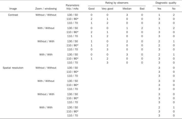
did not present any differences compara-tively with the routine protocols in what concerns spatial resolution and contrast, as shown on Tables 3 to 6. However, it is im-portant to note that in the case of adult skull, there was a trend towards improve-ment in the rating of the contrast quality of the images acquired with the suggested protocol, when the windowing and zoom tools were utilized.
DISCUSSION
Image
Contrast
Spatial resolution
Zoom / windowing
Without / Without
With / Without
Without / With
With / With
Without / Without
With / Without
Without / With
With / With
Parameters kVp / mAs
130 / 70 130 / 75
130 / 80*
130 / 70 130 / 75
130 / 80*
130 / 70 130 / 75
130 / 80*
130 / 70 130 / 75
130 / 80*
130 / 70 130 / 75
130 / 80*
130 / 70 130 / 75
130 / 80*
130 / 70 130 / 75
130 / 80*
130 / 70 130 / 75
130 / 80*
Good 0 0 0 0 1 0 2 1 3 1 1 1 Very good 0 0 1 0 0 0 0 1 0 1 1 1 Median 2 2 2 3 2 3 1 1 0 1 1 1 Bad 1 1 0 0 0 0 0 0 0 0 0 0 Yes 1 1 3 2 2 3 2 3 3 3 3 3 2 3 3 3 3 3 1 2 3 3 3 3 No 2 2 0 1 1 0 1 0 0 0 0 0 1 0 0 0 0 0 2 1 0 0 0 0
* Routine protocol.
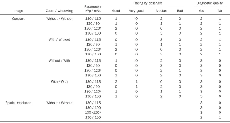
Table 3 Qualitative evaluation of image contrast and spatial resolution of the routine protocol for pediatric skull and the corresponding suggested protocols
with kVp and mAs variations, both with and without utilization of zoom and windowing resources.
Rating by observers Diagnostic quality
Table 4 Qualitative evaluation of image contrast and spatial resolution of the routine protocol for adult skull and corresponding suggested protocols with kVp
and mAs variations, with and without the utilization of zoom and windowing resources.
Rating by observers Diagnostic quality
Image
Contrast
Spatial resolution
Zoom / windowing
Without / Without
With / Without
Without / With
With / With
Without / Without
Parameters kVp / mAs
130 / 115 130 / 90 130 / 120*
130 / 100
130 / 115 130 / 90 130 / 120*
130 / 100
130 / 115 130 / 90 130 / 120*
130 / 100
130 / 115 130 / 90 130 / 120*
130 / 100
130 / 115 130 / 100 130 /120* 130 / 100
Good 1 1 2 0 0 1 2 0 1 0 0 1 2 0 1 1 Very good 0 0 0 0 0 0 0 0 0 0 0 0 1 1 0 0 Median 2 1 0 3 3 1 0 3 2 3 2 2 0 2 1 2 Bad 0 1 0 0 0 1 0 0 0 0 1 0 0 0 1 0 Yes 2 2 2 2 2 2 2 2 3 3 3 3 3 3 3 3 3 3 3 2 No 1 1 1 1 1 1 1 1 0 0 0 0 0 0 0 0 0 0 0 1
246
Table 4.
Rating by observers Diagnostic quality
Image
Spatial resolution
Zoom / windowing
With / Without
Without / With
With / With
Parameters kVp / mAs
130 / 115 130 / 90
130 / 120* 130 / 100
130 / 115
130 / 90 130 / 120*
130 / 100
130 / 115 130 / 90
130 / 120* 130 / 100
Good Very good Median Bad Yes
3 3
3 2
3
3 3
2
3 3
3 2
No
0 0
0 1
0
0 0
1
0 0
0 1
* Routine protocol.
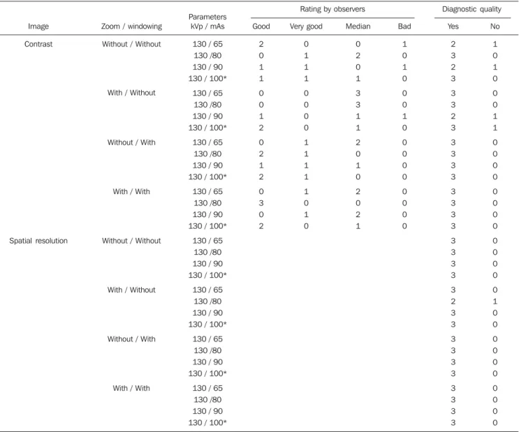
Table 5 Qualitative evaluation of image contrast and spatial resolution of the routine protocol for adult abdomen and corresponding suggested protocols with
kVp and mAs variations, with and without the utilization of zoom and windowing resources.
Rating by observers Diagnostic quality
Image
Contrast
Spatial resolution
Zoom / windowing
Without / Without
With / Without
Without / With
With / With
Without / Without
With / Without
Without / With
With / With
Parameters kVp / mAs
130 / 65 130 /80
130 / 90 130 / 100*
130 / 65
130 /80 130 / 90
130 / 100*
130 / 65 130 /80
130 / 90 130 / 100*
130 / 65
130 /80 130 / 90
130 / 100*
130 / 65 130 /80
130 / 90 130 / 100*
130 / 65
130 /80 130 / 90
130 / 100*
130 / 65 130 /80
130 / 90 130 / 100*
130 / 65
130 /80 130 / 90
130 / 100*
Good
2 0
1 1
0
0 1
2
0 2
1 2
0
3 0
2
Very good
0 1
1 1
0
0 0
0
1 1
1 1
1
0 1
0
Median
0 2
0 1
3
3 1
1
2 0
1 0
2
0 2
1
Bad
1 0
1 0
0
0 1
0
0 0
0 0
0
0 0
0
Yes
2 3
2 3
3
3 2
3
3 3
3 3
3
3 3
3
3 3
3 3
3
2 3
3
3 3
3 3
3
3 3
3
No
1 0
1 0
0
0 1
1
0 0
0 0
0
0 0
0
0 0
0 0
0
1 0
0
0 0
0 0
0
0 0
0
The mean noise values for the routine protocols were within the established threshold of 1%, except for the chest pro-tocol, in which the mean noise value was 1.6%. Such value is explained by the fact that the chest CT utilizes narrow collima-tion in order to obtain thin slices, thus de-termining a smaller quantity of photons incident on the detectors. For this reason, there was no proposal for dose reduction in the chest protocols, as the noise level was already above the established limit.
By observing the quantitative and quali-tative evaluation of the images, it becomes clear that all CT protocols could utilize reduced kVp and mAs parameters while maintaining diagnostic quality, and deter-mining a lower radiation dose in each study, a fact that is in agreement with other stud-ies in the literature(19–23), particularly with respect to mAs reduction. Such a reduction is particularly significant for CT studies in children, which has been a constant preoc-cupation over the last decade(10,13,24,25).
With the methodology utilized, the au-thors observed a reduction in radiation dose between 3.8% and 34.3%. It is possible to achieve further radiation dose reduction with techniques that take anthropometric individual data into consideration, as re-ported by Kalra et al.(22), or by working with less conservative noise levels, for example in the order of 5%. It should be reminded that any level of radiation dose reduction must be pursued as determined by the ALARA principle(3,4,7,8).
Also, it must be highlighted that in prac-tically all subjective evaluations performed by the radiologists, better scores were ob-served with the utilization of the zoom and windowing resources. Such results cor-roborate findings reported in the literature discussing the relation between reduction of radiation dose in CT studies, image qual-ity and reliabilqual-ity of the subjective evalua-tion(26). Considering that in digital imaging method the processes of images acquisition and display are separated, it is possible to
independently optimize each process. Thus, within determined limits, it is pos-sible to compensate for a loss in contrast due to a decrease in the signal/noise ratio by utilizing easily manipulated tools. The optimization of contrast resolution, with the consequential potential for dose reduc-tion, is the main advantage of digital tech-nology as far as the patients radiological protection is concerned.
The main limitation of the present study lies on the fact that the variations of other CT parameters such as pitch, slice thick-ness and rotation time were not ap-proached. It is a known fact that when the pitch is increased the patient is exposed to a higher radiation dose. However such limitation is an expression of reality in sev-eral Brazilian CT centers, where limited resources do not allow an appropriate tech-nological update of relatively old appara-tuses as compared with units equipped with new resources for dose efficiency man-agement, calibration of pediatric images Image
Contrast
Spatial resolution
Zoom / windowing
Without / Without
With / Without
Without / With
With / With
Without / Without
With / Without
Without / With
With / With
Parameters kVp / mAs
130 / 50
110 / 80* 110 / 70
130 / 50
110 / 80* 110 / 70
130 / 50
110 / 80* 110 / 70
130 / 50
110 / 80* 110 / 70
130 / 50
110 / 80* 110 / 70
130 / 50
110 / 80* 110 / 70
130 / 50
110 / 80* 110 / 70
130 / 50
110 / 80* 110 / 70
Good
0
2 1
0
2 1
1
1 0
0
1
Very good
0
1 2
0
1 2
0
2 3
0
2 3
Median
3
0 0
1
0 0
2
0 0
3
0 0
Bad
0
0 0
2
0 0
0
0 0
0
0 0
Yes
1
3 3
1
3 3
2
2 3
2
3 3
3
3 3
3
3 3
3
3 3
2
3 3
No
2
0 0
2
0 0
1
0 0
1
0 0
0
0 0
0
0 0
0
0 0
1
0 0
* Routine protocol.
Table 6 Qualitative evaluation of image contrast and spatial resolution of the routine protocol for adult chest and corresponding suggested protocols with kVp
and mAs variations, with and without the utilization of zoom and windowing resources.
248
quality, measurements of pediatric doses and protocols specifically designed for pe-diatrics(12).
CONCLUSION
Based on the results of the present study, the optimization of CT radiation doses in a university hospital was feasible with the proposed methodology utilizing phantoms and a pencil-type ionization chamber, achieving a radiation dose reduction of up to 34.3% for selected study protocols.
REFERENCES
1. Picano E. Sustainability of medical imaging. BMJ. 2004;328:578–80.
2. Huda W. What ER radiologists need to know about radiation risks. Emerg Radiol. 2009;16: 335–41.
3. Semelka RC, Armao DM, Elias J Jr, et al. Imag-ing strategies to reduce the risk of radiation in CT studies, including selective substitution with MRI. J Magn Reson Imaging. 2007;25:900–9. 4. Frush DP, Donnelly LF, Rosen NS. Computed
to-mography and radiation risks: what pediatric health care providers should know. Pediatrics. 2003;112:951–7.
5. Picano E, Vano E, Semelka R, et al. The Ameri-can College of Radiology white paper on radia-tion dose in medicine:deep impact on the prac-tice of cardiovascular imaging. Cardiovasc Ultra-sound. 2007;5:37.
6. Kalender WA, Buchenau S, Deak P, et al. Tech-nical approaches to the optimisation of CT. Phys Med. 2008;24:71–9.
7. ICRP. 1990 Recommendations of the Interna-tional Commission on Radiological Protection. ICRP Publication no. 60. Oxford, UK: Pergamon; 1991.
8. Kalra MK, Maher MM, Toth TL, et al. Strategies for CT radiation dose optimization. Radiology. 2004;230:619–28.
9. Verdun FR, Lepori D, Monnin P, et al. Manage-ment of patient dose and image noise in routine pediatric CT abdominal examinations. Eur Radiol. 2004;14:835–41.
10. Donnelly LF. Reducing radiation dose associated with pediatric CT by decreasing unnecessary examinations. AJR Am J Roentgenol. 2005;184: 655–7.
11. Berdon WE, Slovis TL. Where we are since ALARA and the series of articles on CT dose in children and risk of long-term cancers: what has changed? Pediatr Radiol. 2002;32:699. 12. Morgan HT. Dose reduction for CT pediatric
im-aging. Pediatr Radiol. 2002;32:724–8; discussion 751–4.
13. Siegel MJ, Schmidt B, Bradley D, et al. Radia-tion dose and image quality in pediatric CT: ef-fect of technical factors and phantom size and shape. Radiology. 2004;233:515–22. 14. McCollough CH, Bruesewitz MR, Kofler JM Jr.
CT dose reduction and dose management tools: overview of available options. Radiographics. 2006;26:503–12.
15. Gerber TC, Kuzo RS, Morin RL. Techniques and parameters for estimating radiation exposure and dose in cardiac computed tomography. Int J Cardiovasc Imaging. 2005;21:165–76. 16. Pina DR, Duarte SB, Ghilardi Netto T, et al.
Con-trole de qualidade e dosimetria em equipamen-tos de tomografia computadorizada. Radiol Bras. 2009;42:171–7.
17. Brasil. Ministério da Saúde. Secretaria de Vigi-lância Sanitária. Portaria nº 453, de 01 de junho
de 1998. Brasília: Diário Oficial da União; 02/06/ 1998.
18. Abramoff MD, Magelhaes PJ, Ram SJ. Image processing with ImageJ. Biophotonics Interna-tional. 2004;11:36–42.
19. Mayo JR, Hartman TE, Lee KS, et al. CT of the chest: minimal tube current required for good image quality with the least radiation dose. AJR Am J Roentgenol. 1995;164:603–7.
20. Heyer CM, Mohr PS, Lemburg SP, et al. Image quality and radiation exposure at pulmonary CT angiography with 100- or 120-kVp protocol: pro-spective randomized study. Radiology. 2007;245: 577–83.
21. Cohnen M, Fischer H, Hamacher J, et al. CT of the head by use of reduced current and kilovol-tage: relationship between image quality and dose reduction. AJNR Am J Neuroradiol. 2000;21: 1654–60.
22. Kalra MK, Prasad S, Saini S, et al. Clinical com-parison of standard-dose and 50% reduced-dose abdominal CT: effect on image quality. AJR Am J Roentgenol. 2002;179:1101–6.
23. Jung KJ, Lee KS, Kim SY, et al. Low-dose, volu-metric helical CT: image quality, radiation dose, and usefulness for evaluation of bronchiectasis. Invest Radiol. 2000;35:557–63.
24. Brenner D, Elliston C, Hall E, et al. Estimated risks of radiation-induced fatal cancer from pe-diatric CT. AJR Am J Roentgenol. 2001;176:289– 96.
25. Donnelly LF, Emery KH, Brody AS, et al. Mini-mizing radiation dose for pediatric body applica-tions of single-detector helical CT: strategies at a large children’s hospital. AJR Am J Roentgenol. 2001;176:303–6.