Extramammary Paget Disease of the Vulva - Case
Report
Relato de Caso: Doença de Paget Extramamária da Vulva
Bianca Ruschel Hillmann
1Amanda Amaro Pereira
2Luiz Fernando Sommacal
31Department of Gynecology and Obstetrics, Residency Program of the Hospital Universitário Professor Polydoro Ernani de São Thiago, Florianópolis, SC, Brazil
2Department of Pathology, Residency Program of the Hospital Universitário Professor Polydoro Ernani de São Thiago, Florianópolis, SC, Brazil 3Department of Gynecology and Obstetrics, Health Sciences Center,
Universidade Federal de Santa Catarina, Florianópolis, SC, Brazil Rev Bras Ginecol Obstet 2016;38:524–528.
Address for correspondence Bianca Ruschel Hillmann, Division of Obstetrics and Gynecology, Hospital Universitário Professor Polydoro Ernani de São Thiago, Campus Universitário, Rua Professora Maria Flora Pausewang, s/n°, Trindade. Caixa Postal 5199, 88036-800– Florianópolis, SC, Brazil (e-mail: [email protected]).
Keywords
►
extramammary Paget
disease
►
vulvar neoplasms
►
pruritus vulvae
►
vulvar Paget disease
Abstract
Extramammary Paget disease (EPD) is a rare malign neoplasm that may affect the vulva
and has manifestations common to benign diseases such as itching, pain and eczema.
This leads to delay in diagnosis and consequent worse prognosis. The de
fi
nitive
diagnosis is obtained by biopsy of the vulva, which shows Paget cells. The treatment
of choice is wide excision with margins, which leads to sequelae, functional and
aesthetic. Recurrence is common. This article reports the case of a 48-year-old female
patient with history of vulvar itching for the past 2 years, who had been submitted to
various treatments for benign pathologies. The patient was biopsied and was
diag-nosed with extensive EPD, being submitted to vulvectomy. This article aims to draw
attention to the need for biopsy of pruritic vulvar lesions that do not respond to usual
treatment.
Palavras-chave
►
doença de Paget
extramamária
►
neoplasias vulvares
►
prurido vulvar
►
doença de Paget
vulvar
Resumo
A doença de Paget extramamária (DPEM) é uma neoplasia maligna incomum que pode
acometer a vulva, e que tem manifestações comuns a doenças benignas como prurido,
dor e eczema. Isto leva a atraso no diagnóstico e, consequente, piora do prognóstico. O
diagnóstico de certeza se dá pela biópsia de vulva que evidencia células de Paget. O
tratamento de escolha é a excisão com margens amplas, que leva a sequela funcional e
estética. A recidiva é frequente. Este artigo relata o caso de uma paciente do sexo
feminino, de 48 anos, com história de prurido vulvar há 2 anos com diversos
tratamentos para patologias benignas. A biopsia da paciente con
fi
rmou diagnóstico
de DPEM extensa sendo submetida a vulvectomia. Este artigo visa chamar a atenção
para a necessidade de biópsia das lesões vulvares pruriginosas que não respondem ao
tratamento habitual.
received March 22, 2016 accepted
September 22, 2016
DOI http://dx.doi.org/ 10.1055/s-0036-1593884. ISSN 0100-7203.
Copyright © 2016 by Thieme Publicações Ltda, Rio de Janeiro, Brazil
Case Report
Introduction
Extramammary Paget disease (EPD) is a rare neoplasm that occurs in areas containing apocrine glands and affects the epidermis and, occasionally, the derm.1–3The most common-ly affected site is the vulva, but it may also affect the axilla, perineum and perianal region.1,2
Vulvar Paget disease is more common from the 5th decade of life on,2with an average age at diagnosis from 61 to 69 years, depending on the study.4–8It is more common in Caucasians and is rare before menopause.6,8Extramammary Paget dis-ease accounts for less than 1% of vulvar neoplasms.6–8
The most common clinical manifestations are vulvar itching, pain and the presence of eczematous plate.5–8Those manifestations are common to several benign diseases such as contact dermatitis, candidiasis, tinea cruris, psoriasis, lichen simplex and seborrheic dermatitis,2,7,9thus, leading to delayed diagnosis. The diagnose occurs on average 20 months after the onset of symptoms, worsening the prognosis of patients, who will have larger lesions and higher recurrence rates.5,6,8It is common that before diagnosis, the patient is subjected to multiple treatments with antifungal and corticoids.8 Therefore it is suggested that any vulvar lesions unresponsive to treatment be biopsied.2,8
Extramammary Paget disease may be associated with bladder, prostate, urethral, or colorectal cancer, so that the investigation of such tumors must be made once the diag-nose of vulvar Paget disease is confirmed.2
Histological diagnosis of EPD is made by identification of Paget cells with abundant pale cytoplasm and large atypical nuclei with prominent nucleoli and vesicular chromatin. Stain-ing of Paget cells is made with colloidal iron, alcian blue, mucicarmine and PAS. Immunohistochemistry can be made with markers such as the epithelial membrane antigen, carci-noembryonic antigen (CEA) and GCDFP-15. Cytokeratin 7 (CK7) and, less frequently, cytokeratin 20, are positive and can help in the diagnosis. Immunohistochemical stains S-100 and HMB 45 are used to differentiate from malignant in situ melanoma.2,8
Extramammary Paget disease secondary to anorectal carcinoma resembles gastrointestinal glands with stratified columnar cells, signet ring cells, necrosis and intraluminal goblet cells.8
Extramammary Paget disease secondary to urothelial carci-noma resembles high-grade urothelial cells with variable shape nuclei, rough and irregular chromatin and not prominent nucleoli. Rarely the cytoplasm contains vacuoles and the signet ring cells are absent. These cells have a high rate of mitose.8
Overexpression of P53 in Paget disease is associated with underlying dermal invasion.8
Histologically, the differential diagnosis is made with Bowen disease and malignant pagetoid melanoma in situ.2 The treatment of choice is excision of the lesions with wide margins, but such treatment leads to aesthetic dissatisfac-tion and sequelae, and does not prevent local recurrence. Therefore, alternative treatments, such as excision without wide margins associated with radiotherapy, laser ablation, and application of imiquimod, are accepted.1–3,10Such alter-native treatments become first choice in the treatment of
recurrence, and primary treatment of patients with severe clinical conditions that do not allow surgery.3,10,11
As recurrence is extremely common, even after aggressive surgery with free margins, it is widely accepted that vulvar Paget disease is multifocal, even if just in a microscopic way.8,9,11Recurrence occurs on average 45 months after the first diagnosis, and is more common when the lesions are multifocal and surgical margins are compromised. Local recur-rence developed in 44% of the patients when followed for 77 months.5,6Still, there are reports of local recurrence 15 years after the diagnosis.8So it is recommended that patients are followed for long periods after the treatment.4,5As for the prognosis, the risk of death from the disease is 3 to 12%.5,9The epidermis restricted disease has an excellent prognosis and death usually occurs by comorbidities, and not by disease.8 Invasive disease has a mortality of 13 to 18%, with survival of 72% after 5 years.8Patients with lymph node metastasis have worse prognosis with a mortality rate of66%.8
Case Report
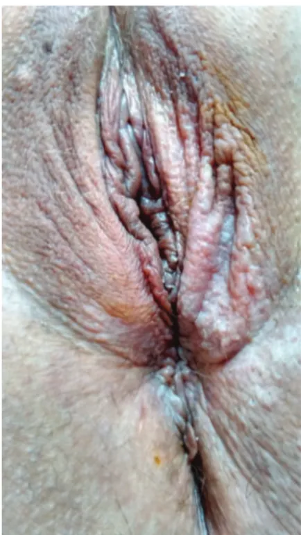
The subjects is a 48-year-old female patient, smoker for the past 35 years, with type 2 diabetes mellitus, in use of Metformin, and premenopausal, without using hormonal contraceptives at the moment. Patient reported vulvar itching for the past 2 years, with various treatments with creams and ointments without success. Physical examination revealed one hardened left ingui-nal lymph node of1cm in size. Cervix had no colposcopy abnormalities. Presence of acetowhite areas in left minora and majora labia, perineal and anterior perianal regions was detected. (►Fig. 1). Collins test was positive (►Fig. 2).
Fig. 1 Examination of the vulva with a lesion difficult to notice in the left majora and minora labia, perianal e perineal regions.
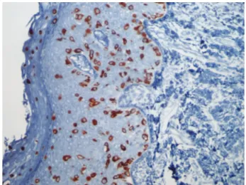
A biopsy of the lesion was performed and showed infi ltra-tion of the epidermis by cells with clear abundant cytoplasm and large nuclei with pleomorphism and prominent nucleoli. These cells were distributed across the epidermal thickness, isolated or in small groups, and were more numerous in the basal layer (►Fig. 3). Immunohistochemistry showed posi-tivity in neoplastic cells for CK7 (►Fig. 4), CEA, and epithelial membrane antigen (EMA). The clinical and histological fea-tures were compatible with primary Paget disease of the vulva.
The disease was clinically staged as T3N1M0, and treated with radical vulvectomy and bilateral inguinal lymphadenec-tomy. The evaluation of the specimen (►Fig. 5) showed no malignancy in the isolated lymph nodes and showed primary
EPD with 9cm of diameter with compromised perianal margin. The patient recovered well from the surgery and will be followed for a long period at our hospital.
Discussion
As in most of the cases reported in the literature, in this one, there was a great delay in diagnosing the disease, and at the moment of diagnosis the lesion was already quite extensive, requiring treatment with radical vulvectomy and leading to aesthetic and functional sequelae.
Other than what is usually described, the patient’s lesion was not eczematous and was very difficult to notice without Fig. 2 Examination of the vulva with toluidine blue (Collins test)
showing an extensive lesion.
Fig. 3 Histological evaluation of vulvar biopsy with Hematoxylin– eosin 400X.
Fig. 4 Immunohistochemistry of vulvar biopsy for Cytokeratin 7.
Fig. 5 Surgical specimen.
Rev Bras Ginecol Obstet Vol. 38 No. 10/2016
the application of toluidine blue. This may have contributed to the delay in diagnosis.
This case reinforces the need for biopsy of all vulvar lesions that do not respond to usual treatment in a short time.
References
1 Jeon MS, Jung GY, Lee JH, Kang KW, No KW. Extramammary Paget
disease of the vulva: minimal excision with adjuvant radiation treatment for optimal aesthetic results. Tumori 2015; [e pub ahead of print]
2 Sardesai VR, Agarwal TD, Sakhalkar SP. Extra mammary Paget’s
disease: a rare case report. Indian J Dermatol 2015;60(1):105
3 Sanderson P, Innamaa A, Palmer J, Tidy J. Imiquimod therapy for
extramammary Paget’s disease of the vulva: a viable non-surgical
alternative. J Obstet Gynaecol 2013;33(5):479–483
4 Nomura H, Matoda M, Okamoto S, et al. Clinicopathologic
features and treatment outcomes of primary extramammary Paget disease of the vulva. J Low Genit Tract Dis 2015;19; (2):145–148
5 De Magnis A, Checcucci V, Catalano C, et al. Vulvar paget disease: a
large single-centre experience on clinical presentation, surgical treatment, and long-term outcomes. J Low Genit Tract Dis 2013; 17(2):104–110
6 Shaco-Levy R, Bean SM, Vollmer RT, et al. Paget disease of the
vulva: a study of 56 cases. Eur J Obstet Gynecol Reprod Biol 2010; 149(1):86–91
7 Gavriilidis P, Chrysanthopoulos K, Gerasimidou D.
Extramam-mary Paget’s disease of the vulva. BMJ Case Rep 2013;2013: [e pub
ahead of print]
8 Delport ES. Extramammary Paget’s disease of the vulva: An
annotated review of the current literature. Australas J Dermatol
2013;54(1):9–21
9 Qi Y, Hu J, Sun C, Zhang J, Liu Q. Extramammary Paget’s disease:
analysis of 17 Chinese cases. Indian J Dermatol Venereol Leprol
2014;80(2):129–133
10 Boulard C, Duval Modeste AB, Boullie MC, Marpeau L, Courville P,
Joly P. [Cryosurgery and photodynamic therapy for the treatment
of Paget’s disease of the vulva: two cases]. Ann Dermatol Venereol
2013;140(4):282–286French
11 Barmon D, Kataki AC, Imchen L, Sharma JD. Extra mammary
Paget’s disease of the vulva. J Midlife Health 2012;3(2):100–102