The Apocrine Pro
fi
le of Triple-negative Breast
Carcinomas in Patients Aged 45 Years or
Younger: favorable but rare features
Per
fi
l apócrino em carcinomas mamários
triplo-negativos de pacientes até 45 anos: característica
favorável, ainda que rara
Sergio Mitsuo Masili-Oku
1,2Carlos Eduardo Bacchi
3Felipe Seabra Fernandes
1José Roberto Filassi
2Edmund C. Baracat
4Filomena Marino Carvalho
11Departament of Pathology, Faculdade de Medicina da Universidade
de São Paulo, São Paulo, SP, Brazil
2Instituto do Câncer do Estado de São Paulo (ICESP), Hospital das
Clínicas da Faculdade de Medicina da Universidade de São Paulo, São Paulo, SP, Brazil
3Consultoria em Patologia, Botucatu, SP, Brazil
4Gynecology Discipline, Department of Gynecology and Obstetrics,
Hospital das Clínicas da Faculdade de Medicina da Universidade de São Paulo, São Paulo, SP, Brazil
Rev Bras Ginecol Obstet 2016;38:512–517.
Address for correspondence Filomena M. Carvalho, MD, PhD, Departament of Pathology, Faculdade de Medicina da Universidade de São Paulo, Av. Dr. Arnaldo 455–sala 1149, 01246-903, São Paulo, SP, Brazil (e-mail:fi[email protected]).
Keywords
►
breast cancer
►
triple-negative
►
apocrine
►
immunohisto-chemistry
►
androgen receptor
►
epidermal growth
factor receptor K
i-67
Abstract
Objective
Triple-negative breast carcinomas (TNBCs) represent a heterogeneous
group of neoplasias, even though they generally exhibit a clinically more
aggres-sive phenotype, and are more prevalent in young women. To date, targeted
therapies for this group of tumors have not been de
fi
ned. The aim of this study was
to evaluate the frequency of the apocrine subtype in TBNCs from premenopausal
patients as de
fi
ned by the immunohistochemical expression of the androgen
receptor (AR) and its association with: histological type; tumor grade; proliferative
activity; epidermal growth factor receptor (EGFR) expression; and a basal-like
phenotype.
Methods
A total of 118 tumor samples from patients aged 45 years or younger were
selected and reviewed according to histological type and grade. K
i-67 expression was
also evaluated. Immunohistochemical expression of the AR, basal cytokeratin
⅚
, and
EGFR expression were analyzed in tissue microarrays. The apocrine subset was de
fi
ned
by AR-positive expression. The basal-like phenotype was characterized by cytokeratin
⅚
and/or EGFR expression.
Results
An apocrine pro
fi
le was identi
fi
ed in 6/118 (5.1%) cases. This subset of cases
also exhibited a lower rate of K
i-67 expression (17.5% versus 70.0%,
p
¼
0.02), and a
trend toward a lower histological grade (66.7% versus 27.9%,
p
¼
0.06).
received May 1, 2016 accepted
September 20, 2016 published online November 3, 2016
DOI http://dx.doi.org/ 10.1055/s-0036-1593854. ISSN 0100-7203.
Copyright © 2016 by Thieme Publicações Ltda, Rio de Janeiro, Brazil
Introduction
Current knowledge: triple-negative breast carcinomas (TNBCs) are, generally, very aggressive neoplasms, more prevalent in young women, with no target for therapy. They proved to be heterogeneous regarding morphology, response to chemotherapy, and genomic changes. There is no doubt that potential targets will work only in subsets of TNBCs. The candidates for these subsets are BRCA-1 mutant, BRCA1-like tumors with underlying defects in homologous recombination-mediated DNA repair, and androgen-recep-tor positive tumors. In this study, we selected 118 consecu-tive premenopausal patients with TNBCs to examine the frequency of the apocrine profile defined by the immuno-histochemical expression of the androgen receptor (AR), which can be performed in diagnostic routine. Apocrine TNBCs were rare in this population; however, ourfindings support a more favorable biology of these tumors based on lower proliferative activity and lower tumor grade.
Triple-negative breast carcinomas correspond to a hetero-geneous group of neoplasias that usually exhibits a clinically more aggressive phenotype. Triple-negative breast carcino-mas commonly exhibit a lack of estrogen and progesterone receptor expression, as well as an absence of overexpression and/or amplification of the epidermal growth factor receptor (EGFR), HER2. Currently there is no targeted therapy for them. Triple-negative breast carcinomas are also more prevalent in young patients.1,2While TNBCs are often associated with a
basal-like genetic phenotype, 21.4% of them correspond to other molecular types, such as HER2-enriched (7.8%), normal (7.0%), luminal B (4.4%), and luminal A (2.2%).3Initially, the basal-like phenotype of TNBCs was defined by the immuno-histochemical expression of basal cytokeratin ⅚ and/or
EGFR.4,5 Other definitions have been proposed, but none have exhibited sufficient concordance with gene expression profiles, consistent with the heterogeneity of the TNBC phe-notype.6Actually, gene expression-based molecular analyses have identified distinct subgroups of TNBCs, such as claudin-low, immunomodulatory, mesenchymal-like, mesenchymal stem cell-like, androgen luminal, and basal-like types 1 and 2.7Moreover, the immunohistochemical profiles correspond-ing to these types are actively becorrespond-ing studied.4,6,7Among the currently identified phenotypes, the best characterized, either by genetic and/or immunohistochemical approaches, is the apocrine-based phenotype, which involves the expression of the AR.7–9Correspondingly, the AR represents a promising therapeutic target for this carcinoma subtype.10
The aim of the present study was to analyze the molecular apocrine profile of TNBCs that were biopsied in women 45 years or younger.
Methods
Institutional Approval
This project was approved by the Scientific Committee of the Department of Pathology and by the Ethical Committee for
Conclusions
The apocrine subtype of TNBCs is rare among premenopausal women,
and it tends to present as carcinomas of lower grade and lower proliferative activity,
suggesting a less aggressive biological phenotype.
Resumo
Objetivo
Carcinomas mamários triplo-negativos representam um grupo
heterogê-neo de heterogê-neoplasias, embora geralmente exibam fenótipo clinicamente mais agressivo e
sejam mais prevalentes em mulheres jovens. Até o presente, terapias-alvo para esses
grupos não foram de
fi
nidas. O objetivo deste estudo foi avaliar a frequência do subtipo
apócrino em carcinomas mamários triplo-negativos de mulheres na pré-menopausa,
de
fi
nido pela expressão imuno-histoquímica do receptor de androgênio, e sua
asso-ciação com tipo histológico, grau histológico, atividade proliferativa, expressão do
receptor do fator de crescimento epidérmico (EGFR) e o fenótipo basal-símile.
Métodos
Foram selecionadas 118 amostras de tecido tumoral de pacientes com até
45 anos de idade. As amostras foram revisadas quanto a tipo e grau histológicos e
expressão do K
i-67. A expressão imuno-histoquímica de receptor de androgênio,
citoqueratina basal
⅚
, e do EGFR foram analisadas em amostras de microarranjos de
tecido. O subtipo apócrino foi de
fi
nido pela positividade do receptor de androgênio. O
fenótipo basal-símile foi caracterizado pela expressão da citoqueratina
⅚
e/ou do EGFR.
Resultados
O per
fi
l apócrino foi identi
fi
cado em 6/118 (5,1%) casos. Este subgrupo
apresentou menor fração de expressão do K
i-67 (17,5% versus 70,0%,
p
¼
0,02) e uma
tendência a menor grau histológico (66,7% versus 27,9%,
p
¼
0,06).
Conclusões
O subtipo apócrino dos carcinomas mamários triplo-negativos é raro em
mulheres na pré-menopausa, e tende a se apresentar como carcinomas de menor grau
e menor atividade proliferativa, sugerindo fenótipo biológico menos agressivo.
Palavras-chave
►
câncer de mama
►
triplo-negativo
►
apócrino
►
imuno-histoquímica
►
receptor de
androgênio
►
receptor do fator
de crescimento
epidérmico
Research Projects of the Hospital das Clínicas da Faculdade de Medicina da Universidade de São Paulo, Brazil (CAPPesq) (protocol n. 311/10). As the study was retrospective, in-formed patient consent was waived, and any form of patient identification was abolished.
Selection of Cases
Formalin-fixed, paraffin-embedded tissue specimens from 118 patients aged 45 years or younger with triple-negative primary breast carcinomas diagnosed between July 2009 and March 2011 that corresponded with consecutive cases with available paraffin blocks from our larger previous study were selected.11In the previous study, samples from 5,687 conse-cutive patients were sent to the Consultoria em Patologia, a private reference surgical pathology laboratory, for routine predictive and prognostic immunohistochemical profiling. Triple-negative breast carcinomas corresponded to 15.7% (894/5,687) of all breast cancer diagnosis. In the subgroup composed by 1,386 patients aged 45 years or younger, the number of TNBCs raised to 19.5% (271/1,386). From the latter, 130 of the more recent cases that had paraffin block specimens available were selected for the present study.
Pathological Study
The same pathologist (FMC) reviewed all of the slides and evaluated their histological subtypes based on World Health Organization criteria.12Tumor grading was assigned accord-ing to the Nottaccord-ingham criteria.13 The following character-istics were also evaluated: microscopic tumor contour (total, partially circumscribed, or infiltrative); fraction of necrosis (absent, focal, or extensive); in situ component (absent,
<25%, or>25%); any tubule formation (yes or no); lympho-cytic stromal infiltration (absent/slight or moderate/in-tense); desmoplastic intratumoral reaction (absent/ discrete or moderate/intense); vascular embolization; and percentage of Ki-67 expression. A representative area of each
tumor was selected for the construction of tissue microarray (TMA) blocks.
TMA Construction
Tissue sections were stained with hematoxylin/eosin, and the corresponding sections in each paraffin donor block were marked. Then, one cylinder of material (2.0 mm in diameter)
was taken from each of these regions, and they were mounted into recipient paraffin blocks at 2 mm intervals using a precision microarray instrument (Beecher Instru-ments, Silver Spring, MD, US). A grid system was established so that each core had an x- and y-coordinate reference for sample identification. The blocks were sealed at 60°C for 10 minutes. Sections (3 µm) from each TMA block were pre-pared using standard techniques, and were mounted on Starfrost® (Light Labs, Dallas, TX, US) slides. Thefirst histo-logical sections that were cut were stained with hematoxy-lin/eosin to ensure that the appropriate sections of the tumor had been obtained.
Immunohistochemistry and Scoring
Epitope retrieval methods and the source and dilution of the antibodies used are listed in►Table 1. Bound antibodies were detected with horseradish peroxidase labeled polymers conjugated to goat anti-rabbit or goat anti-mouse immuno-globulins (DAKO EnVisionTM System, US). Peroxidase activi-ty was visualized with diaminobenzidine staining (DAKO).
Androgen receptor expression was evaluated based on the percentage of cells that exhibited positive nuclear staining in a sample (►Fig. 1). Positive EGFR expression was defined
Fig. 1 Representative image of a breast carcinoma stained for AR expression with nuclear immunostaining (original magnification, 200).
Table 1 Reagents and methods used for the immunohistochemical analyses performed
Antigen Clone Source Dilution Epitope retrieval method
AR F39.44.1 BIOGenex
(Fremont, USA)
1:100 Tris-EDTA buffer (pH 9.0)
20 minutes, 97°C
Ki-67 Mouse, MIB-1 Dako
(Glostrup, Denmark)
1:500 Citrate buffer (pH 6.1)
20 minutes, 97°C
Cytokeratin 5/6 D5/16B4 Dako
(Glostrup, Denmark)
1:4 Tris-EDTA buffer (pH 9.0)
20 minutes, 97°C
EGFR 31G7 Zymed
(South San Francisco)
1:100 Citrate buffer (pH 6.1)
20 minutes, 97°C
based on the presence of complete or moderate/strong membrane staining in10% of cells. For cytokeratin ⅚,
any cytoplasmic staining of moderate to strong intensity in at least 1% of cells was considered positive. The apocrine subtype was defined by the expression of the AR. All of the cases presented with at least 80% AR-positive cells; therefore, no cut-off was defined for the AR staining. A basal-like phenotype was defined by the expression of cytokeratin⅚
and/or EGFR.4
Statistical Analyses
Statistical analyses were performed using SPSS software, version 22.0 (SPSS, Chicago, IL, US). The features of the apocrine and non-apocrine subgroups of the TNBCs were described and compared using Fisher’s exact test (for cate-gorical variables) or the Mann-Whitney test (for patient age and Ki-67). A p-value lower than 0.05 was considered
significant.
Results
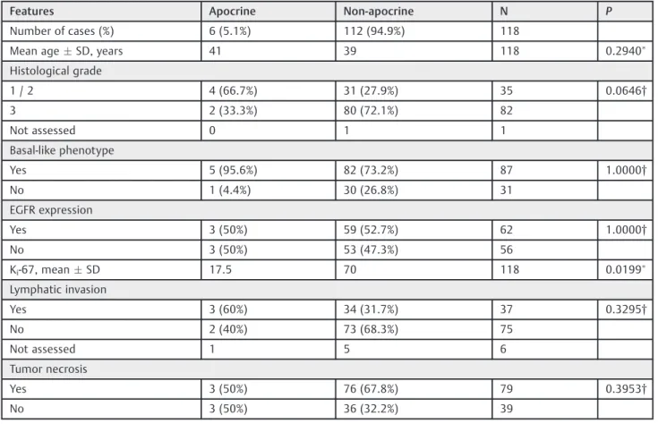
The clinicopathological features of all of the examined cases are summarized in ►Table 2. An apocrine subset was de-tected in 6/118 (5.1%) of the TNBC tissues examined. The median patient age for the apocrine samples versus the non-apocrine samples was 41 and 39 years respectively, and this
difference was not significant. Apocrine tumors also pre-sented a lower rate of Ki-67 expression (17.5% versus 70.0%,
p¼0.02), and a trend toward a lower histological grade (66.7% versus 27.9%,p¼0.06). None of the other studied variables were found to significantly differ between the two groups.
Discussion
Triple-negative breast carcinomas represent a very hetero-geneous group of breast cancers that have a poor prognosis and no targeted therapy available.14Most TNBCs exhibit a basal-like molecular subtype, and have been identified at a higher frequency in women carrying BRCA1.15An important association has been observed between TNBCs, basal-like carcinomas, and BRCA1-associated carcinomas, even though they are not synonymous. In this study, we defined the basal-like phenotype by the immunoexpression of EGFR and/or cytokeratin ⅚, according to the criteria of Nielsen et al.4
Eighty-seven (73.7%) of our 118 cases presented the basal-like phenotype, which was consistent with the rate of cases of previous studies.16,17The basal-like phenotype was pres-ent in 5 out of 6 (95.6%) cases with apocrine phenotype and in 82 out of 112 (73.2%) cases within the non-apocrine group. However, this difference was not significant, probably be-cause of the small number of apocrine cases. The coexistence
Table 2 Clinicopathological features of TNBCs from 118 patients aged 45 years or younger
Features Apocrine Non-apocrine N P
Number of cases (%) 6 (5.1%) 112 (94.9%) 118
Mean ageSD, years 41 39 118 0.2940
Histological grade
1 / 2 4 (66.7%) 31 (27.9%) 35 0.0646†
3 2 (33.3%) 80 (72.1%) 82
Not assessed 0 1 1
Basal-like phenotype
Yes 5 (95.6%) 82 (73.2%) 87 1.0000†
No 1 (4.4%) 30 (26.8%) 31
EGFR expression
Yes 3 (50%) 59 (52.7%) 62 1.0000†
No 3 (50%) 53 (47.3%) 56
Ki-67, meanSD 17.5 70 118 0.0199
Lymphatic invasion
Yes 3 (60%) 34 (31.7%) 37 0.3295†
No 2 (40%) 73 (68.3%) 75
Not assessed 1 5 6
Tumor necrosis
Yes 3 (50%) 76 (67.8%) 79 0.3953†
No 3 (50%) 36 (32.2%) 39
of apocrine and basal-like phenotypes was also described by Choi et al.8These authors studied the immunohistochemical profile of 122 TNBCs, and found the molecular apocrine phenotype in 12 (9.8%) of the cases, the mixed apocrine and basal-like in 5 (4.1%), and the apocrine plus claudin-low in 6 (4.9%) cases. Although their number of apocrine cases was higher than ours, they included older patients (47.512.1 years), and the mean age of the apocrine type was even higher (56.913.7 years).8 The present set of samples was restricted based on younger patients to include a higher percentage of TNBC cases, albeit with a lower frequency of AR expression1,8,18. For example, AR expression was previously detected in 12% of TNBCs,10while only 5.1% of the TNBCs examined in the present study were found to express the AR. Another study found AR expression in 17.7% (87/492) of TNBCs, and the mean age of patients with AR-positive was higher than those with AR-negative tumors (53 years versus 47 years, p<0.001).18 They also observed a significant correlation between AR expression and lower grade tumors. However, they could not explain the worse prognosis they observed in the apocrine group, which dif-fered from the literature.18 The results of our study are consistent with previous observations, despite the limited number of apocrine carcinomas that were examined (n¼6).8,19
Efforts to identify subtypes of TNBCs according to predic-tive variables have been described in recent studies of basal-like carcinomas and/or TNBCs.7,20Less than 30% of women with metastatic triple-negative carcinomas survive more than 5 years, and most do not survive regardless of the chemotherapy regimens they undergo.21 However, it has been reported that basal-like carcinomas are more sensitive to platinum-based therapies,20and drugs acting as poly ADP ribose polymerase (PARP) inhibitors may be effective for the treatment of BRCA1-associated carcinomas once the DNA repair process in these tumors is defective.22 Heretofore, there is a lack of specific and effective therapies for TNBCs. Therefore, a possible role for the AR in defining a distinct subtype of TNBC is of interest.7,8,23 The biological and therapeutic applications of the AR have been widely charac-terized for prostate cancers, but fewer studies have investi-gated the role of the AR in breast cancer.24It is also possible that other molecular events may be involved, such as those involving the microenvironment and epigenetic phenomena. Previously we demonstrated that AR expression in HER2 carcinomas is associated with lower proliferative activity and lower tumor grade, thereby suggesting a less aggressive tumor behavior.25 Mrklić et al26 also observed an inverse
correlation between the Ki-67 index and the AR status for
TNBC, which suggests a possible antiproliferative effect of androgens.
Other studies have analyzed the relationship between AR immunohistochemistry and tumor clinicopathological fea-tures, and an inverse relationship between AR expression and a higher clinical stage, a higher histological grade, and a higher mitotic score was observed.19,25These data suggest that AR-positive TNBCs may be less aggressive tumors. In addition, AR expression appears to predict responses to
anti-androgen therapies. For example, in the phase II study conducted by Gucalp et al10 with AR-positive, metastatic TNBCs, the clinical benefit rate associated with the antago-nist, bicalutamide (150 mg/day), was of 12%.
In spite of the limitations of this study concerning the small number of apocrine cases and the absence of follow-up information, we could confirm the low frequency of the apocrine profile among premenopausal patients and its association with lower grade and lower proliferative activity. Our results reinforce the heterogeneity of TNBCs, a group of different neoplasias that only have in common the absence of estrogen and progesterone receptors, and HER2.
Conclusion
Despite the small number of cases that were analyzed, the present results suggest that the apocrine subgroup of TNBCs is less frequent in premenopausal patients. This subgroup also tends to present as carcinomas of lower grade and lower proliferative activity, which is consistent with a less aggres-sive biological behavior. These results can be considered in strategies for the development of targeted therapies for TNBCs.
This study was supported by the São Paulo Research Foundation (FAPESP) (Process n. 2014/15472–8).
References
1 Carvalho FM, Bacchi LM, Santos PP, Bacchi CE. Triple-negative breast carcinomas are a heterogeneous entity that differs between young and old patients. Clinics (Sao Paulo) 2010;65(10): 1033–1036
2 Malorni L, Shetty PB, De Angelis C, et al. Clinical and biologic features of triple-negative breast cancers in a large cohort of patients with long-term follow-up. Breast Cancer Res Treat 2012; 136(3):795–804
3 Prat A, Adamo B, Cheang MC, Anders CK, Carey LA, Perou CM. Molecular characterization of basal-like and non-basal-like tri-ple-negative breast cancer. Oncologist 2013;18(2):123–133 4 Nielsen TO, Hsu FD, Jensen K, et al. Immunohistochemical and
clinical characterization of the basal-like subtype of invasive breast carcinoma. Clin Cancer Res 2004;10(16):5367–5374 5 Yamamoto Y, Ibusuki M, Nakano M, Kawasoe T, Hiki R, Iwase H.
Clinical significance of basal-like subtype in triple-negative breast cancer. Breast Cancer 2009;16(4):260–267
6 Prat A, Parker JS, Karginova O, et al. Phenotypic and molecular characterization of the claudin-low intrinsic subtype of breast cancer. Breast Cancer Res 2010;12(5):R68
7 Lehmann BD, Bauer JA, Chen X, et al. Identification of human triple-negative breast cancer subtypes and preclinical models for selec-tion of targeted therapies. J Clin Invest 2011;121(7):2750–2767 8 Choi J, Jung WH, Koo JS. Clinicopathologic features of molecular
subtypes of triple negative breast cancer based on immunohisto-chemical markers. Histol Histopathol 2012;27(11):1481–1493 9 McNamara KM, Yoda T, Takagi K, Miki Y, Suzuki T, Sasano H.
Androgen receptor in triple negative breast cancer. J Steroid Biochem Mol Biol 2013;133:66–76
11 Carvalho FM, Bacchi LM, Pincerato KM, Van de Rijn M, Bacchi CE. Geographic differences in the distribution of molecular subtypes of breast cancer in Brazil. BMC Womens Health 2014;14:102 12 Lakhani SR, Ellis IO, Schnitt SJ, Tan PH, van de Vijver MJ. WHO
classification of tumours of the breast. 4th ed. Paris: IARC; 2012
13 Elston CW, Ellis IO. Pathological prognostic factors in breast cancer. I. The value of histological grade in breast cancer: experi-ence from a large study with long-term follow-up. Histopathology 1991;19(5):403–410
14 Gluz O, Liedtke C, Gottschalk N, Pusztai L, Nitz U, Harbeck N. Triple-negative breast cancer–current status and future direc-tions. Ann Oncol 2009;20(12):1913–1927
15 Haffty BG, Yang Q, Reiss M, et al. Locoregional relapse and distant metastasis in conservatively managed triple negative early-stage breast cancer. J Clin Oncol 2006;24(36):5652–5657
16 Thike AA, Iqbal J, Cheok PY, et al. Triple negative breast cancer: outcome correlation with immunohistochemical detection of basal markers. Am J Surg Pathol 2010;34(7):956–964
17 Rao C, Shetty J, Prasad KH. Immunohistochemical profile and morphology in triple - negative breast cancers. J Clin Diagn Res 2013;7(7):1361–1365
18 Choi JE, Kang SH, Lee SJ, Bae YK. Androgen receptor expression predicts decreased survival in early stage triple-negative breast cancer. Ann Surg Oncol 2015;22(1):82–89
19 Ogawa Y, Hai E, Matsumoto K, et al. Androgen receptor expression in breast cancer: relationship with clinicopathological factors and biomarkers. Int J Clin Oncol 2008;13(5):431–435
20 Leidy J, Khan A, Kandil D. Basal-like breast cancer: update on clinicopathologic, immunohistochemical, and molecular fea-tures. Arch Pathol Lab Med 2014;138(1):37–43
21 Dent R, Trudeau M, Pritchard KI, et al. Triple-negative breast cancer: clinical features and patterns of recurrence. Clin Cancer Res 2007;13(15 Pt 1):4429–4434
22 D’Andrea AD. The Fanconi Anemia/BRCA signaling pathway: disruption in cisplatin-sensitive ovarian cancers. Cell Cycle 2003;2(4):290–292
23 Hirshfield KM, Ganesan S. Triple-negative breast cancer: molecu-lar subtypes and targeted therapy. Curr Opin Obstet Gynecol 2014;26(1):34–40
24 Moe RE, Anderson BO. Androgens and androgen receptors: a clinically neglected sector in breast cancer biology. J Surg Oncol 2007;95(6):437–439
25 Lin FdeM, Pincerato KM, Bacchi CE, Baracat EC, Carvalho FM. Coordinated expression of oestrogen and androgen receptors in HER2-positive breast carcinomas: impact on proliferative activi-ty. J Clin Pathol 2012;65(1):64–68