ABSTRACT
http://dx.doi.org/10.1590/1678-775720150156
Static compression regulates OPG expression
in periodontal ligament cells via the CAMK II
pathway
Jianru YI1, MeiLe LI1, Yan YANG1, Wei ZHENG2, Yu LI1, Zhihe ZHAO1
1- Sichuan University, West China School and Hospital of Stomatology, State Key Laboratory of Oral Diseases, Department of Orthodontics, Chengdu, China. 2- Sichuan University, West China School and Hospital of Stomatology, State Key Laboratory of Oral Diseases, Department of Oral and Maxillofacial Surgery, Chengdu, China.
Corresponding address: Yu Li - 14#, 3rd Section - South Renmin Road - Chengdu 610041 - PR - China - Phone: 028-85503645 - e-mail: [email protected]
6XEPLWWHG$SULO0RGL¿FDWLRQ$XJXVW$FFHSWHG6HSWHPEHU
O
bjective: This study aimed to investigate the potential role of CAMK II pathway in the compression-regulated OPG expression in periodontal ligament cells (PDLCs). Material and Methods: The PDL tissue model was developed by 3-D culturing human PDLCs in a thin sheet of poly lactic-co-glycolic acid (PLGA) scaffolds, which was subjected to static compression of 25 g/cm2 for 3, 6 and 12 h, with or without treatment of KN-93. Afterthat, the expression of OPG, RANKL and NFATC2 was investigated through real-time PCR and western blot analysis. Results: After static compression, the NFATC2 and RANKL expression was signi¿cantly up-regulated, while partially suppressed by KN-93 for 6 and 12 h respectively. The OPG expression was signi¿cantly down-regulated by compression in 3 h, started to elevate in 6 h, and signi¿cantly up-regulated in 12 h. The up-regulation after 12 h was signi¿cantly suppressed by KN-93. Conclusions: Long-term static compression increases OPG expression in PDLCs, at least partially, via the CAMK II pathway.
Ke yw or ds: Osteogenesis. Periodontal ligament. Calcium-calmodulin-dependent Protein kinases. Mechanotransduction.
I N TROD UCTI ON
Mechanical loading has long been deciphered to regulate bone metabolism, and plays critical roles in orthodontic tooth movement (OTM)4,10,22.
When teeth are subjected to orthodontic force, the remodeling of the adjacent periodontium is initiated. The static compression induces bone resorption at the pressure side, resulting in a “loose” tooth, followed by bone formation to complete a bone remodeling cycle, which restores the periodontium and strengthens the tooth again29. Lack of this recovery process may lead to
alveolar bone loss followed by gingival recession and tooth removability.
The molecular mechanisms under bone remodeling in OTM has been extensively investigated. Numerous studies have focused on osteoclastogenesis, the rate-limiting step in OTM1,12,16. Particularly, COX-2 has been shown
to induce up-regulation of receptor activator for
nuclear factor-lj % ligand (RANKL), an essential pro-osteoclastogenic factor, in periodontal ligament cells (PDLCs) under compression19. However, little
is known about the pathways to regulate the mechano-induced expression of osteoprotegerin (OPG), the decoy receptor of RANKL, which inhibits bone resorption while promoting bone formation6.
The Ca2+/calmodulin-dependent kinase (CAMK)
family has been recognized as a key mediator in living organisms and various biological processes9.
Recent studies have revealed its critical role in bone development and homeostasis. The CAMK II pathway has been found to regulate the RANKL-induced osteoclast formation via the cAMP-response element binding protein (CREB) pathway3. The involvement of CAMK II in
regulating the RANK-MEK-ERK pathway has been detected21. Moreover, the CAMK II-CREB pathway
was proposed to play an important role in bone homeostasis.
ligament tissue model (PDLtm) to simulate the bioprocess at the pressure side of PDL18.
Interestingly, through microarray screening, we found up-regulation of the CAMK II pathway in the loaded PDLCs17, which is known to respond to
compression and result in OPG enhancement in osteoblast11. Therefore, in the present study, we
aimed to investigate the potential role of CAMK II pathway in the expression of OPG and RANKL in the PDLtm under static compression.
M ATERI AL AN D M ETH OD S
Pr e pa r a t ion of PLGA sca ffold
The poly lactic-co-glycolic (PLGA) polymers (105 g/mol) were used for the synthesis of the
scaffolds as previously described17. BrieÀy, 200 mg
mixture of PLGA polymers and sucrose particulates with a volume ratio of 15:85 was applied with compression to form a thin sheet in a square mould (2 cm × 2 cm). Subsequently, the sheets were placed in a CO2 reaction kettle for 48 h, and the sucrose was then removed by immersing the PLGA sheets in ddH2O for 48 h. After that, the PLGA sheets (2 cm×2 cm×300 Njm) were packaged and sterilized for experimental use.
Est a blish m e n t of t h e PD Lt m
Five lines on human PDLCs were established following a well-documented method17. The
periodontal tissue was collected from the teeth extracted for orthodontic reasons with the donors signing an informed consent form and the approval of the Institutional Review Board in our hospital. The PDLCs of the 3-6th passage were used for this
experiment. The 3-D PDLtm was established by dripping suspension of PDLCs into the PLGA sheet that was put in a 6-well plate, approximate 1×10⁵
cells in 2 ml medium per sheet. After 24 h, the PDLtm was displaced to another 6-well plate. Each cell line (n=5) was used for the experiment in three replicates, two of which were pooled together for western blot and one for real-time PCR.
H ist ologica l obse r va t ion
Four days after the establishment of PDLtm, the growth of PDLCs in scaffolds was investigated by microscopic observations. First, the PDLtm was
stained by acridine orange (0.01%) and observed under a Àuorescence inverted microscope (Leica DMI6000B, Germany). Second, the PLGA/PDLC construct was prepared for scanning of electron microscopy (SEM, Inspect F, FEI, USA)17.
Applica t ion of com pr e ssive for ce
The PDLtms were randomly assigned into three groups, i.e., the control group, the compression group (Cg) and the compression+KN-93 (an inhibitor of the CAMK II pathway) group (CKg). To simulate the pressured periodontium in OTM, a modi¿ed “weight” method was used. BrieÀy, a cover glass and a bottle of granules were placed on the PDLtm to produce a compression of 25 g/ cm2, which has been proved to be the optimal force
level for this model18. In the compression+KN-93
group, 0.01 mM KN-93 (Sigma) was added to the media to suppress the CAMK II pathway.
Re a l- t im e PCR
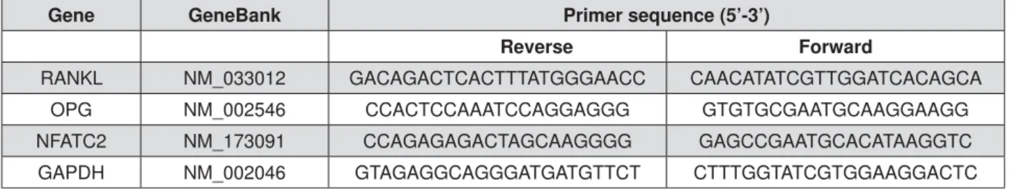
Three, six and twelve hours after applying interventions, total RNA (n=5) was extracted by dissolving the PDLtm using TRIzol reagent (Invitrogen, Carlsbad, USA). The quality and integrity of extracted RNA samples were validated before their use. Real-time PCR was performed with a SYBR Green reaction Kit (Roche Diagnostics, China) in a LightCycler according to the manufacturer’s instruction to investigate the mRNA expression of RANKL, OPG, and Nuclear factor of activated T-cells (NFAT) C2. GAPDH served as the internal control. The sequences of relevant primers were shown in Figure 1.
W e st e r n blot t in g
Twelve hours after force application, the total proteins (n=5) were collected using total protein extraction kit (Keygen Biotech, China). The 15 NjL prepared samples (40 Njg of protein) per lane were separated by SDS-PAGE and then transferred to the polyvinylidene diÀuoride (P9DF) membrane. After that, the PVDF membranes were probed with antibodies to RANKL (1:1000, Santa-Cruz, USA), OPG (1:1000, Santa-Cruz, USA) GAPDH (1:1000, Beyotime, China) overnight at 4°C. Subsequently, the membranes were immersed in secondary antibody (1:5000, Beyotime, China) for
Gene GeneBank Primer sequence (5’-3’)
Reverse Forward
RANKL NM_033012 GACAGACTCACTTTATGGGAACC CAACATATCGTTGGATCACAGCA
OPG NM_002546 CCACTCCAAATCCAGGAGGG GTGTGCGAATGCAAGGAAGG
NFATC2 NM_173091 CCAGAGAGACTAGCAAGGGG GAGCCGAATGCACATAAGGTC
GAPDH NM_002046 GTAGAGGCAGGGATGATGTTCT CTTTGGTATCGTGGAAGGACTC
1 h. The immunoreactive proteins were visualized by a chemiluminescence kit (Millipore). The band intensities were evaluated using Quantity One software (Bio-Rad, Hercules, USA).
St a t ist ica l a n a ly sis
All data was expressed as mean±SD. The comparison among 3 groups was conducted by one-way analysis of variance (ANOVA) followed by LSD post h oc test using SPSS software of version 13.0. Differences with p<0.05 were set as signi¿cant.
RESULTS
Th e 3 - D cu lt u r e d PD LCs
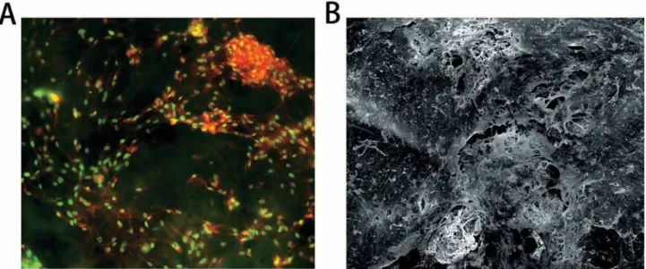
By acridine orange staining, the growth of PDLCs was observed under microscope (Figure
2A). In addition, the secreted extracellular matrix, the PDLCs and the scaffolds were observed to be interconnected under SEM (Figure 2B). More characterization of this PDLtm has been previously shown18.
Th e N FATC2 e x pr e ssion
Under static compression, the NFATC2 expression was signi¿cantly up-regulated, which peaked in 6 h (Figure 3). KN-93 partially suppressed the compression-induced up-regulation of NFATC2 in 6 and 12 h.
Th e OPG/ RAN KL e x pr e ssion
The real-time PCR analysis revealed the signi¿cant up-regulation of RANKL expression under static compression, which peaked in 6 h (Figure 4A). In contrast, the OPG expression was signi¿cantly down-regulated after 3 h, while up-regulated in 12 h (Figure 4B). The OPG/RANKL ratio was reduced in 3 and 6 h while enhanced in 12 h (Figure 4C).
Notably, after 12 h, the up-regulation of RANKL, OPG and OPG/RANKL ratio was partially suppressed by KN-93, suggesting that the CAMK II pathway took part in the up-regulation of RANKL and OPG in PDLCs induced by long-term stimulation of static compression (Figure 4).
Moreover, the western blotting method showed results similar to the PCR assay, indicating that after 12 h of static compression there was an increased protein expression of RANKL and OPG, which was partially suppressed by KN-93 (Figure 5).
Figure 2- The microscopic observations of PDLtm. (A) Spindle PDLCs grew densely with nuclear in green-yellow and cytoplasma in orange-red, 200×; (B) The PDLCs, secreted extracellular matrix (SEM) and PLGA scaffolds were integrated, 300×
D I SCUSSI ON
Orthodontic tooth movement (OTM) has been generally featured with bone remodeling, a fundamental biological process consisting of bone resorption and bone formation23. Particularly, the
initiated bone resorption at compression sites results in tooth movement, and the following bone formation surrounding roots strengthen the new position of the teeth29. Numerous studies
focused on the osteoclastogenesis at the pressure side and sketched the contour of underlying mechanisms12,13, while the subsequent bone
formation was less studied.
The CAMK family contains a series of proteins playing critical roles in bone modeling and remodeling. Afamin enhances osteoclastogenesis
by decreasing intracellular cAMP levels via the CAMK pathways14. The role of the CAMK II pathway
in the parathyroid hormone-related protein (PTHrP)-regulating osteoclast inhibitory lection has been recently identi¿ed32. Though KN-93 was
observed to exert effects on other pathways8, it has
been applied as a classic CAMK II pathway inhibitor in many studies25,27. Recently, by using KN-93,
the enhancement of OPG in osteoblast under mechanical stimuli was suppressed11. However,
limited information concerning its effect on OTM is available till now.
The microarray screening for gene expression pro¿les in our previous study has revealed the potential role of the CAMK II pathway in the mechanoresponse of PDLCs17. In the present study,
PDLCs were embedded in 3-D PLGA scaffolds and Figure 4- The time-course expression of osteoprotegerin (OPG) and RANKL in loaded PDLCs at mRNA level. (A) RANKL; (B) OPG; (C) OPG/RANKL ratio.*p<0.05
cultured under static compressive force. The ratio of OPG/RANKL expression was signi¿cantly down-regulated 3 and 6 h after loading, indicating a potential role of osteoclastogenesis induction24,31.
On the other hand, the OPG expression declined in 3 h, while started to elevate in 6 h and was significantly up-regulated after 12 h. As an important anti-osteoclastic and pro-osteogenic factor, the marked up-regulation of OPG after 12h indicates suppression for bone resorption15.
However, this elevation was greatly impeded by KN-93, a speci¿c CAMK II pathway inhibitor, suggesting that CAMK II pathway takes part in the OPG up-regulation induced by long-term static compression stimulation. Notably, the CAMK II pathway has also been reported to regulate the mechano-induced OPG enhancement in osteoblast11, which is to a great extent similar to
PDL ¿broblast2.
T h e R A N K L– R A N K– O P G a x i s m e d i a t e s osteoclast formation through activation of RANK on the osteoclast precursors by RANKL28.
Although the constitutional expression of OPG in PDLCs is much higher than RANKL26, numerous
studies demonstrated the comparatively slight up-regulation of RANKL in PDLCs induced by compressive force could promote osteoclast formation and the following tooth movement13,18.
Therefore, the large ratio of total OPG vs. RANKL seems meaningless accounting for osteoclastic induction. A reasonable explanation has been given that it could be due to the tight cell–cell contact between PDLCs and osteoclast precursors, which could create a favorable micro-environment for RANKL–RANK binding, thereby preventing the interaction of OPG with RANKL5.
On the other hand, at the sites away from the cell–cell contact area, the expression of OPG could play an important role. In contrast to the study reporting reduced or unchanged OPG expression in PDLCs under compression20, in the present study
the OPG expression was signi¿cantly up-regulated after long-term mechanical stimuli, consistent with our previous data18. The delayed but marked
increase of OPG could account for the subsequent bone formation at pressure periodontium, which prevents from alveolar bone loss and strengthens tooth again after movement. In this sense, targeting the CAMK II pathway might potentially bene¿t the OTM periodontium by stimulating OPG expression.
In the present study, we observed that the expression of NFATC2 in PDLCs was enhanced by compressive force while partially inhibited by KN-93 treatment (Figure 3). NFATC2 is a transcription factor which plays an undisputable role in the mechanoresponse of bone tissue30. As downstream
factors of Wnt-Ca2+ pathways, both NFATC2
and CAMK II are involved in the Wnt-Ca2+ bone
formation regulated by pathways7. Interestingly,
Wnt-Ca2+ pathways were up-regulated when
PDLCs were treated with compressive force in our previous study17. Therefore, the enhancement of
OPG in PDLCs under compressive force might be regulated via Wnt-Ca2+ pathways, which should be
further identi¿ed in future.
Last but not the least, it is interesting to compare the masticatory force and orthodontic force, both of which are transmitted to alveolar bone through PDL, while it results in opposite effects on alveolar bone metabolism. Obviously, the present theory of “compression-PDL-osteoclastogenesis” process cannot interpret this contradictory phenomenon. Based on our results, it is reasonable to speculate that the CAMK II pathways may be much more sensitive to masticatory force (intermittent force) than orthodontic force (static force), which should be further identi¿ed.
CON CLUSI ON
The OPG expression was significantly up-regulated in PDLCs after long-term static compression stimulation, which is at least partially regulated by the CAMK II pathway. These results have enriched the present understanding to molecular mechanisms in bone remodeling modulation in OTM.
ACKN OW LED GEM EN T
This study was supported by NSFC - National Natural Science Foundation of China (11372202 and 31201087), and by Sichuan University Fund for Outstanding Young Scholars (2082604164243).
REFEREN CES
1- Alhashimi N, Frithiof L, Brudvik P, Bakhiet M. Orthodontic tooth
movement and de novo synthesis of proinÀammatory cytokines.
Am J Orthod Dentofacial Orthop. 2001:119(3):307-12.
2- Alves LB, Mariguela VC, Grisi MF, Souza SL, Novaes Junior AB, Taba Junior M, et al. Expression of osteoblastic phenotype
in periodontal ligament ¿broblasts cultured in three-dimensional
collagen gel. J Appl Oral Sci. 2015:23(2):206-14.
3- Ang ES, Zhang P, Steer JH, Tan JW, Yip K, Zheng MH, et al. Calcium/calmodulin-dependent kinase activity is required for
ef¿cient induction of osteoclast differentiation and bone resorption
by receptor activator of nuclear factor kappa B ligand (RANKL). J Cell Physiol. 2007:212(3):787-95.
4- Araújo AS, Fernandes AB, Maciel JV, Netto JN, Bolognese AM. New methodology for evaluating osteoclastic activity induced by orthodontic load. J Appl Oral Sci. 2015:23(1):19-25.
5- Bloemen V, Schoenmaker T, de Vries TJ, Everts V. Direct cell-cell
contact between periodontal ligament ¿broblasts and osteoclast
7- Chen Y, Alman BA. Wnt pathway, an essential role in bone regeneration. J Cell Biochem. 2009:106(3):353-62.
8- Gao L, Blair LA, Marshall J. CaMKII-independent effects of KN93 and its inactive analog KN92: reversible inhibition of L-type calcium channels. Biochem Biophys Res Commun. 2006:345(4):1606-10.
9- HoeÀich KP, Ikura M. Calmodulin in action: diversity in target
recognition and activation mechanisms. Cell. 2002:108(6):739-42. 10- Huiskes R, Ruimerman R, van Lenthe GH, Janssen JD. Effects of mechanical forces on maintenance and adaptation of form in trabecular bone. Nature. 2000:405(6787):704-6.
11- Kaneuji T, Ariyoshi W, Okinaga T, Toshinaga A, Takahashi T, Nishihara T. Mechanisms involved in regulation of osteoclastic differentiation by mechanical stress-loaded osteoblasts. Biochem Biophys Res Commun. 2011:408(1):103-9.
12- Kanzaki H, Chiba M, Shimizu Y, Mitani H. Dual regulation of osteoclast differentiation by periodontal ligament cells through RANKL stimulation and OPG inhibition. J Dent Res. 2001:80(3):887-91.
13- Kanzaki H, Chiba M, Shimizu Y, Mitani H. Periodontal ligament cells under mechanical stress induce osteoclastogenesis by receptor activator of nuclear factor kappaB ligand up-regulation via prostaglandin E2 synthesis. J Bone Miner Res. 2002:17(2):210-20. 14- Kim BJ, Lee YS, Lee SY, Park SY, Dieplinger H, Yea K, et al. Afamin stimulates osteoclastogenesis and bone resorption via Gi-coupled receptor and Ca2+/calmodulin-dependent protein kinase (CaMK) pathways. J Endocrinol Invest. 2013:36(10):876-82. 15- Kong YY, Feige U, Sarosi I, Bolon B, Tafuri A, Morony S, et al. Activated T cells regulate bone loss and joint destruction in adjuvant arthritis through osteoprotegerin ligand. Nature. 1999:402(6759):304-9.
16- Lee YH, Nahm DS, Jung YK, Choi JY, Kim SG, Cho M, et al. Differential gene expression of periodontal ligament cells after loading of static compressive force. J Periodontol. 2007:78(3):446-52.
17- Li Y, Li M, Tan L, Huang S, Zhao L, Tang T, et al. Analysis of
time-course gene expression pro¿les of a periodontal ligament
tissue model under compression. Arch Oral Biol. 2013:58(5):511-22.
18- Li Y, Zheng W, Liu JS, Wang J, Yang P, Li ML, et al. Expression of osteoclastogenesis inducers in a tissue model of periodontal ligament under compression. J Dent Res. 2011:90(1):115-20. 19- Liu L, Igarashi K, Kanzaki H, Chiba M, Shinoda H, Mitani H. Clodronate inhibits PGE(2) production in compressed periodontal ligament cells. J Dent Res. 2006:85(8):757-60.
20- Nishijima Y, Yamaguchi M, Kojima T, Aihara N, Nakajima R,
Kasai K. Levels of RANKL and OPG in gingival crevicular Àuid during
orthodontic tooth movement and effect of compression force on
releases from periodontal ligament cells in vit ro. Orthod Craniofac
Res. 2006:9(2):63-70.
21- Park-Min KH, Ji JD, Antoniv T, Reid AC, Silver RB, Humphrey MB, et al. IL-10 suppresses calcium-mediated costimulation of receptor activator NF-kappa B signaling during human osteoclast differentiation by inhibiting TREM-2 expression. J Immunol. 2009:183(4):2444-55.
22- Ren Y, Maltha JC, Kuijpers-Jagtman AM. Optimum force magnitude for orthodontic tooth movement: a systematic literature review. Angle Orthod. 2003:73(1):86-92.
23- Roberts WE, Goodwin WC Jr, Heiner SR. Cellular response to orthodontic force. Dent Clin North Am. 1981:25(1):3-17.
24- Sa÷lam M, K|seo÷lu S, Hatipo÷lu M, Esen HH, K|ksal E. Effect
of sumac extract on serum oxidative status, RANKL/OPG system and alveolar bone loss in experimental periodontitis in rats. J Appl Oral Sci. 2015:23(1):33-41.
25- Saito A, Miyajima K, Akatsuka J, Kondo H, Mashiko T, Kiuchi T, et al. CaMKIIbeta-mediated LIM-kinase activation plays a crucial role in BDNF-induced neuritogenesis. Genes Cells. 2013:18(7):533-43.
26- Sokos D, Everts V, de Vries TJ. Role of periodontal ligament
¿broblasts in osteoclastogenesis: a review. J Periodontal Res.
2015;50(2):152-9.
27- Souza CF, Carneiro AB, Silveira AB, Laranja GA, Silva-Neto MA,
Costa SC, et al. Heme-induced Trypanosom a cruzi proliferation
is mediated by CaM kinase II. Biochem Biophys Res Commun. 2009:390(3):541-6.
28- Tanaka S. Signaling axis in osteoclast biology and therapeutic targeting in the RANKL/RANK/OPG system. Am J Nephrol. 2007:27(5):466-78.
29- Wise GE, King GJ. Mechanisms of tooth eruption and orthodontic tooth movement. J Dent Res. 2008:87(5):414-34. 30- Wu Y, Yang Y, Yang P, Gu Y, Zhao Z, Tan L, et al. The osteogenic differentiation of PDLSCs is mediated through MEK/ERK and p38 MAPK signalling under hypoxia. Arch Oral Biol. 2013:58(10):1357-68.
31- Yasuda H, Shima N, Nakagawa N, Yamaguchi K, Kinosaki M, Mochizuki S, et al. Osteoclast differentiation factor is a ligand for osteoprotegerin/osteoclastogenesis-inhibitory factor and is identical to TRANCE/RANKL. Proc Natl Acad Sci U S A. 1998:95(7):3597-602.