BIOFUNCTIONAL SILK-FIBROIN MEMBRANES WITH ANTIOXIDANT PROPERTIES FOR THE TREATMENT OF CHRONIC WOUNDS
by
Ana Sofia Ferreira Fernandes
1 BIOFUNCTIONAL SILK-FIBROIN MEMBRANES WITH ANTIOXIDANT PROPERTIES FOR THE
TREATMENT OF CHRONIC WOUNDS
Thesis presented to Escola Superior de Biotecnologia of the Universidade Católica Portuguesa to fulfill the requirements of Master of Science degree in Biomedical Engineering
by
Ana Sofia Ferreira Fernandes
Place: Escola Superior de Biotecnologia and Universidad de Vigo
Supervisor: Prof. Dr.ª Ana Leite Oliveira
Co-Supervisors: Dr.ª Cassilda Cunha Reis, Prof. Dr. Pío González
2
RESUMO
Atualmente, as feridas crónicas são uma patologia cada vez mais recorrente, apresentando uma maior incidência na população idosa, principalmente quando existem fatores de co-morbilidade, tais como Diabetes Mellitus e doenças cardiovasculares. Nos últimos anos, têm sido propostos novos materiais de penso, incorporando uma diversidade de agentes bioativos para o tratamento de feridas crónicas. Entre os biomateriais naturais propostos, a seda ganhou atenção devido à sua biocompatibilidade, biodegradação, por ser facilmente modificada quimicamente e por possuir boas propriedades mecânicas. O objetivo deste estudo foi o desenvolvimento, otimização, e caracterização de membranas biofuncionais feitas à base de fibroína de seda (SF), com a incorporação de dois agentes antioxidantes (ácido cafeico – CA – e ácido tânico – TA) para o tratamento de feridas crónicas superficiais e consequente regeneração da pele. Através da técnica de solvent casting, seguida de um tratamento térmico, produziram-se membranas estáveis, que foram posteriormente caracterizadas relativamente à morfologia da superfície (SEM e perfilometria ótica), à composição química (FTIR-ATR), perfil de degradação, performance mecânica, análise térmica (TG-DTA), capacidade antioxidante e citocompatibilidade. A degradação foi avaliada pela percentagem de peso perdido pelas amostras. A atividade antioxidante foi avaliada pelo método ABTS. Os testes de citocompatibilidade foram realizados, utilizando a linha celular imortalizada de células de fibroblastos de rato (L929), através do método de contacto direto e dos extratos. Todas as membranas desenvolvidas demonstraram ser transparentes, inodoras, apresentando uma superfície homogénea, ainda que com espessura variável. As imagens de SEM mostraram que todas as membranas apresentavam uma superfície lisa e homogénea, sem rugosidade aparente à escala micrométrica. Através da perfilometria ótica concluiu-se que, à escala nanométrica, a adição de glicerol aumentou a rugosidade da superfície e, através dos ensaios mecânicos, verificou-se que este composto reduziu a rigidez das membranas, tornando-as mais maleáveis. Com as membranas no estado seco, os agentes antioxidantes potencializaram um efeito anti-plasticizante, enquanto, no estado hidratado, a água potencializou o efeito plasticizante do glicerol. As membranas de SF apresentaram uma massa constante ao longo de 15 dias, em PBS, enquanto que as restantes membranas apresentaram uma perda de massa inicial de aproximadamente 30%, após as primeiras 2 horas de incubação, maioritariamente relacionada com libertação de glicerol. Com a presença de Protease XIV, a mesma perda de massa inicial foi observada, aumentando drasticamente após 24 horas de incubação, evidenciando a suscetibilidade da seda para a degradaç ão proteolítica. A resistência térmica da SF diminuiu com a introdução do glicerol nas formulações. À concentração de 0.5% de ambos os agentes antioxidantes essa propriedade foi aumentada e o oposto foi observado na concentração de 1%. As membranas de SF mostraram ter capacidade antioxidante, tendo a adição de 0.5% TA reforçado este efeito. Apenas o extrato puro de 1% TA revelou um efeito citotóxico para as células L929. Todas as restantes membranas não apresentaram citotoxicidade. Este sistema demonstrou ser bastante promissor, reconhecendo a capacidade das membranas de SF para incorporar moléculas antioxidantes e o seu possível potencial no tratamento de feridas crónicas. Palavras – chave: antioxidantes, feridas crónicas, membranas de fibroína de seda, pensos para feridas.
3
ABSTRACT
Nowadays, chronic wounds are an increasingly recurrent pathology, presenting a higher incidence in the elderly population, especially when there are comorbidity factors such as Diabetes Mellitus and cardiovascular diseases. In recent years, new dressing materials incorporating a variety of bioactive agents have been proposed for the treatment of chronic wounds. Among the proposed natural biomaterials, silk gained attention due to its biocompatibility, biodegradation, for being easily chemically modified and due to its good mechanical properties. The objective of this study was the development , optimization, and characterization of biofunctional silk fibroin (SF) based membranes, with the incorporation of two antioxidants agents (AA) (caffeic acid - CA - and tannic acid - TA) for the treatment of superficial chronic wounds and consequent regeneration of the skin. Stable membranes were produced using the technique of solvent casting followed by a thermal treatment, which were characterized regarding surface morphology (SEM and optical profilometry), chemical structure (FTIR-ATR), degradation profile, mechanical performance, TG-DTA, antioxidant capacity and cytocompatibility. The degradation was assessed by obtaining the percentage of weight lost by samples. Antioxidant activity was evaluated by the ABTS method. Cytocompatibility tests were performed using the immortalized cell line of mouse fibroblast cells (L929) by direct contact and extracts tests. All the membranes were transparent, odorless and presented a homogeneous surface, although with variable thickness. SEM images showed that all membranes had a smooth and homogeneous surface, wi th no apparent roughness at the micrometer scale. Optical profilometry showed that, at the nanoscale, the introduction of glycerol (Gly) in the formulations increased the surface roughness and, the mechanical tests confirmed that this compound reduced the stiffness of the membranes, making them more malleable. With the membranes in the dry state, the AA potentiate an anti-plasticizer effect, while, in the hydrated state, water has potentiated the plasticizer effect of Gly. SF membranes exhibited a constant mass over 15 days in PBS, while the remaining membranes showed an initial weight loss of approximately 30%, after the first 2 hours of incubation, most likely related to Gly release. In the presence of Protease XIV, the same initial weight loss was observed, increasing drastically after 24 hours of incubation, evidencing the susceptibility of the silk to the proteolytic degradation. The thermal resistance of SF decreased with the introduction of Gly. At a concentration of 0.5%, the AA increased this property and the opposite was observed when the concentration raised to 1%. SF membranes showed to have antioxidant capacity, and the addition of 0.5% TA reinforced this effect. The pure 1% TA extract revealed a cytotoxic effect in L929 cells. All other membranes were non-cytotoxic. This system proved to be quite promising, recognizing the ability of SF membranes to incorporate antioxidant molecules and their potential in the treatment of chronic wounds.
4
ACKNOWLEDGEMENTS
Nenhuma secção foi tão complexa de elaborar como esta. E nenhuma estará tão incompleta quanto esta. Muitas foram as pessoas que me acompanharam ao longo desta etapa da minha formaç ão académica. E muitas foram as pessoas que me acompanharam, não só na minha formação académica, como na minha vida quotidiana e pessoal. Todas, sem exceção, contribuíram para o meu crescimento. A todas estas pessoas deixo, nesta secção, o meu maior e profundo agradecimento, e a maior gratidão que consigo expressar por palavras escritas.
À Professora Doutora Ana Leite Oliveira, pela incomparável orientação. Por todas as oportunidades que me deu ao longo destes quatro anos. Por nunca me ter deixado desistir. Por ter acreditado nas minhas capacidades. Por toda a paciência e disponibilidade, e por toda a ajuda nos momentos bons e menos bons. Foi um prazer enorme poder aprender com a professora e com toda a equipa durante estes anos. Ficar-lhe-ei eternamente agradecida por ter contribuído tanto no meu crescimento científico e profissional.
À Doutora Cassilda Cunha Reis, pela inigualável coorientação, por todos os ensinamentos, por toda a passagem de conhecimento científico, por todo o apoio em todos os momentos, principalmente nos mais difíceis; por todas as palavras de incentivo e por ter sempre acreditado nas minhas capacidades. Fico-te para sempre agradecida, Cassilda, por teres conseguido fazer com que todos os dias se transformasse em dias de esperança e alegria!
Ao Professor Doutor Pío González, à Professora Doutora Júlia Serra e a todo o grupo de investigação Nuevos Materiales da Universidade de Vigo, por tão bem me terem recebido e integrado, por toda a simpatia e amabilidade, pelas oportunidades que me deram e por todo o contributo na minha coorientação e no meu crescimento científico.
Ao Professor Doutor João Paulo Ferreira, coordenador do Mestrado em Engenharia Biomédica, pela oportunidade de frequentar este mestrado, que tanto contribuiu para o meu enriquecimen to científico e académico, assim como por toda a disponibilidade que teve ao longo destes dois anos.
Aos meus Colegas e Amigos de Laboratório – Ana Rita Pinto, Catarina Geão, Cassilda Reis, Gabriela Madanços, Gonçalo Soares, Marcus Vinícius, Nilza Ribeiro, Ricardo Serôdio, Sandra Borges, Sara Baptista e Sara Pérez. Por terem deixado a vossa marca neste meu percurso científico. Por serem todos especiais à vossa maneira. Por cada um me ter passado novos ensinamentos. Por todo o companheirismo, por todo o apoio e por toda a alegria diária! Um agradecimento especial à Doutora Sara Baptista da Silva por todo o apoio, suporte e ajuda na realização do ensaio de ABTS.
5 À Professora Doutora Goreti Sales, à Doutora Manuela Frasco e ao BioMark Research Group do Instituto Superior de Engenharia do Porto pela realização do ensaio de TG-DTA, por toda a disponibilidade e integração.
Ás minhas Colegas e Companheiras da Escola Superior de Biotecnologia – Beatriz Moreira, Francisca Ferreira e Juliana Aguiar – por todos estes anos de amizade. Por todas as conversas. Por todo o apoio. Por todas as gargalhadas. Por todos os momentos que passamos juntas. Por todo o stress partilhado e por serem a melhor equipa de trabalho que poderia alguma vez ter! Sem vocês, esta conquista não teria o mesmo sabor!
Às minhas Colegas e Companheiras de Faculdade – Catarina Loureiro, Carolina Magalhães , Marta Gonçalves e Sofia Sarmento – por toda a amizade que desenvolvemos em cinco anos e que vai ficar para a vida toda! Por, mesmo estando longe, estarem sempre tão perto. Por fazerem a minha vida melhor!
A toda a família dançante da Academia de Dança João Alves, por me acompanharem durante estes cinco anos. Por me terem visto crescer e por terem acompanhado o progresso da minha formaç ão académica. Por cada um de vós, à vossa maneira, sem exceção, ter contribuído, tanto na minha evolução profissional, como na minha evolução enquanto dançarina. Por terem sempre as palavras e os sorrisos mais doces para me dar. Pela vossa camaradagem e companheirismo. Por serem pessoas especiais. E por terem sido um dos motivos que me ajudou a levar este barco a bom porto. A todos vocês, sem exceção, o meu sincero obrigada!
À minha Melhor Amiga – Mariana Silva. Por ter sido o melhor que a faculdade me deu. Por ser um anjo na Terra. Por ser um dos maiores pilares da minha vida. Por ser a melhor amiga que poderia ter. Pelo ser humano lindo que é. Por toda a honestidade e humildade. Por ouvir os meus dramas diários durante horas a fio. Por estar sempre presente em todos os momentos da minha vida. Por nunca me deixar desistir daquilo que ambiciono. Por me mostrar que “a distância não significa nada quando uma pessoa significa muito”. Por me fazer rir e sorrir quando menos espero. Por ter sempre o conselho certo para me dar. Por todos os momentos que já passamos. Por tudo o que ainda vamos passar juntas e por todas as histórias que vamos criar juntas. Por te teres tornado nas pessoas mais importantes da minha vida em tão pouco tempo. Por seres a irmã que nunca tive – o meu maior, mais sincero e eterno obrigada, minha Marianocas!
6 Aos meus Vizinhos – Fernandinha e Sr. Manuel – por me acompanharem nestes meus 23 anos de vida. Por me terem acompanhado nas diversas etapas da minha formação profissional e pessoal, por me verem crescer e evoluir, por acreditarem sempre em mim e por nunca me deixarem desistir de nada. Por me apoiarem em todos os meus projetos, por terem sempre os melhores conselhos e as palavras certas, e por acreditarem sempre no meu sucesso. Muito obrigada por serem os melhores vizinhos (e os melhores avós emprestados) do mundo!
À minha Melhor Amiga de Quatro Patas – Laik a – por saber como me animar nos dias menos bons. Por me compreender sem saber falar português. Por me pôr a sorrir com brincadeiras simples. Pelo amor incondicional e por toda a proteção que me dá sem pedir nada em troca. Por ser a companheira mais fiel e mais leal que poderia ter. Por me ensinar que o melhor da vida está nas coisas mais simples e nos sentimentos mais puros, como o amor e a gratidão. Por ser a cadela mais bonita de sempre. Por me mostrar que os animais são das maiores dádivas que a natureza tem!
Aos meus queridos Pais – Domingos Fernandes e Isaura Fernandes – e aos meus queridos Avós – Manuel Ferreira e Maria da Conceição Ferreira. Os meus anjos da guarda na Terra. Os meus melhores amigos. Os meus heróis. Os meus guerreiros... Os meus exemplos de vida! Nunca hão de haver palavras suficientes para vos agradecer tudo o que fizeram por mim durante estes cinco anos. E muito menos palavras existirão para vos agradecer tudo o que fizeram e fazem por mim desde o meu primeiro segundo de vida. São o maior pilar da minha vida. A minha razão de viver. Os melhores amigos que alguma vez poderei ter e a melhor família que alguma vez poderia desejar ter. Grande parte do que sou a vocês devo. Grande parte do que conquistei e do que serei daqui para a frente a vocês devo. Ser-vos-ei grata para sempre. Por nunca me deixarem desistir dos meus sonhos. Por me apoiarem em tudo o que decido fazer. Por nunca me deixarem baixar os braços. Por fazerem os possíveis e os impossíveis para que nunca me falte nada. Por colocarem a minha felicidade e o meu bem-estar à frente de tudo. Por aturarem todas as minhas frustrações, toda a minha mimalhice e todos os momentos de palhaçadas! Por estarem sempre presentes em todos os momentos da minha vida. Por terem sempre as palavras certas para me dar, nos momentos certos. Por me darem os melhores conselhos. Por celebrarem comigo as minhas vitórias. Por nunca me deixarem cair e por me acompanharem sempre, de mãos dadas, em direção às minhas metas. Por serem a maior luz do meu caminho. Amo-vos com tudo o que tenho e com toda a força que tenho! Para SEMPRE! A vocês, amores lindos do meu coração... Fico-vos eternamente GRATA!
7
TABLE OF CONTENTS
Resumo ...2 Abstract...3 Acknowledgements ...4 List of figures ...9 List of tables ... 10 Chapter 1 ... 11 1. Introduction ... 11 1. Chronic Wounds ... 112. Current clinical approaches to chronic wounds ... 13
3. Membranes in wound healing ... 18
3.1. Bioactive and multifunctional membranes ... 20
4. Main objective... 22
Chapter 2 ... 23
Materials and Methods ... 23
2.1. Materials and Reagents ... 23
2.1.1. Silk ... 23
2.1.2. Glycerol ... 24
2.1.3. Anti-oxidant molecules ... 25
2.2. Preparation of silk fibroin aqueous solution ... 26
2.3. Preparation of SF-based membranes ... 26
2.4. Organoleptic properties ... 27
2.5. Morphological characterization of the surface ... 28
2.5.1. Scanning Electron Microscopy (SEM) ... 28
2.5.2. Optical Profilometry ... 28
2.6. Chemical characterization ... 28
2.6.1. Fourier Transformed InfraRed spectroscopy with Attenuated Total Reflectance (FTIR -ATR) ... 28
2.7. Degradation profile ... 29
8
2.9. Thermal properties ... 31
2.9.1. Thermogravimetry and Different Thermal Analysis (TG-DTA)... 31
2.10. Antioxidant capacity – ABTS assay... 31
2.11. Cytotoxicity tests ... 32
2.12. Statistical analysis ... 33
CHAPTER 3 ... 34
Results and Discussion ... 34
3.1. Organoleptic properties ... 34
3.2. Morphologic characterization of the surface ... 38
3.2.1. Scanning Electron Microscopy (SEM) ... 38
3.2.2. Optical Profilometry (OP) ... 40
3.3. Mechanical Performanc e ... 41
3.4. Fourier transform infrared attenuated total reflectance (FTIR-ATR) spectroscopy ... 44
3.5. Degradation profile ... 45
3.6. Thermal properties ... 48
3.6.1. Thermogravimetry and Different Thermal Analysis (TG – DTA) ... 48
3.7. Antioxidant properties – ABTS assay ... 49
3.8. Cytotoxicity tests ... 50 Chapter 4 ... 52 Conclusions ... 52 Future work ... 53 Comunications ... 54 Referenc es ... 55
9
LIST OF FIGURES
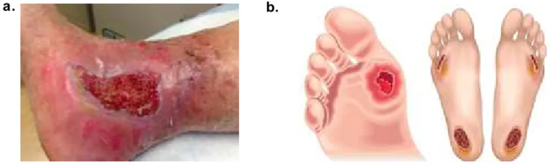
Figure 1.1 – Leg (a) and foot (b) chronic wounds examples. ……….…...………. 12
Figure 2.1.1. – Structural components of silk from Bombyx mori. ……….……….……..……….. 23
Figure 2.1.2. – Glycerol chemical structure ………..…….……… 24
Figure 2.1.3. – Chemical structure of caffeic (a) and tannic (b) acids.……….……….. 25
Figure 2.4.1 – Thickness measurement points in the samples...……… 27
Figure 3.1.1 – Thickness of dry membranes measured in 10 different points of the silk -based membranes (n = 3, Kruskal-Wallis test, P = 0.0061, * – significant differences).……….. 37
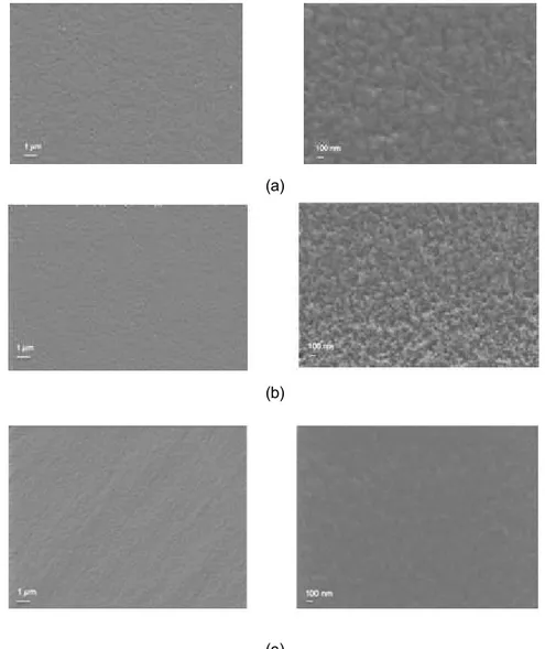
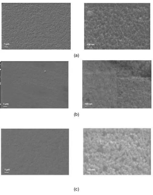
Figure 3.2.1. SEM images of (a) SF, (b) SF/Gly and (c) SF/Gly/CA05 membranes (Magnifications of 500x and 20000x)……… 38
Figure 3.2.2. SEM images of (a) SF/Gly/CA1, (b) SF/Gly/TA05 and (c) SF/Gly/TA1 membranes (Magnifications of 500x and 20000x)……… 39
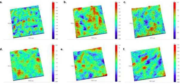
Figure 3.2.3. – OP nanostructure images of SF (a), SF/Gly (b), SF/Gly/CA05 (c), SF/Gly/CA1 (d), SF/Gly/TA05 (e) and SF/Gly/TA1 (f) membranes……….. 40
Figure 3.2.4. – Average roughness of the SF and SF/GLY membranes incorporating 0.5 and 1% of caffeic acid (a), tannic acid (b) obtained by optical profilometry (n = 3, Kruskal-Wallis test, P = 0.0078, * – significant differences)……….. 40
Figure 3.3.1 – Young’s Modulus (E), Ultimate Tensile Strength (UTS) and Elongation at Break (ε) parameters of all membranes, in dry and wet state (n = 10, One-way ANOVA test, P = 0.0078, * – significant differences)……… 42
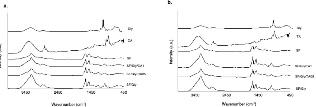
Figure 3.4.1. – FTIR spectra of the SF and SF/GLY membranes incorporating 0.5 and 1% of caffeic acid (a), tannic acid (b)……… 44
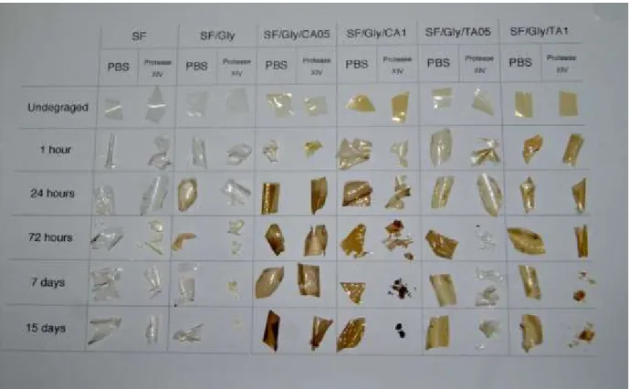
Figure 3.5.1 – Macroscopic evaluation of the samples before and after incubation in either PBS, or protease XIV (derived from Streptomyces griseus, 0.693 U.mL-1), up to 15 days………. 45
Figure 3.5.2 – Degradation profile of the samples incubated in either Phosphate Buffered Saline (PBS), or protease XIV (derived from Streptomyces griseus) (0.693 U.mL-1), up to 15 days………. 46
Figure 3.6.1. – TG and DTA curves for SF, SF/Gly, SF/Gly/CA05, SF/Gly/CA1, SF/Gly/TA05 and SF/Gly/TA1 membranes, from 30 to 330ºC……….. 48
Figure 3.7.1 –Results of (a) mg Ascorbic Acid Eq/mg membrane (n = 6, Kruskal-Wallis test, P = 0.0369, * – significant differences) and (b) mg Ascorbic Acid Eq/cm2 membrane (n = 15, One-way ANOVA test, P < 0.0001, * – significant differences) obtained by the ABTS assay……….. 50
Figure 3.8.1 – Cell viability results for the membranes (a) and for the extracts (b) in L929 fibroblasts after the 24 hours of incubation………. 50
10
LIST OF TABLES
Table 1. Types of modern dressings, their major functions and design.………...………. 14 Table 2. Samples name and formulation……… 27 Table 3. Organoleptic properties and some physical aspects of SF-based membranes………. 35
11
CHAPTER 1
1. Introduction
In this chapter the concept of a chronic wound is introduced, as well as the current clinical approaches available, wound management products available in the market for the different types of wounds and the innovations that are being developed in an attempt to minimize this problem. Important topics, such as bioactive and multifunctional membranes to promote skin regeneration and wound healing will also be discussed.
1. Chronic Wounds
A wound results from the damage or disruption of the integrity of the skin, mucosal surfaces or organ tissues, which can compromise their normal anatomical structure and function (1,2). To restore the injured tissue, our body initiates a complex process of regeneration – the wound healing (2,3). This process is classically divided into four phases: haemostasis, inflammation, proliferation and tissue remodeling. Wounds can be classified according to the time this process takes to occur (1–5). The healing process can be conditioned by several factors, related to the patient: age, body type, patient's nutritional status, alcohol consumption, among others. In addition, the type of wound and some biological aspects may also influence the healing process. However, there are still some factors that condition the successful healing of these wounds which are not related to the patient, such as the lack of prevention, failure in treatment and strategies of management of these wounds by the health professionals (6,7).
Wounds can be classified according to several criteria, as previously mentioned, such as the healing time – this is one of the most fundamental criteria for the treatment and management of wounds (1,2,4). Wounds that pass through the four phases of the healing process quickly are designated acute wounds. However, wounds which have failed the normal reparative process of healing, over extended periods that range from 4 weeks to more than 3 months, often as a result of prolonged pathological inflammation, are classified as chronic wounds (3,8). This type of wound displays some specific characteristics, such as excessive levels of proinflammatory cytokines and reactive oxygen species (ROS), presence of senescent cells and they are stagnant in the inflammatory phase (8,9). In these wounds, the levels of proteases exceed their respective inhibitors, leading to the destruction of extracellular matrix and degradation of growth factors and their receptors. This not only prevents the wound from regenerating at a normal rate, but also attracts more inflammatory cells, amplifying the inflammation cycle. The immune cells of our body produce ROS, which in low concentrations, provide a defense against microorganisms (10). However, in chronic wounds, the predominant inflammatory environment increases ROS production, which causes cell damage. This, consequently, makes all the senescent cells present in chronic wounds – which have an impaired proliferative and secretory capacity – to lose the responsiveness they previously had to the regeneration process, during wound healing.
12 This decrease in proliferative capacity is directly correlated with the failure of wound healing (10–12).
Chronic wounds can be classified as vascular, diabetic and pressure ulcers, and the choice of the appropriate dressing will depend on the type of wound to be treated, mainly on its physiological characteristics (amount of exudate, location, among others) (9,10). This topic will be further discussed in the next chapter.
Chronic wounds have the highest incidence on the elderly population, since wound repair decreases as the body age increases, and the incidence of some diseases that promote the appearanc e of chronic wounds, as diabetes and cardiovascular diseases, increases with age. In 2015, diabetes was the direct cause of the death of 1.6 million people worldwide. In Portugal, in the same year, the prevalence of this disease was about 1 million people between the ages of 20 and 79 (13,14). Adding to this, cardiovascular diseases remains the largest cause of death worldwide (8).
The treatment of a patient with a chronic wound is an economically expensive procedure. According to the World Health Organization, in the United Kingdom, the number of patients developing new ulcers in a year was estimated to be more than 100,000 people, which represents an annual expenditure between £168 million to £198 million, excluding subsequent related problems such as anxiety, depression, among others (15). In this way, it is increasingly important to find sustainable, innovative and viable solutions which are capable of reducing the treatment time, accelerate healing and reduce the physical and psychological suffering of these patients. It is also important to increase the success of treatments, to reduce the costs associated with the current solutions.
Figure 1.1 – Leg (a) and foot (b) chronic wounds examples (16,17).
13
2. Current clinical approaches to chronic wounds
Due to the rise of the morbidity associated to chronic wounds, wound care has become a topic increasingly important nowadays (18). Currently, the standard care for chronic wounds consists of swabbing for infection, leaning the wound, applying a dressing and, in some cases, debridement of the wound (18,19).
Generally, a wound dressing may be a single product or, in some cases, the combination of two or more layers of dressings, consisting of a primary contact layer and a secondary absorpti ve layer – not in direct contact with the wound. An ideal dressing is considered to be the one that maintains adequate humidity, remove the excess of wound exudate, permits thermal insulation, allows gaseous exchange, conforms to the wound surface, facilitates the debridement when necessary, minimizes the scar formation, is impermeable to extraneous bacteria, is non-fiber shedding/non-toxic, is non-adherent, comfortable and conforming (20). The use of wound dressings needs to be integrated into a general management plan, which must consider the different types of wounds and the problem that gave rise to them, and they should also be reviewed regularly with the progress of the treatment (20,21).
Dressings can be classified in several ways, according to their function in the wound, the type of material employed and the physical form of the dressing (22,23). Regarding their nature of action, dressings can be classified as passive, interactive and bioactive products (24). According to Willi Paul and Chandra Sharma (24), traditional primary and secondary wound dressings are included in the passive products classification, which include some components, such as gauze, lint, plasters, natural or synthetic bandages and cotton wool, and they’re used mostly to protect the wound from contaminations. This type of dressings constitute the largest market segment (21,22). Interactive and bioactive products are included in the group of modern dressings and they are developed and designed not only for covering the wound, but also to facilitate their function and to deliver substances which are active in wound healing (24). This type of dressings and their classification is presented in Table 1, according to type, advantages, disadvantages and commercial name. (21).
14 Table 1. Types of modern dressings, their major functions and design.
Type of Dressing Advantages Wound type Disadvantages Commercial
products References
Alginate dressings
Can absorb 15 to 20 times their weight of fluid; suitable for highly exuding wounds; biodegradable; accelerate healing process by activating macrophages to produce TNF-, which initiates inflammatory signals.
Useful in cavities and sinuses; undermining wounds; all wound types with high exudates.
Need for secondary dressing; need to be changed regularly; not suggested for dry wounds (adhere to healing wound surface, causing pain and damaging healthy tissue or removal), third degree burn wounds and severe wounds with exposed bones.
Algisite,Algosteril, Kaltostat,Melgisorb, SeaSorb, Sorbsan, Sorbsan SA, Tegagen,Urgosorb. (21,25,26) Antimicrobial dressings -
Used in all locally colonized or infected wounds.
Drugs may not penetrate well into the wounds (due to poor blood flow and the presence of dead tissue); inappropriate use of systemic antibiotics can be associated with problems of allergy (due to the use of iodine), toxicity and the development of resistance in non-target organisms. Acticoat, Actisorb Silver 200, Aquacel Ag, Arglaes, Avance, Inadine, Iodoflex, Iodosorb, Metrotop Gel. (25–27) Bioactive Dressings Biocompatibility, biodegradability and non-toxic nature. Generally derived from natural tissue or artificial sources.
- - - (21,25,26)
Foam dressings
Primary dressing for absorption; give degree of cushioning; transmit moisture vapour and oxygen and provide thermal insulation to the wound bed;
Flat, shallow wounds (control of exudate depending on type of foam); suitable for lower leg ulcers and
Need secondary dressing; not suitable for low exudating wounds, dry wounds and dry scars.
Allevyn Adhesive, Allevyn Cavity, Allevyn Lite Island, Allevyn Thin, Allevyn Plus Adhesive,
15
highly absorbent; facilitate uniform dispersion of exudate throughout the absorbent layer; protect the area around the wound from further damage; may be left in place for two to three days.
moderate to highly exudating wounds; granulating wounds.
Allevyn Plus Cavity, Biatain Adhesive, Cavi-Care, Lyofoam Extra Adhesive, Tielle Plus, Tielle Lite, Tielle.
Hydrocolloids
Can promote a moist wound healing; permeable to water vapour but impermeable to bacteria; absorbent; conformable; good in “difficult” areas – hell, elbow, sacrum; may be left in place for several days; useful debriding agent; highly absorbent;
non-adherent; promote an
autolytic debridement; reports to reduce wound pain; mostly used as a secondary dressing.
Cavity or flat shallow wounds with low to
medium exudate
(e.g. pressure sores, minor burn wounds
and traumatic
wounds);
recommended for paediatric wound care management (don’t cause pain removal).
May cause maceration;
needs secondary dressing; not indicated for neuropatic ulcers.
Alione, Aquacel, CombiDERM, CombiDERM N, Comfeel Plus, Cutinova Thin, Duo DERM Extra Thin, Granuflex, GranuGel Paste, Tegasorb, Tegasorb Thin, Versiva. (21,25,26) Hydrogels
Supply moist environment to wounds with low to medium exudate; may be left in place several days; promote wound debridement by rehydration of non-viable tissue; soft elastic properties, provides easy application and removal after wound is healed without any damage; suitable for the four phases of wound healing, except
Suitable for sloughy or necrotic wounds; useful in flat wounds, cavities and sinuses; used for dry chronic wounds, pressure ulcers, burn wounds and chronic legs ulcers.
Need secondary dressing; may cause maceration; difficult to handle, because of their low mechanical strength.
Aquaform, Intrasite, GranuGel, Nu-Gel, Purilon, Sterigel.
16
in infected and heavy drainage wounds; not irritant, no reactive, permeable to metabolites.
Low adherent
Allow exudate to pass through into a secondary dressing while maintaining a moist wound bed; reduce adherence at the wound bed.
Useful for patients with sensitive or fragile skin.
- - (21,25,26)
Absorbent Membranes/Films
Absorb small amounts of wound exudate. Allow clinicians to observe a wound’s progress without the necessity for removal due to their transparency.
- - OpSite TM Post-Op Visible, Tegaderm Absorbent Clear Acrylic Dressing (3M). (28)
17
Medicated Membranes/Films
Provides protection against infection and reduces bacterial load by a slowly release of iodine by the iodophor.
Applicable to cover and protect catheter sites and to secure devices to patient’s skin. - Tegaderm Chlorhexidine Gluconate (CHG) IV Securement dressing (3M), Tegaderm TM Plus (3M). (28) Polymeric Membranes/Films
Trap exudates, providing a moist environment. Impermeable to bacteria and liquid and permeable to moisture vapor and air.
Skin of surgical incisions.
Due to their non-absorbent characteristic, exudates can stay accumulate underneath the dressing.
Seeping fluid pressure may cause a break in the environment maintained by the dressing.
Aluderm, Blister, Poly skin II, Silon-TSR, Tegaderm TM,
Opsite.
(29)
Semipermeable Membranes/Films
Promote moist environment (permeable to air and water vapor); used mainly as a transparent primary cover; allow visual checks; may be left in the wound place several days; useful as secondary dressing (due to their high absorbancy and moisture vapour permeability); impermeable to fluids and bacteria.
Suitable for flat, shallow wounds with
low to medium
exudates; good for wounds in “difficult” anatomical sites (e.g. joints).
Adhere to healthy skin (but not to wound); cannot be used for infected or heavily exuding wounds; may cause maceration.
Bioclusive, Mefilm, OpSite Flexigrid, OpSite Plus, Tegaderm.
18
3. Membranes in wound healing
In the last decades, there have been some works proposing new membrane dressings, some of them, presented below, with particular attention for the application in chronic wounds. The first membrane developed was from a form of collagen obtained from the dried bladders of fish, in the 1880s (28). During the Second World War, Bloom (30) described the use of cellophane in burns patients (28). In 1948, Bull et al. (31) developed a nylon film dressing and, in 1950, Schilling et al. (28,32) conducted the first clinical trial with a film dressing. However, the most important assay was the one conduct ed by Winter and co-workers (33), in animals, in 1962. In 1963, Hinman and Maibach (24,28,34) have conducted a study in humans, which provided future support for the clinical use of membrane dressing. Both researches have demonstrated that a moist wound healing environment, created by film dressings, accelerated skin epithelialization twice as on wounds allowed to dry by exposure to air (24). From that year, membrane dressings were introduced under various brands (28).
According to Sussman et al. (28), a membrane dressing is indicated for the management of minor burns and simple wounds, such as scalds, abrasions, lacerations and lightly exudative wounds . The flexibility of these dressings gives them the ability to be applied to sutures and to continue to be used at the incision site, even after removal of the sutures or clips. Film dressings are especially good for reducing skin tension on flexor surfaces, to protect skin from shearing forces and to prevent and treat superficial pressure ulcers (28).
Ahmed and Boateng (35) developed antimicrobial membranes to deliver ciprofloxacin (CIP) for the treatment of bacterial infection in foot ulcers. Calcium alginate films loaded with ciprofloxacin were evaluated in their physico-chemical properties, such as porosity, swelling, equilibrium water content, water absorption, water vapor transmission, evaporative water loss, mechanical strength, adhesion, IR spectroscopy, scanning electron microscopy, X-rays diffraction, drug release, cytological and antimicrobial activity against Escherichia coli, Staphylococcus aureus and Pseudomonas aeruginos a. Different 1% w/v gels were dissolved in different solutions of sodium carbonate (0.005-0.028 M). In addition, different Glycerol (9.1, 20.0, 33.3, 42.8 and 50.0%), based on the total weight of the polymer (w/w), were added to the gels. After this, 20 g of each gel were dispensed into 86 mm diameter Petri dishes and dried (30°C) for 18 hours for obtaining the films. The drug was after loaded onto the optimized film containing 33.3% glycerol (w/w). The obtained films were soft, flexible and uniform, and their transparency was not affected by drug incorporation. They also verified that these membranes had showed potential tensile and bio-adhesive properties, which is required for an easy application of the dressing and to guarantee adherence to the wound bed. An optimal moisture environment and high biocompatibility with human keratinocyte cells was also observed. These results have confirmed that the design of biocompatible and effective dressings was successfully obtained, however, in vivo studies were required to be performed to confirm its effectiveness (35).
Chun-Hsu Yao et al (36) produced a bilayer membrane for wound dressing applications, with keratin extracted from human hair, blended with gelatin, sequentially electrospun onto a commercial polyurethane wound dressing. They verified that a gelatin/keratin blend solution can be successfully electrospun continuously, originating uniform and bead-free nanofibers, when appropriate
19 electrospinning parameters are used. The MTT cell viability assay showed that the residues released from the electrospun gelatin/keratin composite nanofibers enhanced cell proliferation. Fibroblasts showed a more favorable interaction with gelatin/keratin composite when compared to the gelatin mat. Animal studies revealed that the developed bilayer membranes promoted an earlier vascularization and a better skin wound healing. All of these results established the potential of the gelatin/keratin bilayer membranes as a wound dressing (36).
Ye Ma et al (37) proposed the production of a transparent flexible chitosan-based membrane dressing with antibacterial drugs by solvent casting method from suspension of chitosan floccules. Glycerol was used in different percentages in the matrix of the membranes to improve the mechanical properties and they verified that the introduction of this component as a plasticizer had a significant influence on the properties of the membranes. With the increasing of the concentration of glycerol, the swelling rate, water vapour permeability, wettability and tensile strength were improved significantly. The enzymatic degradation in vitro had showed that chitosan membranes had long-term stability and it was not compromised by the glycerol content. Tetracycline hydrochloride (TH) and silver sulfadiazine (AgSD) were added to improve the antibacterial properties of chitosan membranes and it was found that these membranes have a promising future in the treatment of bacterial infection – namely against E. coli and S. aureus – and, with the in vitro dermal fibroblasts seeded in both membranes, a significant higher viability during culture time of 1 to 3 days was observed. With this, it was possible to verify that the chitosan-based membranes with antibacterial agents could give a high therapeutic efficiency as a wound dressing. (37).
One of the demanding aspects when developing membranes for wound healing is the fact that there are no membrane dressings with the ability to combat the ROS produced in chronic wounds , combined with biocompatibility, biodegradability, good mechanical properties and non-cytotoxicity. In this sense, the development of novel multifunctional and bioactive dressings with the ability to aggregate all of these properties and respond to the challenges of a chronic wound is highly demanded.
20
3.1. Bioactive and multifunctional membranes
Recently, new strategies have been proposed to develop innovative wound dressings with the capability to enhance the healing process in chronic wounds. As explained in the previous sections, one of the aspects that characterizes this type of wounds is the excessive levels of ROS, which must be controlled by antioxidant agents, to guarantee the survival of the cells and, consequently, the regeneration of the wounds. Of all the types of dressings which have been investigated, bioactive and biofunctional membranes for wound healing applications are gaining interest.
Kavoosi et al. (38) investigated the improvement of the properties of a gelatin membrane incorporated with Ferula assafoetida essential oil (FAO) as a potential antioxidant and antibacterial wound dressing. These membranes were characterized regarding several physical-chemical properties , such as water solubility, swelling and water vapor permeability, mechanical behaviour and antioxidant and antibacterial activities. The results obtained in this study suggested that the incorporation of FAO in the gelatin membranes caused a significant decrease in swelling and an increase in the water vapor permeability and solubility. The tensile strength and elastic modulus decreased with the incorporation of FAO, while the elongation at break increased. The authors concluded that gelatin/FAO membranes could be used as active membranes for biomedical applications, including as a wound dressing material, since they showed exceptional antioxidant and antimicrobial characteristics, (38).
Rezvanian et al. (39) developed an alginate-based composite membrane for wound dressing applications, which had in its formulation simvastatin, a compound used in cardiovascular diseases for lipid lowering effects. These membranes were prepared and characterized based on their physical properties, as well as surface morphology, mechanical strength and rheology. The cytotoxicity and in vitro drug release results demonstrated that these membranes had appropriate wound dressing characteristics and high mechanical performance, as well as a controlled drug release profile and they were non-toxic for primary human dermal fibroblast cells, which made these membranes a good candidate for bioactive wound dressing. However, further in vivo investigations are required to prove the membrane toxicity and efficiency (39).
Silk-based membrane dressings have been proposed for wound healing. Srivastava et al. (40) developed a flexible silk fibroin membrane with dextrose (5-15% w/w) incorporated. Membranes were obtained by the solvent-casting method followed by crystallization with 80% methanol solution. It could be observed that the flexibility of the membranes increased with the increasing in the dextrose content and the elongation ate break increased from 3.2% to 40% with the increasing of this content in the membrane matrix. With this, it could be concluded that dextrose has acted as plasticizer for those membranes. FTIR and XRD studies showed that the dextrose content did not affect the crystalline structure of the silk fibroin membranes. SEM and AFM analysis showed that the surface roughness of these membranes also increased with increasing dextrose content and that this component enhanc ed the hydrophilicity and swelling capacity of the silk fibroin membranes. Degradation profile of the membranes was evaluated, showing a significantly higher mass loss than pure silk fibroin membranes after 50 days of incubation in Protease XIV. The adhesion, proliferation and viability of L929 fibroblas t cells indicated that they had ability to support cell growth and proliferation when compared with pure silk
21 fibroin membranes. These dextrose modified films presented good potential to be used as dermal wound dressing material (40).
Karahaliloglu et al. (41) developed a nanostructured silk fibroin membrane for wound healing applications. For this, they started to modify the surface of silk fibroin membranes with NaOH alkaline treatment, in order to obtain a biological inspired nanofeatured surface morphology. Surfac e characteristics, such as roughness, energy and chemistry were evaluated. Skin-forming cells (keratinocytes) adhesion and proliferation were also studied to determine the promotion of an epidermal cover on the wound bed to form a new epidermal barrier, as well as dermal fibroblast adhesion and proliferation, in order to assess the capability of these membranes to replace injured dermal tissue in chronic wounds). The obtained results demonstrated that keratinocyte and fibroblast cell density was higher on the novel membranes compared with non-treated silk fibroin surfaces. The improvement in the cellular functions could be associated with a nanotopography induced by silk, wettability and a change in chemistry of this surface due to the NaOH treatment. With the obtained results, the developed nanofeatured silk fibroin membranes were considered a promising alternative for various skin reinforcement and wound dressing applications (41).
Xu et al. (42) improved the mechanical performance and the swelling ratio of chitosan membranes, properties that, until then, had limited their application in wound healing area. Thus, silk microfibers were incorporated in chitosan membranes, and its multiple physical properties were evaluated. By adding silk microfibers in the matrix, it was possible to verify that the mechanical properties were significantly improved, and the swelling ratio had decreased. SEM results have showed embedding of the microfibers and chitosan matrix, as well as connections among the silk microfibers. In vitro cytocompatibility was also evaluated with mouse fibroblasts of cell line L929, and it was showed significant cytocompatibility, demonstrated by cell proliferation and morphology. In vivo healing effects of these membranes were also evaluated on a fill-thickness skin wound rat model and it was possible to verify that the membranes containing silk microfibers exhibited increased wound healing efficienc y when compared with pure chitosan membranes. By examining the histological changes , a higher level of epithelization and collagen formation in the chitosan membranes was verified with silk microfibers after 21-day repair period. These results indicated that the developed membranes might be a potential dressing for wound healing applications (42).
It is notorious the need to develop novel bioactive and biofunctional membranes with natural polymers. This strategy has been explored over the years but commercially, there are only few effective solutions. Being silk a natural biodegradable material with excellent mechanical properties , biocompatibility and antioxidant properties, it is expected to be a promising material to be applied on wound healing membranes. Thus, further studies are necessary for the development of this type of membranes, possibly able to combat the high levels of ROS of chronic wounds and facilitate healing, as well as to promote the regeneration of the damaged skin.
22
4. Main objective
The main aim of this work was the development, optimization and further characterization of new silk fibroin-based biofunctional membranes incorporating anti-oxidant agents to be applied in chronic wound healing, specifically in the stage of inflammation, to provide an efficient solution to combat the excessive levels of ROS, thus contributing to trigger the regeneration process.
A SF/glycerol (GLY) membrane system was produced, incorporating for the first time, caffeic and tannic acids (CA and TA, respectively), two well-known anti-oxidant agents with traditional application in the food sector.
23
CHAPTER 2
Materials and Methods
2.1. Materials and Reagents
2.1.1. Silk
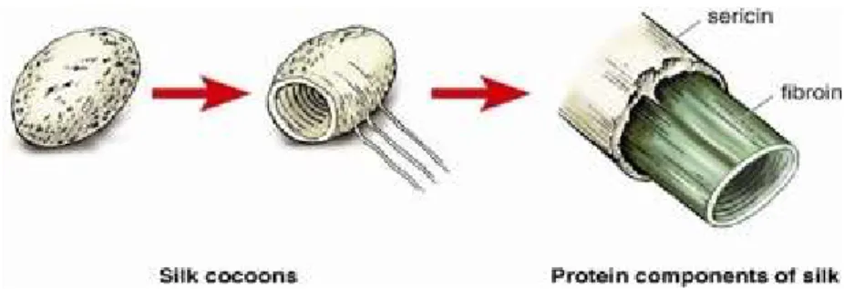
Silk is a natural fibrous protein produced in specialized glands of various arthropods, that is spun into fibers during their metamorphosis phase. The silkworm Bombyx mori is the most extensively studied specie and the silk produced by this arthropod has been used commercially as sutures for biomedical applications and in textile industry for many years (43,44). Silk fibers are composed by two types of proteins: fibroin and sericin. Fibroin has a semi crystalline structure, providing strength and stiffness, and is the inner-core of the fiber. Sericin acts as an adhesive binder, forming an outer protective coating and maintaining the structural integrity of the fiber and cocoon (43,45). The structural components of Bombyx mori silk are illustrated in Figure 1.1.
Figure 2.1.1. – Structural components of silk from Bombyx mori (43).
Silk fibers spun by silkworms hold excellent mechanical properties, such as high tensile strength, elongation at break and energy absorption (45). Silk is insoluble in most solvents (including water) and detailed structural analysis on silk proteins has yielded information on the orientation and organization of the small numerous -sheet crystals in the fibers (43,46). This conformation of silk is commonly called Silk II – it’s a mixture of noncrystalline and crystalline domains and provides a basis for the outstanding mechanical properties of silk. Nevertheless, the structure inside the middle silk glands is different and is named Silk I, which is the solid structure of silk fibroin (47).
24 In fact, the biological responses to the fibroin fibers appeared to be comparable to most other commonly used biomaterials (44). In the literature there is a clear evidence that silk is susceptible to proteolytic degradation in vivo and, over longer time periods, will slowly be absorbed (44). Due to the unique mechanical properties, biocompatibility and low immunogenicity advantages, these fibers have a large application in biomedical field, such as in the development of scaffolds for tissue engineering, coatings and drug delivery (45,48). Therefore, it constitutes a very interesting raw material for the development a functional wound healing dressing.
In the present work, cocoons from Bombyx mori were supplied by the Portuguese Association of Parents and Friends of Mentally Disabled Citizens (APPA-CDM, Portugal). All the remaining reagents were purchased from Sigma-Aldrich, unless otherwise stated. Lithium bromide was purchased from Honeywell, UK.
2.1.2. Glycerol
Glycerol, a derivative of natural and petrochemical raw materials. It is a viscous, colorless, odorless and water-soluble liquid with a wide range of applications, namely in medical and pharmaceutical preparations, due to its plasticizing action and low toxicity (49,50). Gly is systematically incorporated in film-forming solutions to prevent film brittleness, increase flexibility, workability and distensibility, and was used as a plasticizing agent in the developed formulations in this work (50,51).
25
2.1.3. Anti-oxidant molecules
The excessive level of ROS in chronic wounds is a serious problem that interferes with the healing process of these wounds. In this way, introducing antioxidant agents into these membranes may be favorable for the treatment of this type of wounds.
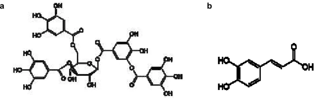
Tannic and Caffeic acids, two well-known molecules with anti-oxidant properties, were included in SF-based formulations. Tannic acid (TA) is a water-soluble polyphenolic compound which can be extracted from some fruits (e.g. grapes, pears and bananas), drinks (e.g. red wine, beer, coffee and black and green tea), lentils and chocolate (53,54). TA has shown antibacterial properties and UV-resistant activities, due to its polyphenolic structure. This compound has also shown high antioxidant activity, especially in the prevention of lipid oxidation, which makes it very useful in the biomedical area (55,56). Caffeic acid (CA), also a phenolic compound, is the result of the secondary metabolism of plants and is the main hydroxycinnamic acid present in the human diet. This acid is commonly found in various fruits and coffee beans, as well as in wine (57,58). Several studies have shown that CA has antioxidant capacity, as well as anti-inflammatory, antibacterial and anticancer properties which makes it attractive for use in treatment of chronic wounds (57).
Due to the natural origin and the high antioxidant potential of these two compounds, its introduction into the formulations of the membranes was seen as an added value.
Figure 2.1.3. – Chemical structure of caffeic (a) and tannic (b) acids (59,60). a
.
b .
26
2.2. Preparation of silk fibroin aqueous solution
SF solution was prepared using a previously developed procedure (61). Briefly, for each extraction, 5 g of clean cocoons were cut into small pieces and boiled in 2 L of a 0.02 M sodium carbonate (Na2CO3) solution for 1 hour, under constant stirring. During this process, the outer layer of
sericin was dissolved in the sodium carbonate solution, leaving a SF mesh available for further processing. In order to ensure that all the remnants of sericin were removed, the obtained fibroin was rinsed in 1 L of distilled water, under constant stirring, for 30 minutes. Lastly, the extracted fibroin mesh was dried, at room temperature, for 48 hours.
5 g of the dry pure silk-fibroin was then dissolved in 25 mL of a 9.3 M lithium bromide solution (LiBr) at 70ºC, with constant magnetic stirring, until complete dissolution. After that, the SF/LiBr solution was dialyzed for 48 hours against 5 L of distilled water, using a benzoylated dialysis tubing (length ~ 30 cm; molecular weight cut-off: 2000), in order to remove the salt. In the first day, the water was changed 1 hour, 2 hours and 4 hours, after starting the dialysis. In the second day, the water was changed 3 times, after regular time intervals. The purified SF solution was filtered using Whatman Filters (grade 1:11 m – medium flow filter paper) and used in the same day. The concentration of SF after dialysis, using the described methodology, was previously determined by dry weight analysis (data not published) and is approximately 7% (w/w).
2.3. Preparation of SF-based membranes
Polymeric membranes have a huge applicability in different areas, especially in regenerative medicine (28,62). There are several techniques for producing membranes, such as solvent casting (63), layer-by-layer assembly (64) and electrospinning (65). However, solvent casting is the most used technique, namely for the production of silk fibroin membranes, because it enables the production of transparent films with uniform thickness distribution, at low cost and operational simplicity (63,66,67). This technique consists in dissolving a polymer on a solvent, casting this solution in a mold with the required geometry and then allowing for the solvent to evaporate under adequate conditions, leaving behind a membrane or a film with the same shape as the mold (63).
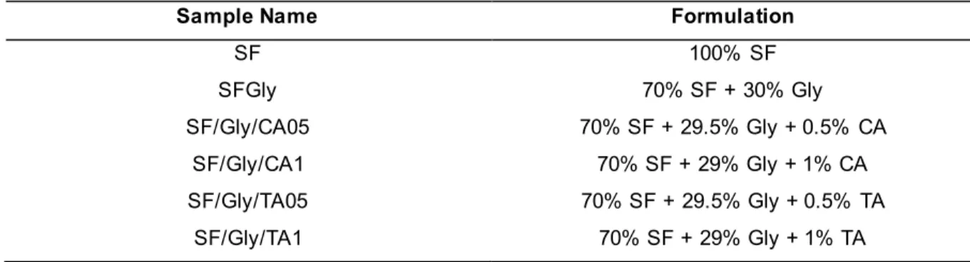
A control group of membranes containing exclusively SF was produced by casting 8 mL of the original solution into Petri dishes (100mm x 15mm) and drying at 85ºC, during 8h. Another control group of membranes, composed of 70% (w/w) SF and 30% (w/w) glycerol was prepared, by mixing the appropriate amount of both components, and leaving under constant stirring until the solution became homogenous. Then, 8 mL of that solution were casted onto Petri dishes and dried under the same conditions. Four experimental groups containing SF at 70% (w/w), glycerol at 29.5 % or 29 % (w/w) and either caffeic, or tannic at 0.5% and 1% (w/w) acid were produced following the same methodology. The respective formulations and sample designations are summarized in Table 2.
27 Table 2. List of the different formulations that were used and corresponding designation.
Sample Name Formulation
SF 100% SF SFGly 70% SF + 30% Gly SF/Gly/CA05 70% SF + 29.5% Gly + 0.5% CA SF/Gly/CA1 70% SF + 29% Gly + 1% CA SF/Gly/TA05 70% SF + 29.5% Gly + 0.5% TA SF/Gly/TA1 70% SF + 29% Gly + 1% TA
2.4. Organoleptic properties
The organoleptic properties of the membranes, such as color, transparency, smell, texture, and malleability, as well as their thickness and mass per unit of area, were evaluated. The organoleptic properties were evaluated by visual, olfactory and manual inspection. Mass and thickness were determined using an analytical scale (Mettler AE 200) and a digital micrometer (Adamel Lhomargy). Thickness was measured in 10 different points of the membrane, as suggested in Figure 2.4.1.
28
2.5. Morphological characterization of the surface
2.5.1. Scanning Electron Microscopy (SEM)
The morphology of the films was analysed on a Leica Cambridge S360 (Wetzlar, Germany), by Scanning Electron Microscopy (SEM). The samples were fixed with mutual conductive adhesive tape on aluminium stubs and covered with gold/palladium using a sputter coater (Fisons Instruments, Sputter Coater SC502, UK) prior analysis and the micrographs were taken at an accelerating voltage of 15 kV at different magnifications (68).
2.5.2. Optical Profilometry
In order to assess and compare the surface roughness of the produced SF, SF/Gly, SF/CA and SF/TA membranes, the non-contact topographic characterization technique White Light Optical Interferometry (WLOI) was used. 3D surface maps were obtained using an optical profiling system Wyko-NT1100 (Massachusetts, USA), operating in Phase-Shifting Interferometry (PSI) mode and Vertical Scanning Interferometry (VSI) mode. PSI used a measurement range of 160 nm, while VSI used a measurement range of 2 mm. All images were analyzed using the WyconVision 32 software package and the average roughness (Sa) was obtained. The results were expressed as the average ±
standard deviation of 3 samples.
2.6. Chemical characterization
2.6.1. Fourier Transformed Infra-Red spectroscopy with Attenuated Total
Reflectance (FTIR-ATR)
In order to assess the chemical profile of the developed membranes, their spectra, as well as those of glycerol, caffeic acid and tannic acid were acquired using a FTIR spectrometer Nicolet 6700 (Thermo Scientific, United States of America) equipped with an Attenuated Total Reflectance (ATR) device. The software was programmed to record each spectrum between 4000 and 400 cm-1 at a
resolution of 4 cm-1. Samples and background (air) measurements were made by co-adding 32 scans.
29
2.7. Degradation profile
The degradation profile of the SF-based membranes was conducted in vitro, for 15 days, based on the standard BS EN ISO10993-13:2009. Since chronic wounds are characterized by high levels of proteases and decreased protease inhibitors levels (69), two conditions were tested to assess the degradation of SF-based membranes: during incubation in PBS (negative control) and incubation in a solution of a protease in PBS, both at pH 7.4. Many proteolytic enzymes are used to digest silk -fibroin. However, protease XIV was contemplated to show high activity against beta-sheet structures in fibers, films and scaffolds of silk (70). Samples measuring 1.5 cm x 1.5 cm (n=3, weight ~ 14.86 mg) were weighted and immersed in either 1.5 mL of PBS, or in the same volume of a 0.01879 mg/mL of protease XIV solution in PBS (0.693 U mL-1, derived from Streptomyces griseus), in order to obtain a ratio of 1U
of protease/mg of silk (71). They were then incubated under orbital shaking (100 rpm) at 37°C, under sterile conditions, obtained by adding 0.2% of sodium azide (w/v) in the PBS solution. The samples were collected at predetermined time points (1, 3, 6, 8, 24, 48 and 72 hours, 7 and 15 days). The degradation medium was replaced every 2 days to ensure the activity of the enzyme. In each timepoint, the samples were removed from de solution, rinsed with distilled water and dried at 40°C overnight, until they reached a constant weight. After cooling at room temperature for 1 hour in a desiccator, the specimens were weighed (7). The degradation was expressed in terms of weight loss, and was determined following Equation 1:
(1) weight loss (%) = (wi-wf) wi
×100
where, wi is the initial weight of the sample and wf is the weight of the sample after incubation in PBS or
protease XIV solutions. The results were expressed as the average of three measurements ± standard deviation.
2.8. Mechanical Performance
A TA XT Plus Texture Analyzer (Stable Micro Systems Ltd., Surrey, United Kingdom) with tensile grips was used to determine the mechanical performance of SF-based membranes under uniaxial tensile stress. Films specimens were tested as suggested by Q. Sun et al. (2014) with some adaptations. SF, SF/Gly, SF/CA and SF/TA films were cut into strips (3 x 1 cm). Then, the thickness of each sample was measured with a Micrometer MI 20 (Adamel Lhomargy, France). The tests were conducted in dry and hydrated samples. In the latter case, the membranes were immersed in PBS for at least 3 hours, in order to assure that hydration equilibrium was achieved. In order to avoid the rupture of the membranes near the grips, or slippage, the edges of the samples were sheathed with two strips of paper held together with double sided adhesive tape and fixed between the grips. The distance between the grips was set to 10 mm and the crosshead speed was. 5 mm.min-1. Young's Modulus (E)
30 and elongation at break (ԑ) were obtained from the stress vs strain plots (72). The ultimate tensile strength (MPa) was calculated according to Equation 2:
(2) UTS (MPa) = Maximum load (N) Cross sectional area (mm2)
The percentage of elongation at the break was obtained according to equation 3:
(3) Ԑ (%) = Sample length at break (mm) - Initial sample length (mm) Initial sample length (mm)
31
2.9. Thermal properties
2.9.1. Thermogravimetry and Different Thermal Analysis (TG-DTA)
Thermogravimetry (TG) and Differential Thermal Analysis (DTA) were used to study the thermal properties of the films.
TG is a measurement of the rate of mass loss plotted against temperature and it is used for degradation evaluation, while DTA allows to measure the differences between the temperature of the sample and a reference, and it is used to ascertain phase changes in a sample. However, although these two techniques can be applied separately, each of the methods does not always give sufficient information. In this way, both techniques can also be applied simultaneously, in the same sample, at the same time. They work, in this way, as complementary techniques, but the optimal conditions may be different for both methods – e.g. the highest sensitivity for DTA experiment is achieved at high heating rates while, in TG, the best resolution is reached at low rates (73).
Simultaneous TG/DTA of each sample was carried out under a nitrogen flow rate of 50 mL/min, with the temperature ranging, from 30 to 350ºC, at a heating rate of 10ºC/min, using a Scansci-Hitachi TG/DT 7200 Exstar equipment. Each sample was cut so as to present a minimum volume that could fill the aluminum crucible (n = 3, weight ~ 3 mg). Three samples were analyzed for each condition under study.
2.10. Antioxidant capacity – ABTS assay
In order to determine the antioxidant activity of the produced films, an improved ABTS-based assay was performed. This method was adapted from Gião et al. (74). The ABTS solution was prepared by adding 7 mmol. L-1 of ABTS (2,2-azinobis (3-ethylbenzothiazoline-6-sulfonic acid) diammonium salt
(Sigma-Aldrich, Germany) to 2.45 mmol. L-1 potassium persulfate (Merck, Germany) solutions. The
obtained solution was stored overnight in the dark for 16 hours, for the reaction to occur. This solution was subsequently diluted in ultrapure water to provide a final absorbance in the range of 0.700 ± 0.020. The absorbance of this solution was evaluated in a UV spectrophotometer.
For the analysis of the samples, a specific volume was added (v = 10 L) to 1 mL of ABTS, in order to obtain an inhibition percentage between 20 and 80%, during 6 minutes of reaction. The average of 3 replicates was used. The total antioxidant capacity was obtained according to the following equation, expressed in percentage of inhibition (PIAC):
(5) PIAC = AbsABTS- Abssample
AbsABTS × 100
Where AbsABTS is the inicial absorbance of diluted ABTS and Abssample is the absorbance of the sample
32 standard) solutions with known concentrations. The results were expressed as equivalent concentration of ascorbic acid, I g.L-1. From equation (5), the results were normalized relative to the membrane mass
present in the 10 microliters.
2.11. Cytotoxicity tests
For the evaluation of the cytotoxicity of the produce film dressings, an assay based on the international standard ISO 10993-5 was conducted, in vitro, by direct contact and extract tests.
Cell culture and seeding
Immortalized mouse fibroblast cell line (L929), with 39 passages, were maintained at 37ºC in a humidified atmosphere containing 5% CO2 in culture medium supplemented with 89% of MEM-alpha,
2mM of glutamine, 10% of fetal bovine serum and 1% of antibiotic. Culture medium was removed, and 5 mL of pre-warmed PBS was added to wash the cells, in order to remove dead cells and cellular debris. To detach the cells from the culture flaks, an enzymatic digestion with of trypsin was made. 2 mL of TrypLE Express (Gibco) was added, and the flask was incubated for 5 minutes at 37ºC in 5% CO2
atmosphere. After this time, 5 mL of culture medium was added to neutralize the effect of trypsin. The flask was observed under the microscope to guarantee that cell detachment occurred. The obtained cell suspension was centrifuged (5 minutes, 1200 rpm) and the obtained pellet was resuspended in 10 mL of culture medium, adjusting cell density to 1x10⁵ cells/ml. The cells were subsequently seeded (2x10⁴
cells/well) in a 96-well tissue culture plate and incubated for 24 hours (37ºC, 5% CO2 atmosphere), to
ensure cell recovery, adherence and progression to exponential growth phase. After 24 hours of incubation, culture medium was removed, and cells were washed twice with 200 L of PBS, in order to remove dead cells and cellular debris. After this, direct contact and extract tests were performed at the same time.
Materials preparation
Membranes were sterilized by ethanol sterilization method. Samples were placed in ethanol solutions with different concentrations (90%, 50%, 10% and 0%) for 10 minutes in each solution. The use of a decreasing concentration of ethanol is to assure the elimination of traces of ethanol from the samples, in order to prevent cell death.