Mestrado Integrado em Medicina Veterinária
Ciências Veterinárias
ECONOMIC LOSSES FROM MASTITIS: INTENSIVE DAIRY FARM CASE STUDY FROM
VALENCIA, SPAIN
RYAN VIEIRA VICENTE
Orientador: Professor Dr. Filipe Silva
UNIVERSIDADE DE TRÁS-OS-MONTES E ALTO DOURO Vila Real, 2011
"Learning is what most adults will do for a living in the 21st century."
-- Perelman
To my family and everyone that has stood beside me during this fascinating life.
Keywords: Mastitis, Clinical mastitis, Sub-clinical mastitis, Etiological agents, Infectious agents, Contagious agents, Environmental agents, Chronic mastitis, Acute mastitis, Hyper-acute mastitis, Economical value, Economical loss, Economical factor, Milk quality programs.
ABSTRACT
Mastitis infections are a pathologic entity of great importance to the milk industry. It is the most common disease in an operating dairy-oriented farm and provides the largest amount of losses to the farm. The aim of this thesis is to organize and analyze the amount of money lost due to various categories that mastitis affects and compare it with other authors who have made economical studies about the losses provoked by this disease. The study provides information about the losses at the farm, including the production loss, drug costs, veterinary fees, extra labour fees and replacement costs. In conclusion, the prevention and treatment have slowly become better throughout the world and studies like these help to monitor the hygienic state of farms. Further investigation will always be needed to develop new therapeutic techniques that would increase of life quality and expectancy of the animals and lower economical losses.
RESUMO
Infecções causadas por mastite são uma entidade patológica de grande importância para a indústria do leite. É a doença mais comum em explorações de gado leiteiro e provoca a maior quantidade de perdas para a industria. O objectivo desta tese é organizar e analisar a quantidade de dinheiro perdido devido às várias categorias que a mastite afecta e comparar com outros autores que fizeram estudos economicos sobre as perdas provocadas por esta doença. O estudo fornece informações sobre as perdas na exploração, incluindo a perda de produção, custos de medicamentos, taxas veterinárias, extra taxas de trabalho e os custos de substituição. Em conclusão, a prevenção e tratamento vem lentamente a melhorar em todo o mundo e estudos como este ajudam a monitorizar o estado de higiene das explorações. Outras investigações serão sempre necessários para desenvolver novas técnicas terapêuticas que aumentam a qualidade de vida e as expectativas dos animais, bem como a diminuição das perdas economicas.
TABLE OF CONTENTS
CHAPTER I – INTRODUCTION……… 1
CHAPTER II – OBJECTIVE………... 4
CHAPTER III – LITERATURE REVIEW……… 5
3.1 – MASTITIS………. 5
3.2 – ANATOMY OF THE MAMMARY GLAND……… 5
3.3 – PATHOGENESIS………. 6
3.4 – THE INFLAMMATORY RESPONSE……….. 7
3.5 – MASTITIS AND THE PRODUCTION OF MILK………... 9
3.6 – ETIOLOGY………... 11 3.6.1 – Streptococcus agalactiae………. 11 3.6.2 – Staphylococcus aureus………... 12 3.6.3 – Mycoplasma spp……….. 14 3.6.4 – Corynebacterium bovis………... 15 3.6.5 – Streptococcus dysgalactiae………. 15 3.6.6 – Coliforms……… 15
3.6.7 – Pseudomonas spp……… 17 3.6.8 – Coagulase-Negative Staphylococcus………. 17 3.6.9 – Streptococccus uberis………. 18 3.6.10 – Prototheca spp………... 18 3.7 – TYPES OF MASTITIS……… 19 3.8 – DETECTION OF MASTITIS………. 19
3.8.1 – Observation of the animal………. 19
3.8.2 – Changes to milk……….. 20
3.8.3 – California Mastitis Test (CMT)……… 20
3.8.4 – pH indicators……….. 21
3.8.5 – Electric conductivity……….. 21
3.9 – DIAGNOSING MASTITIS………. 21
3.9.1 – Bacteriological culture………... 21
3.10.1 – Risk factors for the animal……….. 23
3.10.2 – Environmental risk factors………. 25
3.11 – ECONOMIC IMPACT OF MASTITIS………... 26
3.12 – MILK QUALITY CONTROL PROGRAMS……….. 28
3.13 – ESTABLISHED OBJECTIVES FOR UDDER HEALTH………. 30
3.14 – ANIMAL WELFARE……… 31
3.15 – MILKING PROCEDURES AND TECHNIQUES……….. 33
3.16 – MILKING MACHINE AND OTHER EQUIPMENT MAINTENANCE 34 3.17 – REGISTRY SYSTEM………. 35
3.18 – MANAGEMENT OF CLINICAL MASTITIS DURING LACTATION 35 3.19 – DRY COW MANAGEMENT……….. 36
3.20 – BIOSAFETY MEASURES FOR INFECTIOUS AGENTS AND THE IDENTIFICATION OF CHRONICALLY INFECTED ANIMALS………….. 36
3.22 – MILK QUALITY PROGRAM REVISION……… 37
CHAPTER IV – FARM STUDY: ECONOMIC LOSSES FROM MASTITIS.. 38
4.1 – INTRODUCTION AND OBJECTIVES OF THE STUDY………. 38
4.2 – METHOD AND MATERIALS……….. 40
4.2.1 – SAMPLES………. 40
4.2.2 – SURVEY……… 41
4.2.3 – DATA ANALYSIS……… 42
4.2.4 – PROCEDURES………. 42
4.2.4.1 Procedures for calculations……….. 43
4.3 – RESULTS………. 45
4.4 – DISCUSSION – ECONOMIC LOSSES FROM MASTITIS……….. 50
4.5 – THE IMPORTANCE OF A REGISTRY SYSTEM……… 54
4.6 – HYGIENE AND PREVENTION TO CONTROL MASTITIS………….. 55
4.8 – IMPORTANCE OF THE SENSITIVITY TO THE ANTIBIOTIC
TREATMENT……….... 57
4.9 – IMPORTANCE OF DRY COW MANAGEMENT... 58
4.10 – CONCLUSION... 59
CHAPTER V – BIBLIOGRAPHY………. 61
CHAPTER VI – ANNEX 1... xix
ANNEX 2………..………. xx
TABLE INDEX
Table 1: Classification of EU milk producing countries and production
prices………... 2
Table 2: Components and their % in total revenue from operating a dairy
oriented farm……….. 3
Table 3: Decrease in milk production compared to the increase in Somatic
Cell Count………..………. 8
Table 4: Alterations to milk composition when an increase of somatic cells
exists……… 9
Table 5: Contagious and environmental etiological agents………... 11
Table 6: Californian Mastitis Test result corresponding to the Somatic Cell Count………... 20
Table 7: Annual milk production losses in litres (L) from sub-clinical mastitis related with the nº of cows in lactation and Bulk Milk Somatic Cell Count………...
29
Table 8: Average amount of profit per cow in the European Union (EUR
€).………. 38
Table 9: Average Bulk Milk Somatic Cell Count and milk loss...……… 41
Table 10: Average milk produced during Days In Milk intervals for infected
and non-infected animals……….……. 43
Table 11: Prices for various antibiotics in Spain and Portugal………. 44
Table 13: Cows diagnosed infected by mastitis through Californian Mastitis
Test control methods………..……... 49
Table 14: Economical losses from mastitis……….. 50
Table 15: Average milk production of Days In Milk groups and Somatic
Cell Count………... 51
Table 16: Prices for various antibiotics and costs per dose in Spain and
Portugal (EUR €)………... 52
Table 17: Mastitis occurrence related to the amount of lactations…………... 55
FIGURE INDEX
Figure 1: Anatomy of the udder and teat……… 5
Figure 2: Difference of the teat canal diameter and form before and after milking………. 6
Figure 3: A representation of the cellular migration when inflammation occurs………... 7
Figure 4: Staphylococcus aureus confirmation with a bacteriological culture in blood agar…...………... 13
Figure 5: Streptococcus agalactiae confirmation………. 22
Figure 6: Classification of hyperkeratosis lesions at the teat……… 24
Figure 7: Classification of teat health……….. 32
Figure 8: Plaque given to the visited farm from the council of agriculture, fishing and food products………. 40
ABREVIATIONS AND SYMBOLS
% - Percentage
ACMDPL – Average Clinical Mastitis Dairy Production Loss
ADIT – Average Days In Treatment
ADITWP – Average Days In Treatment and Waiting Period
BMSCC – Bulk Mastitis Somatic Cell Count
BP – Bottle Price
CDC - Centers for Disease Control and Prevention Group
CDP – Cost per Dose Price
CCS - Cow Cull Sales
CE – Council Directive/ Regulation
Cl+ - Chlorine ion
CMT – California Mastitis Test
CNS – Coagulase-negative Staphylococcus
CRC – Cow Replacement Costs
CV – Closing Valuation
DARD - Department of Agriculture and Rural Development Group
DC – Drug Costs
DGV – Direcçao Geral Veterinária
DIM – Days In Milk
DIMUD – Days In Milk Until Diagnosis
DNA – Deoxyribonucleic Acid
EEC - European Economic Council
EU – European Union
EUR – European Euro (€)
EX – Extra Labour
FAO –Food and Agriculture Organization
Fe2+/ Fe3+ - Iron oxidative states Fig. – Figure
FLD – Fatty Liver Disease
HACCP – Hazard Analysis Critical Control Point
HADP – Healthy Animal Dairy Production
ISO – International Standardization Organization
LP – Litre Price
LPS – lipopolyssaccharides
mL - Mililitre
Na+ - Sodium ion
NMC – National Mastitis Council
OV – Opening Valuation
pH – Acidity/Alkalinity scale
PR – Purchased Replacements
SCC – Somatic Cell Count
TCMDPL – Total Clinical Mastitis Dairy Production Loss
TDPL – Total Dairy Production Loss
TI – Transfers In
TSCMDPL - Total Sub-Clinical Mastitis Dairy Production Loss
USD – United States Dollar ($)
USJ – United States Jersey Group
ACKNOWLEDGEMENTS
My sincerest thanks to my coordinator, Dr. Filipe Silva, for accepting orienting me throughout this thesis.
I thank Professor Jorge Antonio Colaço and Antonio Mario Silvestre for giving me advice and helping at the right time.
To Paco and Miguel who had the patience to teach and train me during the time spent at the farm. Without you allowing me to conduct this study, providing the means and materials needed over time all would be lost. Thank you also for the support, sympathy, trust and friendship.
To the workers at the farm for all the sympathy, interest and motivation demonstrated
by all.
A very special thanks to João Santos, João Guimaraes, João Dias , António Machado, Rui Mota, Marta Ruperez, Natalia Nyrka, Dave Cassar, Javi Fernández, Jason Calleja, Mark Williams and Mark Bazylewicz for the availability and friendliness throughout
and especially at the end of this project.
To my family, Carmen, Carlos and Jay, for their courage and support they have always shown, which allowed me to finish the thesis with the spirit, dedication and all the other
motivation that I recieved from them.
To my aunt, Zelia Carvalhal, for putting up with me and helping during these months, and these years! Without your availability it would not have been possible ...
INTRODUCTION
CHAPTER I – INTRODUCTION
1 – INTRODUCTION
Concerns and expectations of consumers about the safety of purchased products are growing with the capacity to detect and relate the problems of health security with the processing, the producers and/or private identities (Jonhson, 2003).
Products of animal origin are incorporated and/or used in a large quantity of food goods. Combined with vegetables, animal products play a key role in human consumption as providers of basic elements for growth and maintenance of life. Specifically for animal origin goods, as well as other food items, consumption is influenced by the quality available to consumers. The quality of products depends on the production methods used and sanitary conditions for the origin, processing and marketing of these products. The disease represents a negative factor in the conversion of resources or factors in processing of products, goods and/or services available to the population (Otte et al, 2000).
Mastitis is considered the most expensive disease in the dairy industry. The management of dairy cows, like many other businesses, is the transformation of many factors of production or resources in one or more product(s) that could be used as final products: i) for consumers, ii) the producer for his own use, iii) reused materials for production, or even iv) intermediaries in other production processes, where it creates a new product and with a superior economic value (Radostits et al, 1994).
Currently there is a greater concern with the food consumers buy. The products from organic production methods have increased, including normal production and not enough to meet national needs. The rules and regulations of HACCP (Hazard Analysis Critical Control Point) and biosecurity start to become accepted in the industry with an objective to implement/adapt this to all dairy farms so they are all in the accordance to the rules established (FAO, 2004).
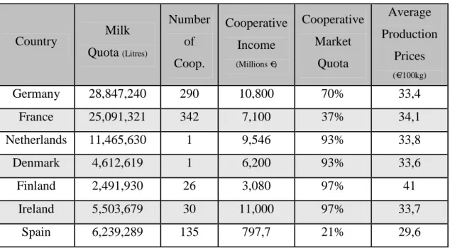
The agents responsible for the occurrence of mastitis vary according to geographic region, and within the same geographical region, there is variability between each operation (Garrido, 2005). Spain is a large contributor to the dairy produced in the European Union (EU) as shown in table 1 (Noche, 2010).
INTRODUCTION
This study was performed in the Valencian Community (Betera, Spain), at an intensive style dairy farm with over 1100 cows in lactation throughout the year.
Table nº 1 – Classification of EU milk producing countries and production prices (adapted from EU Commission, 2010 cit in Noche, 2010).
Country Milk Quota (Litres) Number of Coop. Cooperative Income (Millions €) Cooperative Market Quota Average Production Prices (€/100kg) Germany 28,847,240 290 10,800 70% 33,4 France 25,091,321 342 7,100 37% 34,1 Netherlands 11,465,630 1 9,546 93% 33,8 Denmark 4,612,619 1 6,200 93% 33,6 Finland 2,491,930 26 3,080 97% 41 Ireland 5,503,679 30 11,000 97% 33,7 Spain 6,239,289 135 797,7 21% 29,6
Diseases affecting the livestock in any area have a direct effect in reducing the conversion efficiency of livestock into final products, or livestock productivity. Besides the direct effect of animal conditions, there may be indirect effects, including repercussions in terms of commercial transactions, such as materials used in feed made for other domestic animals (Philpot et al, 2000).
The types and extent of economic losses sustained by the dairy industry due to mastitis were described in a number of papers during this century (Brodauf, 1963). In the region of Entre-Douro e Minho, the losses from mastitis are around EUR $249 per cow/year (Aires et al, 2007).
The final balance of processing resources into income can be assessed very simply compared to the costs of all the steps until the final product is sold. If the difference is positive, it is justified that there is money to be made from by livestock producers (Henriques et al, 2004).
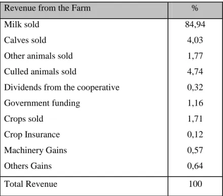
According to Radostits et al (2001), the decrease in milk production per cow due to the prevalence of clinical or sub-clinical mastitis, contributes about 80% to the total revenue of dairy cattle farms and economic losses (Table 2).
INTRODUCTION
Table nº 2 – Components and their % in total revenue from operating a dairy-oriented farm (adapted from Radostits et al, 2001).
Revenue from the Farm %
Milk sold Calves sold
Other animals sold Culled animals sold
Dividends from the cooperative Government funding Crops sold Crop Insurance Machinery Gains Others Gains 84,94 4,03 1,77 4,74 0,32 1,16 1,71 0,12 0,57 0,64 Total Revenue 100
Nearly 70% of the total losses attributable to mastitis (Kleinschrot et al, 1989), results from decreased production by alterations to the mammary gland and the quantity of discarded milk during treatment period and its withdrawal period, during which the residues of the pharmaceuticals administered are expelled.
A dairy farm without disease does not exist, but a controlled operation from the viewpoint of hygiene, has an elevated index. This creates a higher quality product and therefore can produce higher revenues. The existence of milk quality programs for the dairy industry, its implementation and adequacy, may translate into an increase of profits for the producer. It is therefore important to see the quality of milk as an economic factor for the farm. Thus, the quality of milk is no longer a clinical problem, which implies treatment costs, loss of milk, etc., and becomes a tool to increase cost-effectiveness between the measures implemented and results achieved (Halasa et al, 2007).
OBJECTIVE
CHAPTER II – OBJECTIVE
2 - OBJECTIVE
The principal cause of alterations to the udder is mastitis. It produces changes to the milk and can lead to critical clinical infections. The detection of mastitis has a double interest: economic and sanitary, as its destination is for human consumption and subject to the regulations of the Council Directive 92/26/EEC (1992).
With consumers increasingly informed and concerned about the food they buy, it’s important to ensure that in primary production the producer is aware to deliver quality milk, free of pharmaceutical residues while increasing the maximum income to their business (Halasa et al, 2007).
The main objective of the thesis is to characterize the main mastitis-causing agents in the Valencian Community of Betera, Spain and to evaluate the quantity of money lost after treatments commonly utilized to combat the disease. Moreover, the intention is to highlight the role of the veterinarian in the European community, in milk quality programs that are used to provide superior quality milk and the prevention of pathologies in dairy farms, in order to maximize the amount of final product and consequently achieve a higher profit.
LITERATURE REVIEW
CHAPTER III – LITERATURE REVIEW
3.1 – MASTITIS
A complex disease that can be defined simply as an inflammation of the mammary gland (De Oliveira et al, 2000; Riffon et al, 2001; Tollersrud, 2001; Menzies et al, 2001; Kerr et al, 2001; Zadoks, 2002; Boulanger et al, 2003; Romero, 2004; Soca et al, 2005; Gill et al, 2006; Gonen et al, 2008). Inflammation is most commonly caused by intra-mammary infection with a pathogen, but can also be caused by an injury (wound) and less frequently, by allergy and neoplasms (Menzies et al, 2001).
Mastitis occurs when the udder becomes inflamed because leukocytes are released into the mammary gland in response to invasion of the teat canal, usually by bacteria. These bacteria multiply and produce toxins that cause injury to milk secreting tissue and various ducts throughout the mammary gland. Elevated leukocytes, or somatic cells, cause a reduction in milk production and alter milk composition. These changes in turn adversely affect quality and quantity of dairy products (Jones et al, 2009).
3.2 – ANATOMY OF THE MAMMARY GLAND
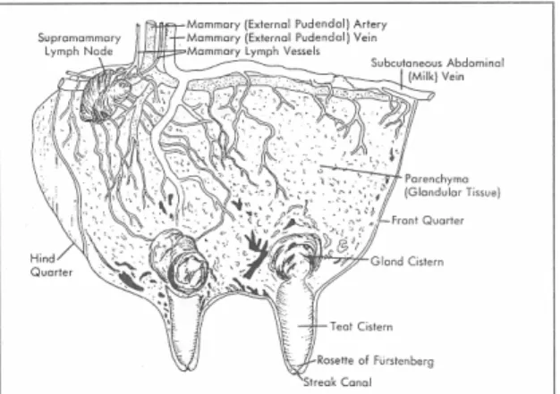
The mammary gland shown in figure 1, composed of lobules consisting of secretory tissue (alveoli), collecting ducts, the cistern and the teat canal. The teat canal is usually closed by a muscle ring in the period between milkings and is blocked by a keratin plug secreted by the cell walls. In bovine, the utter is formed by four quarters. Each one is independent to the other with its own gland for secretion (Hurley, 2010).
LITERATURE REVIEW
3.3 – PATHOGENESIS
Mastitis is the single most common disease in adult dairy cows, accounting for 38% of all morbidity. Mastitis may result following the introduction of micro-organisms through the teat sphincter. The clinical course varies with the ability of bacteria to colonize and thrive in the udder, virulence, and host response (Narayanan, 2009).
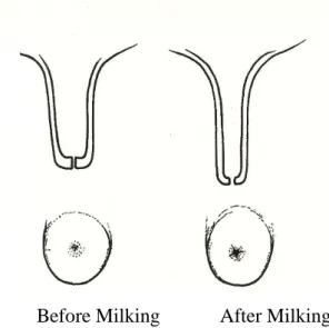
The infection of the mammary gland occurs mainly by the invasion of micro-organisms through the teat canal, which ascend to the cistern. This invasion usually occurs during milking by the milking machine, the milkers hands or other contaminated objects. It can also be provoked in the parks or beds that are not cared after in the time between milkings. After milking, the teat canal maintains open for the next 1-2 hours (Fig. 2) which simplifies the entrance of micro-organisms into the cavity (Meseguer et al, 2005).
Fig. nº 2 – Differences of the Teat Canal diameter and form before and after milking (Blowey et al, 2000).
Before Milking After Milking
The development of mastitis can be divided into five phases: 1) the organisms enter the teat duct. 2) the organisms multiply in the teat duct, streak canal, and mammary gland. 3) organisms progress upward into the lactiferous sinus, collecting ducts and alveoli. 4) organisms localize, resulting in leukocytic attraction, edema, and in some cases, abscess formation. 5) healing often results in fibrous connective tissue formation with some resolution (Narayanan, 2009).
LITERATURE REVIEW
The inflammatory process generally leads to a decrease in milk production, leading to macroscopic changes and consequently changes to the composition of the milk. To understand the changes that occur to milk it is essential to understand the mechanisms involved in the inflammatory response (Radostits et al, 2007).
3.4 – THE INFLAMMATORY RESPONSE
One factor that is exhibited is an increase in somatic cell counts (SCC) when the animal is with clinical or sub-clinical mastitis. Intra-mammary inflammation is associated with an increased count of somatic cells in milk. It is now used to detect mastitis which produces physical and chemical changes to the milk composition. However, the magnitude of the increase in SCC varies according to the bacteria involved in the intra-mammary infection (Djabri et al, 2002).
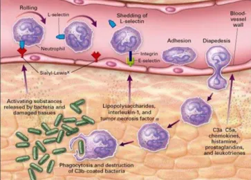
The term somatic cells are referring to all cells present in milk, including blood cells, such as leukocytes, and cells of the squamous secretory epithelium. The leukocytes are mobilized from the bloodstream into the mammary tissue due to changes in capillary permeability, which occur after the increase in blood supply in the area affected (Smith, 2010).
Fig. nº 3 – A representation of the cellular migration when inflammation occurs (Delves et al, 2000).
For a healthy cow, the quantities of somatic cells present in milk are: i) neutrophils (<11%), ii) macrophages (66-88%), iii) lymphocytes (10-27%) and iv) epithelial cells (0-7%). In case of infection of the mammary gland there is an increase of leukocytes in the milk. The majority are neutrophils that migrate from the bloodstream to the milk and
LITERATURE REVIEW
represent more than 90% of the leukocytes in mastitic milk. Figure 3 exemplifies the cellular movement in case of infection. Once in the infected area, the neutrophils exert their anti-bacterial functions (Radostits et al, 2007).
Some gram-negative bacteria release substances such as endotoxins, which serve as markers for leukocytes. If the inflammation response is quick, neutrophils move rapidly from the bloodstream to the interior of the mammary gland and eliminate the stimulus. However in situations which the bacteria survive, there is a continuous inflammatory stimulus, with continued neutrophil migration between the secretory cells and the alveolar lumen, producing a high amount of somatic cells. Prolonged inflammation causes glandular tissue damage and consequently decreased milk secretion (Hogan et al, 2003).
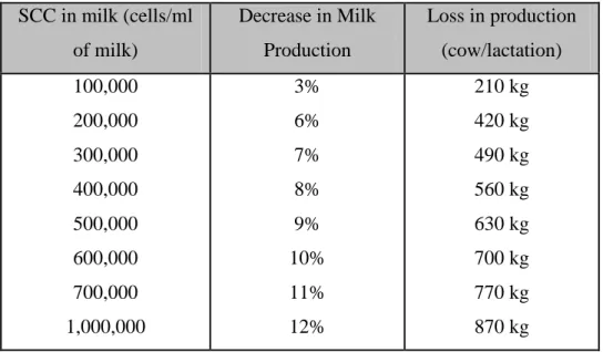
During the period of infection, the somatic cells increase from an average of 100,000 cells/ml of milk in a healthy cow to 1,000,000 cells/ml of milk in just hours (Radostits et al, 2007), as a result of the increase in neutrophils, combined with the decrease in milk production, as illustrated in table 3.
In total, the number of somatic cells is influenced by various factors. Mastitis is the most important factor but also the season of the year, the breed, the lactation phase, the amount of production, the number of lactations, stress, nutritional problems, climatic conditions along with others affecting the SCC (Ostrenksy, 1999).
Table nº 3 – Decrease in milk production compared to the increase in Somatic Cell Count (adapted from Delaval, 2006).
SCC in milk (cells/ml of milk) Decrease in Milk Production Loss in production (cow/lactation) 100,000 200,000 300,000 400,000 500,000 600,000 700,000 1,000,000 3% 6% 7% 8% 9% 10% 11% 12% 210 kg 420 kg 490 kg 560 kg 630 kg 700 kg 770 kg 870 kg
LITERATURE REVIEW
In another study, mastitic milk alterations vary, with the phase of lactation in which it occurs. Stage of lactation was found to affect mastitis prevalence significantly. Early stages of lactation and the involution period of the mammary glands were the most susceptible stages with prevalence of 45.8% and 38.7%, respectively. The figure was lower for cows at mid lactation (25.8%).The largest loss of milk is normally associated if the mastitis occurs during the beginning of the lactation, before reaching the peak of lactation (Biffa et al, 2005).
3.5 – MASTITIS AND THE PRODUCTION OF MILK
The more extensive the damage to the mammary tissues coincides with greater alterations to the components of milk and SCC (Delaval, 2006).
The reduction in milk production is due to the damage to the secreting epithelial cells of the infected teat and thus reduces the production and secretion of the gland as a whole. The increased vascular permeability also causes a decrease in volume because of the passage of water back into the bloodstream (Pyörälä, 2008).
Table nº 4 – Alterations to milk composition when an increase of somatic cells exists (adapted from Schällibaum, 2001).
SCC x 10 cells/ mL Milk Components <100 250 -5000 500 - 1000 >1000 Cause and Alterations Lactose Casein Fat 4,90 2,81 3,74 4,74 2,79 3,69 4,60 2,65 3,51 4,21 2,25 3,13 Decreased because of less production Plasmatic Proteins Soroalbumins Immunoglobulins Chloride Sodium Potassium 0,81 0,02 0,12 0,91 0,057 0,173 0,82 0,15 0,14 0,096 0,062 0,18 1,10 0,23 0,26 0,121 0,091 0,135 1,31 0,35 0,51 0,147 0,105 0,157 pH 6,6 6,6 6,8 6,9 Increase due to vascular permeability
LITERATURE REVIEW
Apart from the loss of production, mastitis causes, among others, changes in three major components of milk; protein, fat and lactose (Schällibaum, 2001). Table 4 summarizes the changes in milk composition and the origins of these changes.
In mastitic milk, there is a reduction of synthesized proteins at the glandular level. There is a higher quantity of alpha-caseins, caseins, alpha-lactoglobulin, beta-lactoglobulin, and an increase in proteins commonly in the bloodstream, like plasmatic albumin and immunoglobulins. This is provoked by the secondary vascular permeability caused by the infection, which translates into a higher concentration of proteins in milk (Hurley, 2010).
During clinical mastitis the concentrations of lactoferrin are increased possibly because of the role it plays in the chelation of iron (Fe2+/ Fe3+), making it unavailable for the bacteria. During the cows dry period, lactoferrin also increases, which helps in the prevention of mammary infections during this period (Kawai et al, 1999).
In general the fat content has a tendency to drop in concentration as SCC increases but, in cases where there is little milk production, the concentration may be higher. The type of fatty acids present in the mastitic milk changes greatly, and is caused by the increased activity of the lipases, provoking a higher concentration of free fatty acids and of the short-chained unsaturated fatty acids. Furthermore, the long-chained fatty acids decrease, diminishing the nutritional and organoleptic qualities of milk (Hurley, 2010).
Lactose has a key role to play in the osmotic regulation of milk. It is unanimously accepted that mastitic milk has less quantity of lactose as the SCC increases. This decrease is provoked by the destruction of the epithelial cells and the consequent decrease in production, but also the fermentation of lactose by micro-organisms is much lower (Petrovski et al, 2006).
Another important aspect of increased SCC is its negative effect on technological properties of milk; the yield is lower in protein-based products, with an increased coagulation time (very important in cheese production) and causes changes in product flavour. It can be bitter and rancid due to increased proteolytic and lipolytic enzymes (Mazal et al, 2007).
LITERATURE REVIEW
3.6 – ETIOLOGY
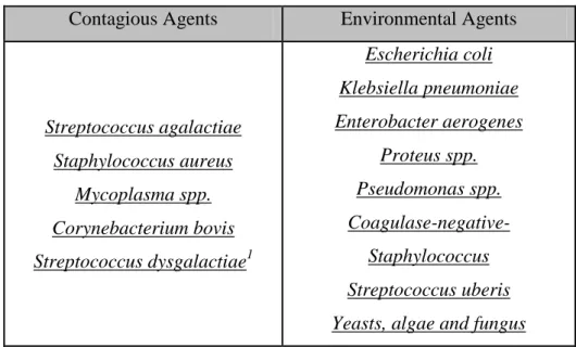
In bovine mastitis is associated with different infectious agents, generally divided into sub-groups as shown in table 5. Contagious agents, disperse through the infected teat through the milking machine or the hands of handler. Environmental agents, normally don’t colonize the mammary gland but when the environment, the milking machine, or the exterior of the teat is contaminated it takes advantage and enters the teat canal until the cistern of the mammary gland where it infects and multiplies, causing mastitis (Radostits et al, 2007).
Table nº 5 – Contagious and environmental etiological agents (adapted from Blowey et al, 2000).
Contagious Agents Environmental Agents
Streptococcus agalactiae Staphylococcus aureus Mycoplasma spp. Corynebacterium bovis Streptococcus dysgalactiae1 Escherichia coli Klebsiella pneumoniae Enterobacter aerogenes Proteus spp. Pseudomonas spp. Coagulase-negative- Staphylococcus Streptococcus uberis Yeasts, algae and fungus
1 considered by some authors as an environmental (Radostits et al, 2007)
The knowledge of infectious agents for mastitis is essential to implement the most effective strategies to combat this disease. In the next section is a brief description of the most frequent causative agents of mastitis (Buxton, 2005).
3.6.1 – Streptococcus agalactiae
A gram-positive and catalase negative cocci. It survives a short time in the environment, but can persist in the mammary gland for extended periods and is the largest source of infection for the udder. The environment also plays a role in the propagation of the agent. It is very contagious and is transmitted during milking, either by machine or via the milkers hands and tools. Bettering hygienic conditions during the
LITERATURE REVIEW
milking, the use of post-dipping techniques after milking and a correct method of applying dry cow therapy will ultimately help in the control of this micro-organism (Maroney et al, 2005).
Most cases of mastitis caused by this agent are sub-clinical, alternating with sporadic cases of clinical mastitis presenting obvious macroscopic changes (watery) and a drastic reduction in production of milk. Thus, the greatest loss resulting from the presence of the agent on a farm is the decrease in production. The decrease in production is estimated at 25% for one cow during lactation with Streptococcus agalactiae and if present in the herd, the milk production will drop 10-15% compared to the expected quantity (Radostits et al, 2007).
Streptococcus agalactiae is sensitive to most anti-microbial agents available and is normally successful in most cases. All sub-clinical cases that appear in any phase of lactation should be treated immediately since the response to the therapy is effective. Moreover it reduces the risk of spreading to the rest of the herd. Studies have shown that the response to this therapy is 90% successful with penicillin, erythromycin, cloxacillin and cephalosporins(Maroney et al, 2005).
The implementation of therapeutic protocols during the cow drying period is critical to control Streptococcus agalactiae in dairy farms. Isolation of chronically infected animals is done to prevent and reducing the chance of spreading through the herd. The treatment of sub-clinical mastitis results in increased production, a decrease in somatic cell counts (SCC) and the benefit of its implementation on infected farms (James et al, 2002).
3.6.2 – Staphylococcus aureus
A highly contagious agent, ubiquitous in the environment and the infected udder tends to contaminate the whole tank. They are gram-positive, catalase positive and coagulase positive cocci (Petersson-Wolfe et al, 2010).
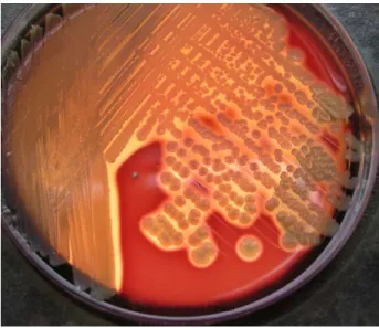
Most Staphylococcus aureus infections (Fig. 4) are sub-clinical, showing signs of high SCC´s repeatedly over various periods throughout the lactation. This agent is present in animal skin, teats, tonsils, beds, the milkers hands and vectors, multiplying in these places. Milking is the most usual route of infection through contaminated teats, hands or cups from pre/post-dipping. Staphylococcus aureus first secures and colonizes
LITERATURE REVIEW
by entering the epithelium of the mammary gland, making it harder for antibiotics to reach the agent (Green et al, 2004).
The infected glands produce less amounts of milk, caused by permanent destruction of the cellular parenquima. This causes fibrosis of the teat and may be palpable at examination. The fibrosis also protects the bacteria from the normal defense mechanisms of the udder by reducing phagocytosis of neutrophils (Petersson-Wolfe et al, 2010).
Fig. nº 4 –Staphylococcus aureus confirmation with a bacteriological culture in blood agar (Buxton, 2005).
The cure rate of mastitis caused by Staphylococcus aureus is very low: i) due to inadequate penetration of the antibiotics at the infected area (inside the cells or micro-abscesses) ii) due to the “L” forms of Staphylococcus aureus that are resistant to antibiotics and then change back when the treatment has ended and iii) due to the formation of the enzyme beta-lactamase which also helps the bacteria resist antibiotics like penicillin, pentamate, amplicilina and amoxicillin (Radostits et al, 2007). The association of amoxicyclin with clavulanic acid or cloxicyclin-ampicyclin counters the resistance of the micro-organisms that produce beta-lactamase. Zafalon et al (2007) states, the treatment of sub-clinical mastitis cases with this micro-organism are economically counterproductive. For animals in lactation the bacteriological cure rate is between 43-65% depending on the antibiotic used (erythromycin, cloxicyllin, amoxicillin and cephapyrin), (Wilson et al, 1999).
LITERATURE REVIEW
During the dry cow phase, the treatment should be chosen according to the profile of sensibility of the anti-biogram. Isolation is recommended for any animal presenting chronic infection from Staphylococcus aureus to prevent further contamination of the herd (Meseguer et al, 2005).
3.6.3 – Mycoplasma spp.
Mastitis normally begins in one animal of the herd and spreads, when infected with the genus, Mycoplasma spp. A concerning matter because it is ranked as an infectious agent. Mycoplasma bovis is the most common organism being isolated (Ruegg, 2000).
Endemic outbreaks, initially cause clinical mastitis that progress to chronic mastitis generally occur after the introduction of an infected animal into the herd. Transmission normally occurs during milking and especially in farms with poor hygiene. It is an ubiquitary agent that survives in the respiratory tract and reproductive system. In calves the symptoms suggest respiratory problems, arthritis and reproductive problems (Whitford et al, 2009).
Thus the transmission can occur, aside from milking, horizontally in any area with contact between animals (especially in poorly ventilated spaces) through coughing, contaminated semen, contaminated milk and objects (USJ Group, 2011).
In lactating cows the principal symptoms are very obvious and easily diagnosed as clinical mastitis. Symptoms include a large inflammation of the udder, reduced milk production, more than one teat quarter affected and a highly visible alteration to the milk. Chronically infected animals don’t have many macroscopic differences at the udder that can help to distinguish this micro-organism (Whitford et al, 2009)
The confirmation of this agent should be confirmed by micro-biological cultures. When found these animals should be culled, as antibiotic therapy has proven to be ineffective. In vitro, susceptibilities to fluoroquinolones, florfenicol and tiamulin were reported (Rosenbusch et al, 2005).
The main measure of control for this agent passes by limiting the entry of new animals to the herd through the application of biosecurity methods. In the presence of positive animals, the milk should not be used to feed other animals and isolation is recommended (Ruegg, 2000).
LITERATURE REVIEW
3.6.4 – Corynebacterium bovis
A gram-positive, catalase negative bacillus, which is classified as a contagious agent because it has the mammary gland as its primary source of infection. It is relatively common in mastitic cases with a moderate increase of the SCC and a slight reduction in production and therefore minor agent and minor problem of mastitis (NMC, 1999).
It spreads rapidly during milking, in situations where the disinfection of the teats before and after milking is not done with a germicidal product. Corynebacterium spp. needs to be diagnosed by bacterial culture and is sensitive to penicillin, ampicillin, amoxicillin and erythromycin. Drying treatments help to control this agent between lactations and the use of pre\post-dipping techniques during milking allows the control of dispersion for this micro-organism (Leslie et al, 2004).
3.6.5 – Streptococcus dysgalactiae
A gram-positive, catalase negative cocci, that adheres to the epithelium cells and can persist during long periods of time without losing its viability as an agent (Calvinho et al, 1998).
The symptoms of infection are unspecific, producing changes to the milk, with appearances of inflammation of the udder and fever. A bacterial culture is needed to confirm the diagnosis. It disperses easily through the herd during milking, especially animals with lesions on the udder or at farms with poor hygiene (including poor milking routines). There is a good response when using penicillin, cephalosporins and pirlimicine in sub-clinical cases (Boeringer, 2007). Controlling this agent is done by improving hygienic conditions of the milking machine, bedding and feed to promote better udder health (Noordhuizen et al, 2005).
3.6.6 – Coliforms
Bacilli that are gram-negative, oxidase negative, with capacity to ferment lactose (Fig. 5). This group, coliforms, contains various agents with different serotypes from Eschericia coli, Klebsiella sp. and Enterobacter sp., amongst others. Classified as environmental agents, they appear frequently in farms with low SCC levels. Known,
LITERATURE REVIEW
due to the severity of mastitis and may provoke death to some animals. The heat from summer, high humidity and long times of tight confinement all contribute to the multiplication and the persistence of the environmental agent. Coliforms primarily provoke clinical mastitis with a higher rate of incidence at the beginning of lactation and the two weeks before lactation (Herrera, 2011).
Animals that expel milk between milkings have a greater probability of receiving these environmental agents due to the teat canal remaining open for sometime and the bacteria having access to cistern of the mammary gland. The occurrence of metabolic disorders such as hypocalcaemia and Fatty Liver Disease (FLD) also contribute to the appearance of coliform mastitis because they diminish the activity of the neutrophils and other defense mechanisms of the udder. Levels of lactoferrin are also important to compete against the bacteria for the iron (Fe2+/ Fe3+) they search for in the blood. Levels are lowest after giving birth and during the drying phase increasing the free iron (Fe2+/ Fe3+) levels for the coliforms (Rebhun, 1995).
After invasion of the teat canal, the normal defense mechanisms of the udder are triggered and in the case of coliform mastitis, it may contribute to the onset of clinical signs of the disease. The rapid destruction of bacteria by neutrophils leads to a lipopolyssaccharides (LPS) released from the endotoxins present in the walls of these bacteria and triggers an inflammatory reaction stronger and more evident, which is manifested systemically and can lead to the death of animals. Animals with coliform mastitis may develop a severe reaction, with inflammation of the udder and exuberant changes in the milk, “watery or beer coloured” milk, systemic changes with high temperature (40.0 º C to 41.67 º C), ruminal stasis, amongst other symptoms (Maroney et al, 2005).
The diagnosis is confirmed by clinical signs present and through bacteriological cultures of the milk. The bacterial culture may be negative in some cases since, despite the clinical signs being evident. There is a rapid phagocytosis and destruction of bacteria by neutrophils and phagocytic cells of the udder (Nickerson et al, 2004).
The initial approach to treatment is to control the signs of toxaemia from the inherent infection with intense fluidotherapy, milking the affected quarters with more frequency and the use of antibiotics should only be used as support to ensure the elimination of infection and cases of septicaemia. Due to the milk becoming more
LITERATURE REVIEW
alkaline the correct type of antibiotic should be chosen carefully. Gentamicin, trimetroprim-sulphanimida, fluoroquinolones have shown to be effective in vitro against coliforms. Monitoring involves the improvement of hygienic conditions at the farm and milking environment, reducing moisture, promoting ventilation and adequate space available to the animals (Boeringer, 2007).
There are vaccines available on the market to control the coliform infection. Animals vaccinated exhibit lower SCC in milk and less systemic changes than animals not vaccinated (Costello, 2010).
3.6.7 – Pseudomonas spp.
Bacilli that are gram-negative, aerobic, oxidase-positive, catalase-negative and found commonly in the environment. Associated with contaminated water used to clean the teats before milking, low quality bedding and poor hygiene. Simultaneous cases of infection appear frequently in the herd. These etiological agents provoke cases of acute mastitis with a mortality rate estimated at 17% of the infected cows. The udder shows signs of inflammation, the milk has macroscopic alterations (serous or blood-tinged), also a decrease in milk production (Radostits et al, 2007).
Treatments with antibiotics are ineffective, but third generation cephalosporins, ceftiofur, aminoglycosides and fluoroquinolones have been used to try and resolve these cases. Improving milking procedures and equipment, plus control of the water quality are most important to control this type of mastitis (Strateva et al, 2009).
3.6.8 – Coagulase-negative Staphylococcus
Coagulase-negative Staphylococcus (CNS) are a heterogeneous group of bacteria of different species including S. epidermis, S. simulans, S. chromogenes, S. xylosus and S. haemolyticus, amongst others. They are considered minor agents and cause less changes to the udder and milk but can cause large amounts of losses in situations of high prevalence (Thorberg, 2008).
Spontaneous healing can occur in most cases, but treatment is normally done with penicillin, ampicillin, and amoxicillin with or without clavulanic acid, cephalsporins and tetracyclins because they have high cure rates (Wilson et al, 2007).
LITERATURE REVIEW
3.6.9 – Streptococcus uberis
Gram-positive, catalase negative cocci, ubiquitous in livestock farms, and appears in situations of bedding and environmental contamination. Most infections occur early in lactation or at the end of the drying period. Infected animals exhibit quarters with edema, hard and painful udders, occasionally with inapetance and fever. The milk appears altered and “watery” (Zadoks, 2002).
A definitive diagnosis requires a micro-biological culture for identification of this agent. The therapy is based on the local intra-mammary injections with penicillin, amoxicillin, erythromycin and cephalosporins, although spontaneous healing may occur. Dry cow therapy proves effective in reducing the incidence at the beginning of the drying phase. Control is done by reducing the exposure to toxic agents, improving the hygiene of the environment surrounding the animal, reducing the exposure and increasing their resistance to intra-mammary infections through diets supplemented with vitamins A, E, beta-carotenes and minerals such as selenium, copper and zinc (Hogan et al, 2003).
3.6.10 – Prototheca spp.
Prototheca spp. is a unicellular alga without chlorophyll that binds inside the animal cells and is universally distributed. In dairy cattle, it can cause clinical mastitis, or more often sub-clinical mastitis, with consequent production implications (Bexiga, 2003).
They are often found in mud, faeces, still water and damp places. It can also be passed through the incorrect application/dosage of intra-mammary antibiotics to the teat. The Prototheca spp., where Prototheca zopfi stands out, is usually resistant to antibiotic therapy, but susceptible to some anti-fungal pharmaceuticals. To control this condition, separation and culling of infected animals should be practiced, also improving animal husbandry techniques, identifying the locations of contamination and treatment of affected areas (Ostrensky, 1999).
LITERATURE REVIEW
3.7 – TYPES OF MASTITIS
Mastitis can be classified according to clinical signs of clinical mastitis cases. Aside from the changes in milk, edemas, inflammation and pain at the udder appear. Sub-clinical mastitis can have no apparent alterations to the udder and in most cases the milk as well. Sub-clinical mastitis is normally diagnosed through indirect methods, evaluating the somatic cell count (SCC) with the CMT (California Mastitis Test), or evaluating the electrolyte concentration of the milk and then checking its conductivity (Blowey et al, 2000).
In terms of immune responses, mastitis can be classified into hyper-acute mastitis where the animals have a sudden onset, showing a severe inflammation and a marked systemic reaction, which may lead to the death of the animal. Acute mastitis also has a sudden onset and severe clinical manifestations but without systemic reactions. Sub-acute mastitis may show some signs of inflammation but the milk has persistent changes. Chronic mastitis is a situation with long periods of high cell discharges in the milk (increasing the SCC) and normally don’t put in cause the animal’s life. As already mentioned, according to the source of infection, mastitis can also be classified as environmental mastitis and infectious mastitis (Philpot et al, 2000).
3.8 – DETECTION OF MASTITIS
To detect mastitis, it should be noted that mastitis is an inflammation of the mammary gland. The first approach is to observe the clinical signs for inflammation of the udder (Meseguer et al, 2005).
3.8.1 – Observation of the animal
In cases of clinical mastitis (hyper-acute or acute), we can see alterations to the normal udder appearance. It can be hard, with edema and hot to the touch. These clinical signs are due to vascular changes and inflammation already presented the last chapters. Scars, warts, knotty tissue and asymmetries should all be taken into account as clinical signs. In many cases the udder becomes extremely painful and manifests with kicking or moving at palpation. A disadvantage to palpation is the difficult detection of
LITERATURE REVIEW
fibrosis often associated with sub-clinical mastitis infections caused by infectious agents such as Staphylococcus aureus (Klaas et al, 2004).
3.8.2 – Changes to milk
During milking, changes in milk should be observed by the worker and should be made attention to the owner, such as clots, discolouration and/or watery aspect. The handlers while milking visualize the changes to the milk and it is the most common way on farms to detect clinical mastitis. The first portion of milk from every quarter is checked by the handler before connecting any milking machine tubes to make sure the contaminated milk isn’t mixed with healthy milk. Most of the mastitis that occurs on a farm is sub-clinical, with no significantly clear changes to the milk. Therefore other tests were developed based on cell counts of milk to detect these cases (Meseguer et al, 2005).
3.8.3 – California Mastitis Test (CMT)
The CMT is an indirect test that measures the amount of deoxyribonucleic acid (DNA) from the nucleated cells in milk. It is the most common method used in the detection of sub-clinical mastitis on dairy farms. The CMT reagent is a detergent with a pH indicator. When mixed with milk in equal parts, it dissolves the cell walls and nuclear membranes of the leukocytes, releasing the material of the nucleus. The free DNA forms a gelatinous substance that increases proportionately with the number of leukocytes present in milk (Mellenberger, 2001). The degree of gelation formed between the milk and reagent can be read subjectively, according to table 6.
Table nº 6 –Californian Mastitis Test result corresponding to the Somatic Cell Count (Adapted from Rebhun, 1995).
CMT Approximated SCC (x103) Negative (-) (1) (2) (3) 0-200 150-500 400-1000 800-5000 >5000
LITERATURE REVIEW
Classification 1, 2 and 3 are considered positive cases of mastitis. The classification (-) is considered a doubtful case. If all teat quarters are considered to be doubtful the animal is not deemed as having the infection (Mellenberger, 2001).
There are several tests available for detection of mastitis based on the SCC, easy to use and fast enough for the dairy farmer to monitor the animals at the time of milking (Boeringer, 2007).
3.8.4 – pH Indicators
As mastitis progresses, the pH of the milk is increasing due to changes in the permeability of the blood vessels and therefore resembling more the blood pH (Table 4). This data can serve as a clue in the detection of mastitis, using pH indicator paper. This technique is not used as much in reality, possibly because it is not as efficient as seeing the foremilk against a black surface before milking (Philpot et al, 2000).
3.8.5 – Electric Conductivity
The analysis of the milks electrical conductivity is widely used by modern milking machines, which have a built-in-system that allows the analysis of milk from each cow, or even a specific teat quarter. This test is based on the increase in electrical conductivity of mastitic milk due to increased concentrations of Na+ and Cl- (Meseguer et al, 2005)
In experimentally infected animals with Staphylococcus aureus and Streptococcus uberis there was a change in the milk conductivity, detected two milkings before any other signs of change in the milk, which shows the benefits of this test for early detection of mastitis (Radostits et al, 2007).
3.9 – DIAGNOSING MASTITIS
After identification of infected animals it is important to know the agents responsible for mastitis on the dairy farm (Ruegg, 2000).
3.9.1 – Bacteriological Culture
One method for diagnosing mastitis is to make a bacterial culture using the milk, noting which and how many bacterial colonies grow. Milk collected can be from all
LITERATURE REVIEW
four quarters, but it is recommended to milk every quarter individually, thus identifying which quarter is affected. This way the treatment can be chosen properly and avoids unnecessary expenses with pharmaceuticals. The milk can be collected from the milk tank. The bacteria present in the milk tank results from infected mammary glands, the surfaces of the udder and teat and varied sources from the environment surrounding the animals. Milk from the tank is taken principally when wanting to identify infectious agents at the farm, such as Staphylococcus aureus and Streptococcus agalactiae (Boeringer, 2007).
Fig nº 5 - Streptococcus agalactiae confirmation (Buxton, 2005).
The number of micro-organisms found varies with the number of cows infected, with the amount of milk production and the severity of infection. These tests have a low sensitivity but high specificity (Nickerson et al, 2004).
In general, for milk quality control, it is necessary to identify which animals are responsible for the changes to the milk in the tank. It is imperative to identify the infected cows and treat these animals in particular. Knowledge of the agents responsible for mastitis, contagious or environmental, is essential for developing therapeutic protocols and to develop control programs (Buxton, 2005).
In either situation sampling must be done to reduce the maximum amount of contamination outside. The tip of the teat should be cleaned with cotton and 70% alcohol after already being cleaned normally before milking. The first two or three squirts of foremilk must be discarded, since the cells and bacteria represent a general
LITERATURE REVIEW
idea of the contamination of the teat canal and not the milk from the cistern of the mammary gland. The collection bottle must be sterile and immediately refrigerated after collecting (Nickerson et al, 2004).
3.10 – RISK FACTORS FOR THE OCCURANCE OF MASTITIS
For the occurrence of mastitis it is necessary that the causing agent of mastitis, with greater or lower virulence, penetrates the teat canal and reaches the mammary glands cistern as long as the conditions are suitable for the bacteria to multiply and cause mastitis. Several factors, either in the animal(s) or the environment that surround it, predispose the occurrence of mastitis (Narayanan, 2009).
3.10.1 – Risk factors for the animal
The main risk factors to the animal, which increase the occurrence of mastitis, are: - Age and number of lactations: the prevalence of incidences of mastitis increase with age, peaking at age 7. The heifer may show signs of mastitis during the pregnancy, especially during the development of the udder and at birth already presenting mastitis until 2 weeks after calving. This infection happens when the environment is contaminated, with the presence of flies and/or other animals suckling the udder (Jones et al, 1998).
- Phase of lactation: the majority of infections occur during the early drying period and during the first two months of lactation, especially with environmental agents (Rosen, 2011).
- Physical characteristics of the udder, teats and milking procedure: the milking frequency and higher diameter of the teat canal are associated with higher SCC scores and a higher risk of intra-mammary infection (Slettbakk et al, 1995). There is also a greater incidence of mastitis in animals that liberate milk between milkings by having the teat canal open longer and/or with over-sized udders (Philpot et al, 2000).
- Physical conditions of the teat quarter: the teat is the first barrier against the entry of the micro-organisms. The keratin present in the teat canal serves both as a physical barrier to the agent, preventing progression, or through chemical defense composed of anti-microbial lipids and proteins (Rebhun,
LITERATURE REVIEW
1995). Another factor to consider is the milking machine and all products and materials involved in milking, which is expected to preserve the integrity of the teat tissue. Cases of hyperkeratosis of the teat (Fig. 6) occur when there is an exaggerated production of keratin in the teat canal. This may be provoked by the milking machine or inadequate products and increase the chance of mastitis (Sousa, 2008).
- Udder hygiene: cows that live in moist/humid or very dirty environments are more predisposed to having a high SCC. Therefore more likely to develop mastitis by increased contamination of the teat and easier passage for the etiological agents through the teat canal into the mammary glands cistern (Jones et al, 1998).
Fig. nº 6 – Classification of hyperkeratosis lesions at the teat (Hulsen, 2008).
- Nutritional status: deficiencies of selenium and vitamins A and E in the diet are associated with increases in mastitis. Supplementing the diet with anti-oxidants like selenium and vitamin E have proven to be beneficial for udder health, reducing incidence and duration of mastitic cases, by increasing the immunity of the animals (Leblanc et al, 2004; Rodenburg, 2005).
- Genetic resistance: aspects such as udder conformation, length and morphology of the teat quarter should be considered as factors that enhance the occurrence of mastitis. Also the production of keratin and their physical and biochemical factors are important for increased resistance to mastitis. Shook (1994) and
LITERATURE REVIEW
Radostits et al (2007) noted in a study that the rate of heritability of clinical mastitis cases is only 0.05%, but that the environment exerts a strong influence on their appearance. Therefore the morphological characteristics of the udder and teats should be considered in the selection of animals (Slettbakk et al, 1995). 3.10.2 – Environmental risk factors
The environment that surrounds the animals is where we can find most of the micro-organisms causing mastitis (Rosenbusch et al, 2005). The main environmental risk factors are:
- Management of the animals and quality of the facilities: the cows which are producing milk are between 45% and 60% of the time lying down, so it is essential that the beds and resting places are clean and are often disinfected to prevent spread of micro-organisms (Pritchard, N/A). Factors such as humidity, temperature, nutrient availability and pH of the bedding should be controlled to prevent the proliferation and survival of these agents. Studies show that beds made of inorganic materials, such as sand, have lower cell counts compared with beds made of straw or sawdust (Pritchard, N/A). Moreover, the size of the beds should be ideal for the animals on the farm, to avoid more contamination of the bedding, rugs or materials used. The size of the farm is also important, noting an increase in incidence of clinical mastitis in larger herds, since there are greater difficulties in controlling the spread of agents among cows, especially infectious agents. The ventilation of stables/parks can also lead to changes in moisture and lead to an increased animal susceptibility to mastitis (Radostits et al, 2007). - Techniques and procedures of milking: milking must be performed by
informed people and done methodically. The milking machine should be always clean, with the vacuum pressure levels and teat cleanliness being constantly monitored (FAO, 2004). The milking parlour should be organized and the entire surrounding environment must be quiet to avoid stressful situations for the animals. The use of clean gloves by the handler is a method that reduces the contamination of infectious agents (Reneau, 2001). Another example is to use clean cloths or wipes on each individual teat before milking in combination with the pre-dipping and post-dipping techniques. The path in which the animals
LITERATURE REVIEW
travel to and from the milking area should also be cleaned between milkings with the appropriate products and adequate temperature to ensure maximum hygiene (Philpot et al, 2000).
- Season the year: in temperate climates, the incidence of mastitis tends to increase in the autumn and winter months due to the higher amount of time spent inside stables. This provides a greater contact between cows and eases transmission of agents between them (Radostits et al, 2007). The warmer periods of the year bring the animals to look for more humid places to settle down which also increases mastitis (Morse et al, 1988). The seasonal variation is due to the increased number of bacteria in the parks and pastures, so it is necessary to guarantee resting areas with the minimum amount of contamination possible (NMC, 1999).
3.11 – ECONOMIC IMPACT OF MASTITIS
Mastitis is a highly prevalent disease in dairy cattle, and is one of the major diseases affecting the industry worldwide milk, for heavy economic losses caused to all milk producers in the world (Wellenberg et al, 2002; Rabello et al, 2005) due to the decrease in the milk yield and increased the number of early clinical treatment and disposal of cows (Ceron- Muñoz et al, 2002). It has been recognized for some time, as the most costly disease in dairy herds (Correa et al, 2002; Boulanger et al, 2003).
The occurrence of mastitis in the herd is reflected in costs and possible losses for the producers. In assessing the impact of a disease and strategies to combat it, it is necessary to consider all benefits and losses. A direct reduction in production due to disease is immediately visible to the producer, but there are also losses such as the additional costs of treatment, reduction in movement by the presence of disease which are not as obviously related as a problem to the producer (Seegers et al, 2003).
The impact of mastitis on a farm has to be quantitatively calculated to estimate the losses and additional costs incurred by the disease on the farm (Aires et al, 2007).
According to Seegers et al, (2003) the losses are: less production of milk; the decrease in value of milk because of changes to quantity of proteins and lipids, apart from the increase in SCC; longer intervals between births and therefore less calf births;
LITERATURE REVIEW
lower carcass weight due to the cows being sick and increased mortality rates among others. According to these authors, the cost expenditure corresponds with veterinarian services, including drugs and treatments applied to animal patients (Seegers et al, 2003).
Aires et al (2007) developed an economic model with the main objective to calculate the costs and benefits for mastitis in collaboration with SEGALAB S.A., with reference to other economic models in literature (Fetrow et al, 2000; Seegers et al, 2003; Bennett et al, 2003). In addition, it was assessed which parameter contributes most to the losses and costs from mastitis in order to intervene in each individual farm. The economic model from SEGALAB S.A. to estimate losses and costs evaluates the following parameters:
1. Costs and losses in cases of clinical or sub-clinical mastitis: which considers the cost of drugs, manpower, losses from discarded milk and treatment during the interval security;
2. Losses by devaluation of milk: measured by the difference in, price per litre, according to the SCC;
3. Losses in milk not produced: which includes milk that is no longer produced due to the occurrence of clinical or sub-clinical mastitis;
4. Losses from diseased animals and deaths due to mastitis: which reflects the additional cost per animal that dies or is prematurely infected with the disease;
5. Losses from discarded milk: quantifies the discarded milk with the objective to control the SCC in the milk tank;
6. Loss of lactation due to clinical cases: the animals that have mastitis five weeks before starting lactation don’t reach the normal peak of the lactation; 7. Costs from laboratories and/or milk quality program.
With each individual analysis of each parameter it was possible to infer on which was the primary source responsible for mastitis. The model was applied to different farms. The average value of losses per cow per year on farms analyzed was 249€, compared with the studies of other authors, 208€/ cow/ year (Yalçin, 2000) and between 100 and 216 €/ cow/ year (Gil et al, 1990) shows that there is still much work to do among producers, to research about the mastitis problem. The study also