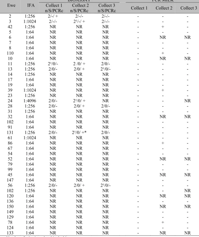
Toxoplasma gondii in milk of naturally infected dairy ewes on west
Texto
Imagem
Documentos relacionados
The aims of this study were to construct a plasmid containing the RABV G-gene insert (pSH-G), to transfect this plasmid into eukaryotic cells and to establish the stability
Para Maggie (1988, 1996) as gradações de cor ou continuum de cor são utilizadas pelas pessoas de uma forma que parece “encobrir” ou “escurecer” as polarizações, uma vez
Para este estudo foi escolhida a cidade de Benguela, Província de Benguela, sendo estudados, especificamente os seguintes bancos: BPC (Banco Poupança e Crédito; primeiro
Curso de Linguagem Computacional C/C++ Uma classe que não pode ser utilizada para criar objetos é dita classe abstrata e existe somente para ser usada como base
disputas por hegemonia no âmbito das políticas educacionais, debate-se sobre os embates em torno de interesses e correlações de força entre as classes sociais. O que está em
Embora a idéia não fosse do agrado de Padre José, dado que não queria modificar tão radicalmente a edificação de Monsenhor Tombrock e, afirmam alguns, contava
Resumo Este estudo investigou a variação intra-individual do lactato sLac, alfa-amilase sAA e cromogranina A sCgA salivares com relação ao acúmulo de lactato sanguíneo bLac durante
