UNIVERSIDADE DE LISBOA
FACULDADE DE CIÊNCIAS
DEPARTAMENTO DE BIOLOGIA ANIMAL
Understanding how Capping Protein cooperates with
aPKC to maintain epithelial integrity in Drosophila
imaginal disc
Cláudia Carolina de Almeida Mendes
MESTRADO EM BIOLOGIA EVOLUTIVA E DO
DESENVOLVIMENTO
UNIVERSIDADE DE LISBOA
FACULDADE DE CIÊNCIAS
DEPARTAMENTO DE BIOLOGIA ANIMAL
Understanding how Capping Protein cooperates with
aPKC to maintain epithelial integrity in Drosophila
imaginal disc
Cláudia Carolina de Almeida Mendes
Dissertação de mestrado orientada por: Doutora Florence Janody
1Orientador interno: Doutor Gabriel G. Martins
21
Instituto Gulbenkian de Ciência, Oeiras. 2
Faculdade de Ciências da Universidade de Lisboa, Lisboa
MESTRADO EM BIOLOGIA EVOLUTIVA E DO
DESENVOLVIMENTO
I
ACKNOWLEDGMENTS
This thesis would not have been possible without the support and encouragement of several people that I would like to show my gratitude:
Foremost, I would like to thank to my external supervisor, Florence Janody, for giving me the opportunity to be part of the Actin Dynamics Group. She quickly became for me the role model of a great researcher and a great woman. One of the many essential guidelines I learned with her was the importance of good controls!
To my internal supervisor Gabriel Martins for being the first person who taught me how to work with confocal microscopy.
To Beatriz, an exceptional person that I had the pleasure to meet. She essentially taught me everything I know about flies and gave me a huge help and lots of good ideas during my lab work and thesis-writing.
To actins: Catarina for the enlightening discussions; Basia for the good music when we had to worked late; Rita for helping me in my Western Blot attempt; João for making my lab work more fun; and Gaspar for having the results that led to my project!
To the Drosophila community at the IGC, especially to Gastón for the amazing discussions we had about science and movies.
To my teachers, Élio Sucena and Eduardo Crespo, who persuaded and got me interested in evolution and development.
To my friends for being part of my life.
To my family for encouraging me to follow my dreams. To Rafael for being always there.
II
LIST OF ABBREVIATIONS
ABPs Actin Binding Proteins
A-P Anterior-Posterior
AJs Adherens junctions
Arm Armadillo (β-catenin) aPKC Atypical protein kinase C
Baz Bazooka
CP Capping protein
Cpa Capping protein α
Cpb Capping protein β
Crb Crumbs
Dlg Discs large
Dlt Discs lost
E-Cad E-Cadherin
F-actin Filamentous actin
Lgl Lethal giant larvae
Par Partitioning defective
RNAi RNA interference
SAR Sub apical region
Scrib Scribble
SJs Septate junctions
Std Stardust
TSGs Tumor supressor genes
III
ABSTRACT
Epithelial integrity generally depends on the balance between the stability and remodelling of adherens junctions (AJs). Growing evidence indicates that endocytosis has a central role in maintaining AJs in a state of dynamic equilibrium. However, it is not currently understood how AJs remodelling is achieved without losing epithelial integrity. In this work, I found that Capping Protein (CP), which prevents extension of the barbed ends of actin filaments, interacts genetically with atypical protein kinase C (aPKC), a major effector of the Par-aPKC complex, to maintain epithelial integrity in Drosophila wing imaginal disc. This genetic interaction appears to have differential requirements along the wing disc, since decreasing CP and aPKC levels induce extrusion and death of wing blade cells, whereas cells in the distal hinge region seem to overproliferate. Interestingly, loss of CP results in a significant reduction of aPKC from the apical membrane, suggesting that CP may restrict apical localization of aPKC, which in turn, is required to control AJs dynamics. In addition, similar to apkc mutant tissues, loss of CP leads to decreased apical levels of the AJs components, E-Cadherin (E-Cad) and Armadillo (Arm), suggesting that both CP and aPKC promote AJs stability by regulating early endocytic events.
Therefore, by promoting the formation of a highly branched actin network, CP could have a putative role in AJs stability and remodelling by restricting aPKC to the apical membrane and by promoting early endocytic events. Taken together, the presented data highlight the crucial synergy between polarity complexes and actin cytoskeleton in regulating AJs dynamics, which is intimately linked to the maintenance of epithelial integrity.
Keywords: Capping Protein, atypical protein kinase C, adherens junctions, endocytosis,
IV
RESUMO
Os tecidos epiteliais possuem duas propriedades contraditórias: têm uma arquitectura robusta, necessária para a sua estabilidade, porém exibem uma extensa plasticidade de modo a permitir eventos celulares tais como divisão e morte celular [1, 2]. Quando ocorre uma perda na regulação deste balanço entre estabilidade e plasticidade, as células epiteliais perdem a adesão entre elas e adquirem a capacidade de sobreproliferar. De facto, a maioria dos tumores malignos surge apartir de epitélios com defeitos no controlo da adesão e proliferação [3, 4].
A adesão entre células epiteliais é em parte devida à acção coesiva das junções aderentes (AJs), cuja principal função é ligar o citoesqueleto de actina entre células vizinhas. As AJs circundam a região apical das células e são principalmente compostas pela proteína transmembranar E-Caderina (E-Cad). Através do seu domínio citoplasmático, E-Cad associa-se a β-catenina (em Drosophila codificada por armadillo, arm) e α-catenina, que por sua vez, promove a ligação ao citoesqueleto de actina [5]. Apesar de terem um papel crucial na manutenção da integridade epitelial, recentes estudos têm revelado que as AJs são constantemente remodeladas através de endocitose [6-8], processo celular pelo qual moléculas extracelulares ou associadas à membrana são internalizadas para o interior das células [9, 10]. No entanto, ainda não é compreendido como é que a remodelação das AJs é estabelecida sem que a integridade do epitélio seja perdida.
Na região apical às AJs existem dois complexos proteicos que interactuam entre si de modo a promover a polaridade dos tecidos epiteliais: o complexo composto por Crumbs (Crb), Stardust and Discs lost, denominado por complexo Crb, e o complexo constituído por Bazooka/Par3, proteína cinase C atípica (aPKC) e Par6, designado por complexo Par-aPKC [11]. O complexo Par-aPKC é activado através da associação entre Par6 e a Rho-like GTPase Cdc42. Após esta activação, aPKC fosforila Crb, que por sua vez, promove a estabilização das Ajs [12, 13]. Curiosamente, foi demonstrado que a remoção de cdc42 ou apkc em tecidos epiteliais de Drosophila, resulta na acumulação
V de proteínas apicais associadas à membrana, tais como Crb, em vesículas endocíticas e à redução dos níveis apicais de E-Cad e Arm. Análises genéticas adicionais sugerem que aPKC actua como efector de Cdc42, regulando a endocitose de proteínas apicais, que por sua vez, tem um efeito estabilizador nas AJs durante processos dinâmicos de reorganização celular [14, 15].
Nos tecidos epiteliais, a região apical das células contém uma banda de filamentos de actina, essencial para o suporte das Ajs [2]. O citoesqueleto de actina desempenha um papel fundamental em numerosos processos celulares, tais como regulação da morfologia celular e manutenção da polaridade celular [16]. Dada a sua dinâmica, é necessária uma regulação eficaz, que é desempenhada por proteínas que se ligam à actina (ABPs) [17, 18]. Uma dessas proteínas é o heterodímero Capping Protein (CP), composto pelas subunidades α (Cpa) e β (Cpb), que se ligam à região terminal dos filamentos de actina, prevenindo a perda ou adição de monómeros de actina e, desta forma, a sua polimerização excessiva [19]. A remoção de CP no disco imaginal da asa de Drosophila leva a diferentes fenótipos celulares que são dependentes da região do disco. Enquanto clones de células mutantes para CP mantêm a polaridade e sobrevivem nas regiões do hinge e notum, células mutantes na wing blade sofrem extrusão basal e morrem por apoptose [20]. Semelhante ao fenótipo observado em epitélios mutantes para apkc, diminuição dos níveis proteicos de CP usando a técnica ARN de interferência (RNAi), resulta na acumulação de Crb em estruturas vesiculares [21]. Isto sugere que, tal como aPKC, CP parece regular a endocitose de proteínas apicais. Curiosamente, foi postulado que CP interactua geneticamente com aPKC no disco imaginal da asa [21]. No entado, a ferramenta genética usada para diminuir a activade de aPKC pode ter efeitos inespecíficos [22] e portanto, a possível interacção genética entre CP e aPKC foi novamente analisada.
Desta forma, o presente trabalho tinha como objectivo compreender o mecanismo pelo qual CP pode cooperar com aPKC na manutenção da integridade epitelial do disco imaginal da asa de Drosophila. De modo atingir este objectivo, a técnica RNAi sob o controlo do sistema UAS-Gal4, foi usada para diminuir os níveis proteicos de CP em regiões específicas do tecido (García Fernández, B. and Janody, F.
VI et al., unpublished data), e um alelo mutante termosensível para aPKC (apkcts) foi usado para modular a actividade cinásica de aPKC consoante a temperatura (Martinho, R. et al., unpublished data).
Quando comparado com o fenótipo anteriormente descrito para células com níveis reduzidos de CP, ao diminuir ambos os níveis de CP e aPKC no disco de asa, uma maior quantidade de células sofreram extrusão basal e morreram por apoptose. Pelo contrário, células mutantes para apkcts encontraram-se mantidas no epitélio, sem nenhum indício de morte celular. Estes resultados confirmam que CP interactua geneticamente com aPKC na manutenção da integridade epitelial do disco da asa. Esta interacção genética parece ter diferentes requerimentos ao longo do disco da asa. Enquanto que a diminuição dos níveis de CP e aPKC levou à morte das células da wing blade, as células na região distal do hinge pareceram proliferar. Isto sugere que CP e aPKC mantêm a integridade epitelial e previnem a morte celular na wing blade, ao passo que na região do hinge, CP e aPKC parecem restringir o crescimento, possivelmente através da regulação da sinalização Hippo [23].
Curiosamente, a diminuição dos níveis de CP resultou numa redução significativa dos níveis apicais de aPKC, sugerindo que, através da sua função como regulador da dinâmica do citoesqueleto de actina, CP restringe a localização de aPKC na membrana apical das células epiteliais. Além disso, semelhante ao fenótipo observado em epitélios mutantes para apkc, diminuição dos níveis de CP levou a um decréscimo acentuado dos níveis apicais de E-Cad e Arm. Isto sugere que CP tem um papel putativo na dinâmica das AJs. Por último, o efeito nos níveis apicais de E-Cad após diminuição dos níveis de CP foi semelhante ao observado após sobreexpressão de Src64B, um importante regulador da estabilidade das AJs. Uma vez que a actividade de Src parece ser regulada por CP (García Fernández, B. and Janody, F. et al., unpublished data), este resultado sugere que o efeito de CP nas AJs pode ser dependente da actividade de Src. Baseando nestas observações, são apresentadas no presente trabalho diferentes hipóteses que pretendem explicar o papel de CP na manutenção da estabilidade das AJs, e de como esta função putativa de CP pode estar interligada com a função conhecida de aPKC na regulação da dinâmica das AJs.
VII É importante referir que as diferentes hipóteses não são mutuamente exclusivas e por isso, de um modo geral, através da sua principal função, prevenir a polimerização dos filamentos de actina, CP parece ter um papel putativo na manutenção da estabilidade das AJs por: (a) restringir a localização de aPKC na membrana apical; (b) regular eventos iniciais de endocitose; e (c) regular a actividade de Src. No conjunto, os dados apresentados neste trabalho apontam para uma crucial sinergia entre complexos de polaridade e o citoesqueleto de actina na regulação da dinâmica das AJs, o qual está intimamente ligado à manutenção da integridade dos tecidos epiteliais.
Palavras-chave: Capping Protein, proteína cinase C atípica, junções aderentes,
VIII
TABLE OF CONTENTS
ACKNOWLEDGEMENTS ... I LIST OF ABBREVIATIONS ... II ABSTRACT ... III RESUMO ... IV TABLE OF CONTENTS ... V I. INTRODUCTION 1 1. Epithelial tissues 11.1 Epithelial architecture: polarity and cell-cell adhesion 1
1.2 The Par-aPKC complex 3
1.3 The development of epithelial tumours 3
1.4 Drosophila wing imaginal disc as an epithelial model system 4
2. Endocytosis 6
2.1 Mechanisms of Endocytosis 6
2.2 Remodelling of cell-cell adhesion through endocytosis 8 2.3 Endocytic genes define a novel class of Drosophila tumour suppressor genes 8
3. The actin cytoskeleton 9
3.1 Actin dynamics and its regulation by actin binding proteins 9
3.2 Capping Protein: a highly conserved heterodimer 11
3.2 CP prevents cell extrusion within restricted regions of Drosophila epithelia 11 3.3 CP prevents accumulation of apical membrane-associated proteins 12 3.4 CP appears to interact genetically with aPKC to maintain epithelial integrity 13
3.5 CP cooperates with Src signalling 15
II. AIMS 16
III. MATERIAL AND METHODS 17
1. Fly strains and genetics 17
1.1 Fly husbandry 17
1.2 Fly Stocks: mutant allele and transgenic lines available for this study 17
1.3 Fly Stocks generated 18
1.4 Genetic tools 19
1.4.1 The UAS-Gal4 system 19
1.4.2 Generation of clones by mitotic recombination 21
2. Immunohistochemistry 23
IX
3. Image Acquisition and Analysis 23
4. Classification of cell death observed in mosaic analysis 24
5. Statistical analysis 25
IV. RESULTS 26
1. Capping Protein cooperates with aPKC 26
1.1 apkcts induces extrusion and death of Cpa-deleted cells in mosaic clones induced in the wing
blade 26
1.2 apkcts enhances the Cpa-depletion phenotype in the posterior compartment of the wing disc 30 1.3 Capping Protein acts synergistically with aPKC during adult wing morphogenesis 33
2. aPKC apical localization is affected by knocking-down CP levels 37
3. Capping Protein maintains AJs components in the apical surface 40
VI. DISCUSSION 44
1. Capping Protein interacts genetically with aPKC 44
1.1 The interaction between CP and aPKC has differential requirements along the proximal-distal axis 45 1.2 The interaction between CP and aPKC is dosage dependent 46
2. CP appears to restrict aPKC localization to the apical membrane 47
3. CP has a putative role in AJs stability 49
VII REFERENCES 54
1
I. INTRODUCTION
1. Epithelial tissues
A great variety of epithelia line the walls of cavities, separating two different chemical milieus or, in the case of the epidermis, serve as protection from the external environment [2]. The structure and function of epithelial tissues generally depend on apicobasal polarization, which is achieved and maintained by linking asymmetrically distributed intercellular junctions to the cytoskeleton of individual cells [11]. During aberrant epithelium development, cells often lose adhesion between each other, disrupting the polarity and normal growth of the tissue. In fact, the majority of human tumours arise from epithelial tissues with abnormal cell adhesion and proliferation [3, 4].
1.1 Epithelial architecture: polarity and cell-cell adhesion
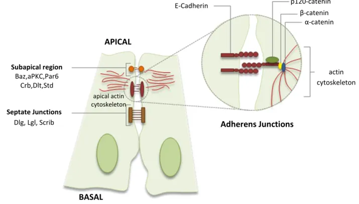
Simple epithelia generally consisted of a laterally coherent sheet of cells with an apical and a basolateral domain. The apical surface is divided into the free apical domain and a domain where a neighbouring cell is contacted. Similarly, the basolateral domain is further subdivided into a basal domain mediating cell-matrix adhesion and a lateral domain involved in cell-cell adhesion [2, 11] (Fig.).
In Drosophila epithelial cells, cell-cell adhesion is mainly accomplished by three junctional complexes: adherens junctions (AJs), septate junctions (SJs), the functional equivalent to the vertebrates’ tight junction, and the sub-apical region (SAR) [2].The AJs are a generally circumferential junction located just basal to the apico-basolateral boundary, which major function is to link the actin cytoskeleton of neighbouring epithelial cells. They are composed of the transmembrane protein E-cadherin (E-cad), which is linked on its cytoplasmic side to β-catenin (in Drosophila encoded by armadillo) and α-catenin. In turn, α-catenin binds to actin filaments, forming a continuous band, the zonula adherens (ZA) [5]. In addition to β-catenin, the Armadillo family also includes several other related molecules, including p120-catenin. Although not directly involved in coupling the cadherins to the cytoskeleton, p120 has emerged
2 as a critical regulator of cadherin adhesive activity and cytoskeletal organization [24, 25] (Fig.).
The SJs act as permeability barriers between cells and contain a complex of three proteins: Discs large (Dlg), Lethal giant larvae (Lgl) and Scribble (Scrib) (the Scrib complex). These proteins have been proposed to form a biochemical complex that controls and refines the segregation of apical and basolateral membrane domains [26]. SAR is the most apically localized junctional complex and it is defined by the accumulation of two protein complexes: the Crumbs (Crb), Stardust (Std), Discs lost (Dlt) complex (the Crb complex) and the Bazooka/Par3, atypical Protein Kinase C (aPKC), Par6 complex (the Par-aPKC complex) (Fig.1) [2, 14]. Mutations in genes coding for the Lgl, the Crb, and the Par-aPKC complex all disrupt epithelia formation but appear to do so because they are required for the initial generation of epithelial polarity and junctions.
Fig. 1 - Schematic representation of Drosophila epithelial cells. The major junctional complexes are
represented: the apically localized subapical region (SAR), the adherens junctions (AJs) and the septate junctions (SJs). Epithelial polarity complexes are located in stratified regions along the apico-basal axis of cells. The Crb complex along with the Par-aPKC complex is located in the SAR, while the basolateral Scrib complex forms the SJs. Adapted from Shock, F. & Perrimon, N. (2002) [2] and Mosesson, Y. et al. (2008)[27]. APICAL BASAL E-Cadherin p120-catenin β-catenin α-catenin actin cytoskeleton Adherens Junctions Subapical region Baz,aPKC,Par6 Crb,Dlt,Std Septate Junctions Dlg, Lgl, Scrib apical actin cytoskeleton
3
1.2 The Par-aPKC complex
The apico-basal polarity and the early phases of ZA assembly in epithelial cells depends on the Par-aPKC complex, which is composed of two scaffold proteins, Par3 (Bazooka, Baz in Drosophila) and Par6, and an atypical protein kinase C (aPKC) [28-30]. In Drosophila epithelia, aPKC physically interacts with Par6, and although this interaction tethers aPKC to the apical membrane, it is also though to inhibit aPKC kinase activity. It has been shown that a key event that activates aPKC at apical domain is the binding of the Rho-like GTPase Cdc42 [31] to the CRIB (Cdc42/Rac interactive binding) domain of Par6 [12, 13]. Although Baz, Par6, and aPKC are often assumed to function as a complex, it was recently reported that Baz is excluded from the apical domain by aPKC phosphorylation. This apical exclusion of Baz was shown to be required for the establishment of apical and basolateral membranes [32].
Similar to its interaction with Baz, aPKC-mediated phosphorylation is required for the correct apical localization of Crb and its cytozolic associates, Sdt and Dlt, whose activity is required to further recruit and concentrate sparsely distributed E-Cad into the AJs [22, 33]. Concomitantly, the interaction with the Crb complex upregulates aPKC activity and inhibits the function of basolateral regulators such as the Scrib complex at the apical space [12, 13]. In addition, aPKC phosphorylates Lgl to exclude it from the apical membrane and restricts it to the lateral domain, where it interacts with Dlt and Scrib [34].
1.3 The development of epithelial tumours
Epithelial tumour progression involves the stepwise acquisition of a number of neomorphic traits by tumour cells. These traits include disruption of epithelial adhesion and apicobasal polarity, self-sufficiency in growth signalling, resistance to apoptosis, inability to differentiate and acquisition of migratory and invasive abilities [35]. The development of epithelial tumours is thought to involve cooperation between tumour suppressor genes (TSGs) and oncogenes, as well as between tumour and its microenvironment [36].
4 TSGs are genes that, when mutated, can act cell-autonomously and cause excessive tissue overgrowth. In Drosophila, TSGs are generally divided into two categories: hyperplastic whenever they show increased proliferation, although epithelial architecture is maintained, whereas they are considered to be neoplastic if tissue overproliferation occurs concurrently with disruption of epithelial structure [37]. Interestingly, the components of the Par-aPKC complex were identified as neoplastic TSGs [38]. Indeed, the invasive potential of ovarian and breast cancer cells is associated with loss of aPKC from the apical membrane and its recruitment as a positive mediator for proliferation and invasiveness [38-40]. Additionally, aPKC can be regulated by other TSGs. For example, mutations in the TSG von Hippel-Lindau (VHL) results in mislocalization and degradation of aPKC and subsequent loss of epithelial architecture [41]. Thus, failure to restrain aPKC to the apical domain and deregulation of its activity are key factors for cancer progression [38].
One of the oldest and most investigated oncogenes is the non-receptor protein tyrosine kinase Src. In addition to the well-established role of Src in regulating cellular proliferation, there is accumulating evidence that Src also acts to affect adhesion, invasion and motility events, particularly in epithelial cells and in cancer cells during later stages of cancer progression [42]. Src interacts directly with the AJs components, E-Cad and Arm, and its overactivity promotes AJs disassembly, reducing adhesion and promoting motility and invasiveness [42, 43].
1.4 Drosophila wing imaginal disc as an epithelial model system
The fruit fly, Drosophila melanogaster, has been used as a model organism in diverse fields since the turn of the previous century, and today is one of the most widely-used and genetically best-known of all multicellular organisms. Drosophila imaginal discs are considered bona fide epithelia and thereby, they are an excellent model to study epithelial integrity. The imaginal discs can be seen in the newly hatched larvae as local invaginating epidermal cell clusters. Whereas most of the larval cells have very limited mitotic capacity, imaginal disc cells proliferate rapidly
5 and give rise to adult cuticular structures, such as the wings and legs. In this work, I used the wing imaginal disc as an epithelial model due to its well characterized patterning events and ease of dissection in 3rd instar larvae.
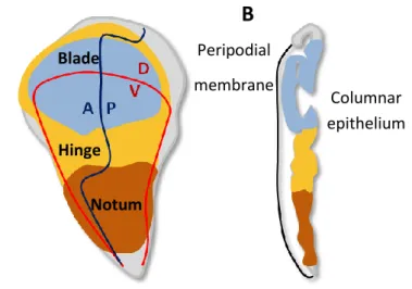
The wing imaginal discs are bilayered epithelial tissues consisting of a columnar monolayer epithelium, whose cells are polarized along the apicobasal axis, being covered by a thin layer of squamous epithelium called the peripodial membrane. As pupation begins, the cells at the centre of the disc telescope out to become the most distal region of the wing – the wing blade - , the surrounding cells give rise to the wing hinge and the outmost cells become the proximal structures – the notum and the pleura (dorsal thorax structures) (Fig.2). Early in their development, discs are subdivided into spatially distinct, stable units called compartments, which are developmental fields of cells sharing common ancestry and adhesive properties. Signalling between compartments establishes the anterior-posterior (A-P) and dorsal-ventral (D-V) boundaries [44, 45].
Fig. 2 – Drosophila wing disc and fate maps. (A) Fate map of 3rd instar wing imaginal disc showing the anterior-posterior (A-P) and dorsal-ventral (D-V) compartment boundaries and major regions of the disc: the wing blade (blue), which gives rise to the proper wing, the hinge (yellow), that constricts to form a mobile link to the body wall of the fly and the notum (orange), which gives rise to dorsal thorax structures. (B) Cell layers of the wing disc. There are two main cell layers: the peripodial membrane and the columnar epithelium. Adapted from Butler, M.J. et al., (2003) [46].
The A-P boundary is defined during the 1st instar larvae and requires engrailed (en) expression in the posterior compartment for its maintenance. The Engrailed (En)
Peripodial membrane
B
Columnar epithelium Notum Hinge BladeA
6 transcriptor factor promotes hedgehog (hh) expression in the posterior compartment and it acts as a short-range morphogen to specify the expression of decapentaplegic (dpp) in a narrow stripe of cells adjacent to the A-P boundary [30]. Additionally, Dpp is also required for proximal-distal patterning [30]. During the 2nd instar larvae, an antagonist relationship between the epidermal growth factor (EGF) and Wingless (Wg) signalling establishes the D-V boundary and divides the disc into a dorsal region that gives rise to the notum, and a ventral region that forms the wing [31]. The wing is further subdivided into distal and proximal regions by expression of the selector genes vestigial (vg) and scalloped (sd) in the wing blade and homothorax (hth) and teashirt (tea) in the wing hinge [45].
2. Endocytosis
Growing evidence has revealed an important role for vesicle trafficking in maintaining epithelial architecture. Exocytosis, in which recently synthesized proteins are released from the trans-Golgi network to apical or basolateral membrane domains, have received primary emphasis among trafficking routes responsible for epithelial polarity, but accumulating evidence now reveals that endocytosis plays an equally important role in regulating polarity and adhesion of epithelial tissues [8, 47].
2.1 Mechanisms of Endocytosis
Endocytosis is a cellular process used by all eukaryotic cells to internalize extracellular or membrane-bound cargoes from the plasma membrane into the cell interior through a series of vesicle compartments. Endocytosis occurs by various mechanisms, which can be divided into those that are clathrin-dependent and those that are clathrin-independent, such as phagocytosis and macropinocytosis [9, 10].
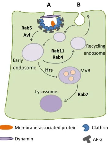
In clathrin-dependent endocytosis, with the help of the Adapter Protein-2 (AP-2), clathrin triskelia form a cage structure around invaginated membrane and the activity of the GTPase dynamin (in Drosophila encoded by shibire, shi) results in vesicle scission. Newly formed vesicles fuse with other endocytic vesicles to form an early
7 (sorting) endosomal compartment [9]. These fusion events are regulated by the endocytic syntaxin Avalanche (Avl) and the GTPase Rab5 [48]. The primary function of the early endosome is the sorting of internalized cargo to different intracellular destinations. Cargo destined for degradation remains in the early endosome as it matures into a multivesicular body (MVB). Ultimately, when the endosome reaches its maturity, Rab7 GTPase mediates its fusion with a lysosome, where proteolytic degradation occurs. Alternatively, cargo destined for recycling is sorted into a specialized recycling endosome and then returned via vesicles to the plasma membrane [48, 49]. The GTPase Rab11 mediate slow endocytic recycling through recycling endosomes, whereas Rab4 mediates fast endocytic recycling directly from early endosomes (Fig.3) [48].
Fig. 3 - Simplify view of the endocytic pathway. Clathrin-dependent (A) and clathrin-independent (B)
vesicle formation is shown. Internalized membrane-associated proteins either are sent back to the membrane by the recycling pathway or else are degraded in the lysosome. Adapted from Fisher, J.A. et al. (2006) [50]. Membrane-associated protein Dynamin Clathrin AP-2
A
Early endosome Recycling endosome MVB Lysossome Rab5 Avl Rab11 Rab4 Hrs Rab7B
8
2.2 Remodelling of cell-cell adhesion through endocytosis
Although all junctional complexes are functionally significant, recent evidence has shown that AJs play an essential role in regulating the dynamics of epithelial tissues. In fact, the redistribution of E-Cad through regulated endocytosis is crucially important to mediate dynamic adhesion and epithelial integrity during processes of cell division, cell death and cell rearrangement [6-8]. Even though AJs remodelling plays a key role in epithelial integrity, its regulation is still poorly understood.
Studies in mammalian cells and Drosophila have pointed to the Rho-like GTPases Cdc42 and Rac as one group of AJs regulators [14, 51]. Knocking-out cdc42 function causes a disruption of AJs during dynamic cell rearrangements in Drosophila neuroectoderm. Interestingly, when cell rearrangement is blocked, neuroectoderm integrity is restored, indicating that epithelia undergoing extensive adhesion remodelling are more susceptible to endocytic defects. Further genetic interaction studies suggested that, the failure to maintain AJs in cdc42 mutant tissues is a possible secondary consequence of the loss of apical polarity proteins, such as Crb, from the plasma membrane [14]. Interestingly, constitutively active aPKC partially suppressed effects of Cdc42 reduction, strongly implicating aPKC as the key effector in regulating endocytosis of apical proteins, which in turn, has a stabilizing effect on AJs during cell rearrangement [14, 15].
The complete disassembly of AJs in epithelial tissues results in loss of adhesion and the acquisition of a migratory or mesenchymal-like phenotype [52]. For instance, upon Src activation, E-Cad is ubiquitinated and then shuttled to the lysossomal degradation instead of following its normal trafficking route of recycling back to the apical membrane. This results in loss of E-Cad at apical junctional sites, leading to loss of adhesion and disruption of epithelial integrity [53, 54].
2.3 Endocytic genes define a novel class of Drosophila tumour suppressor genes
With the introduction of genetic mosaic screening techniques, several neoplastic TSGs have been characterized that are key regulators of cargo entry into
9 the early endosome and endosomal maturation. Among these new endocytic TSGs are the genes shibire (shi), rab5 and avalanche (avl) [35, 37]. Mosaic clones mutant for shi, rab5 or avl are slow-growing and usually eliminated from the disc [35, 55] through a process of cell competition [56]. When the wild-type contribution is removed, mutant imaginal discs for shi, rab5 or avl accumulate apical proteins, such as Crb and the Notch receptor, at the plasma membrane, disrupt AJs and undergo massive overproliferation, resulting in the development of neoplastic overgrowths [35, 55]. Interestingly, Notch signalling is downregulated in shi, rab5 and avl mutant tissues, suggesting that overgrowth in this subset of endocytic TSGs mutants is not a result of increased Notch signalling [57]. In fact, it appears to be due to mislocalization of Crb, since increasing Crb levels in imaginal discs causes neoplastic transformation, whereas interfering with Crb activity through co-expression of a dominant negative form of aPKC (aPKCCAXXDN) ameliorates the phenotype [55].
3. The actin cytoskeleton
The cytoskeleton comprises a network of protein filaments including microtubules, actin filaments and intermediate filaments, which are held together and linked to subcellular organelles and the plasma membrane by a variety of accessory proteins. A key feature of epithelial cells is the polarized actin cytoskeleton, which plays a central role in numerous cellular processes, such as generation and maintenance of cell shape and polarity, cell division, contractility and motility. A continuous band of actin filaments localized apically and links epithelial cells to each other, forming the ZA. This continuous band of actin filaments is extremely important for the maintenance of the epithelia, since it provides adhesive strength between epithelial cells [2].
3.1 Actin dynamics and its regulation by actin binding proteins
Actin is present in cells in two main forms: the monomeric globular actin (G-actin) and the polymeric filamentous actin (F-(G-actin). The filamentous actin is a
10 polarized structure, in which the fast-growing barbed (+) end is the preferred site for ATP-monomeric-actin addition, whereas depolymerization occurs by loss of ADP-actin subunits from the slow-growing pointes (-) end. This ATP-hydrolysis-driven filament polymerization is a very dynamic process, which requires a tight regulation in vivo.
Fig. 4 – Actin dynamics is controlled by a large array of ABPs. The actin nucleators, such as Arp2/3
complex, promote de novo actin filament nucleation and branching. ADF/Cofilin factors severe the actin filaments and promote dissociation of the actin monomers from the pointed (-) end. Cyclase-associated proteins (CAP) sequester actin monomers, preventing their incorporation into filaments. Capping proteins (CP) restrict the access to the barbed (+) end, forming a protein cap that prevents further addition of actin monomers. Adapted from Disanza, A. et al. (2005) [18].
Actin dynamics and structure are modulated by several actin binding proteins (ABPs), which in turn are under the control of specific signalling pathways (Fig.4) [17, 18]. For instance, the nucleation of actin filaments is an energetically unfavourable event, being promoted by certain ABPs, such as the Arp2/3 complex and formins. Once activated by a member of the evolutionary conserved Wiskott - Aldrich syndrome protein (WASp) family, the Arp2/3 complex nucleates new actin filaments by binding to the side of a pre-existing filament. Importantly, the activity of WASp family is regulated by the Rho-like GTPases Cdc42 and Rac [18, 58]. Among the ABPs that prevent actin polymerization, the actin depolymerizing factor (ADF)/Cofilin family, the Cyclase-associated protein (CAP) and Capping Proteins (CP) are some of the most important. Cofilin severs filaments and enhances dissociation of actin
Capping Protein Arp2/3 complex ADF/Cofilin CAP
11 monomers from the pointed end [19], CAP sequesters actin monomers, preventing their incorporation into filaments [59] and CP restricts accessibility of the barbed end, inhibiting addition or loss of actin monomers [60].
3.2 Capping Protein: a highly conserved heterodimer
CP is a highly conserved heterodimer, composed of α (Cpa) and β (Cpb) subunits, which binds the barbed end of actin filaments with high affinity, thereby preventing excessive actin polymerization [61]. Interestingly, in vitro studies showed that CP shunts actin monomers away from the barbed ends and onto Arp2/3 complex, promoting more frequent filament nucleation by the Arp2/3 complex [62]. Thus, functional CP, along with the Arp2/3 complex, support the assembly of a dominantly branched network of small actin filaments, generating protruding force and giving rise to the lamellipodia of migrating cells [58]. When capping of actin filaments by CP is inhibited by PIP2, CARMIL or Melanotrophin/V-1, long actin filaments can become bundled, giving rise to filopodia [63]. Moreover, besides having a critical role in the termination of filaments, CP has been suggested to mediate actin filament attachment to the plasma membrane [64, 65].
Although the mechanism and regulation of CP have been studied, most of the available data results from in vitro biochemical studies and cell culture. Since in vitro analysis do not exactly reproduce the complexities within a tissue, it is crucial to understand the role of CP and its regulation in vivo.
3.2 CP prevents cell extrusion within restricted regions of Drosophila epithelia
As expected, in vivo studies showed that CP prevents excessive actin filaments polymerization, confirming its already known role in actin filament termination [20]. In addition, loss of either cpa or cpb in Drosophila imaginal discs gives rise to identical developmental phenotypes [20, 66], consistent with its role as part of a functional heterodimer [61].
12 disc. While cpa or cpb mutant clones maintain cell polarity and survive in the hinge and notum regions, mutant clones in the wing blade mislocalize Arm and E-Cad to basolateral domains and undergo cell extrusion and apoptosis [20]. By expressing a HA-tagged form of Cpa, it was observed that Cpa mostly accumulates at the apical regions of the cell [20], suggesting that CP regulates a specific pool of apical actin filaments. However, excessive actin filament accumulation may not be the main cause of extrusion and death of CP mutant clones, since mutations in the genes encoding the Cyclase-associated protein Capulet (Capt) and the Cofilin homolog Twinstar (Tsr) also induce strong accumulation of actin filaments, but do not cause cell extrusion [20, 66].
3.3 CP prevents accumulation of apical membrane-associated proteins
It has been recently pointed that actin cytoskeleton has a crucial role in invagination and scission of endocytic vesicles [67, 68]. For instance, in epithelial cell lines, the use of Cytochalasin D, an actin-disrupting agent, inhibits endocytosis of membrane-associated proteins and the formation of coated vesicles [69]. Similarly, in budding yeast, when actin polymerization is blocked, the initial movement of endocytic vesicles away from the plasma membrane, which presumably corresponds to vesicle scission and release, is completely blocked [70, 71].
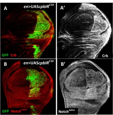
Previous data from F. Janody’s lab revealed that RNA interference (RNAi) leading to CP depletion in the posterior compartment of Drosophila wing disc leads to accumulation in punctuated structures of the membrane-associated proteins, Crb and Notch (Fig.5) [21]. In particular, similar to shi, rab5 and avl mutant tissues [57], Notch accumulation is not due to transcriptional upregulation, since Notch target genes, such as cut and m8-lacZ, are downregulated in CP depleted cells [21]. In this sense, Notch accumulation in CP depleted cells was suggested to be due to an early endocytic defect, blocking progression to early endosomes where activation of Notch takes place [57]. Accordingly, accumulation of Crb in CP depleted cells could also be due to an endocytic defect.
13 Fig. 5 - CP depleted cells accumulate the Notch receptor and Crb in the wing imaginal disc. All panels
show apical Z-projections sections of third instar wing discs. (A-B’) Wings discs expressing UAS-cpbIRC10 and UAS-GFP (green) under en-Gal4 control are stained with either anti-Crb or anti-Notch extracellular domain (red in A and B or grey in A’ and B’, respectively). Adapted from Gaspar, P. (2008) [21].
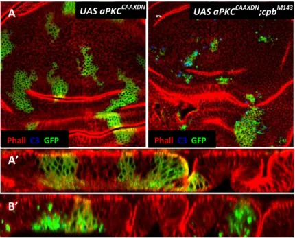
3.4 CP appears to interact genetically with aPKC to maintain epithelial integrity
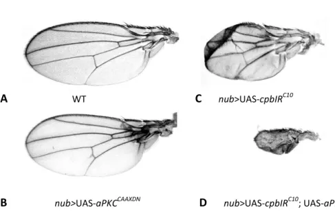
As mentioned before, loss of CP in Drosophila wing discs leads to accumulation of Crb in punctuated structures. In the same study, it was shown that clones overexpressing the intracellular domain of crb extrude and die mainly in the wing blade epithelium [21]. This clonal behaviour is reminiscent to the CP mutant phenotype [20], suggesting that extrusion and death of CP mutant cells could be the result of excessive Crb signalling activity [21]. To investigate this possibility, a ‘kinase-dead’ form of aPKC (aPKCCAXXDN), that acts as a dominant negative form and is known to reduce the activity of overexpressed crb [22, 55], was expressed in cells mutant for cpa or cpb [21]. Unexpectedly, expression of aPKCCAXXDN induced extrusion and death of CP mutant cells not only in the prospective wing blade, but also in the hinge and notum epithelia (Fig.6) [21]. Consistent with an enhancement of the CP depletion phenotype, driving both aPKCCAAXDN and cpb depletion using nub-Gal4, which drives the expression of the UAS-target gene in the wing blade and distal region of the hinge,
A’ A GFP; Crb; en>UAScpbIRC10
Crb B’ en>UAScpbIRC10
B Notchextra GFP; Notchextra
14 results in a severely enhanced adult wing phenotype, when compared to the phenotype of wings depleted of cpb alone (Fig.8) [21]. Given that expressing aPKCCAXXDN in either clones of cells (Fig.6A-A’) or using nub-Gal4 (Fig.7B) has no visible effect on wing disc, this suggested that CP and aPKC interact genetically to maintain epithelial integrity of wing disc [72].
However, some problems associated to the use of aPKCCAXXDN as a proper kinase dead are raised. Although this construct harbours similar modifications to a Xenopus aPKCλ kinase inactive protein that acts as a dominant negative [73], no biochemical analysis were performed to investigate the kinase activity of Drosophila aPKCCAXXDN. Additionally, the presence of the CAAX sequence enables aPKCCAXXDN to be targeted to the cell membrane. Therefore, the possibility that the genetic interaction between aPKCCAXXDN and CP results from unspecific effects of aPKCCAXXDN, which might trap random kinase substrates, cannot be excluded.
Fig. 6 - CP interacts genetically with aPKC to prevent loss of epithelial cell polarity and extrusion. All
panels show third instar wing discs with clones positively labelled with GFP (green in A-B’). (A and B) Basal Z-projections. (A’ and B’) Reconstructed XZ sections through the wing disc epithelium. (A-A’) Clones expressing aPKCCAXXDN, (B-B’) cpbM143 mutant clones expressing aPKCCAXXDN are stained with anti-Arm to outline apical cell membrane (red in A-B’) and anti-activated Caspase 3, which identifies Caspase-dependent cell death (blue in A-B’). Adapted from Gaspar, P. (2008).
A Phall C3 GFP A’ B B’ Phall C3 GFP
15 Fig. 7 - CP genetically interacts with aPKC during wing morphogenesis. (A) wild-type (WT), (B) nub>cpbRIC10, (C) nub>UAS-aPKCCAAXDN and (D) nub>UAS-cpbIRC10; UAS-aPKCCAAXDN adult wings. From Gaspar, P. (2008) [21].
3.5 CP cooperates with Src signalling
In the wing blade epithelia, exponential decrease of Cpb levels induces a switch from neoplastic growth to cell death (García Fernández, B. and Janody, F. et al., unpublished data). Interestingly, overexpression of Src64B (Drosophila possess two Src kinases, Src64B and Src42A) mimics the effects of decreasing Cpb levels: slight increase induces tissue overgrowth, while strong overexpression promotes cell death (García Fernández, B. and Janody, F. et al., unpublished data) [74, 75]. Moreover, when Cpb depleted tissues also contained a slight increased in Src64B, this results in massive cell death, associated to a strong decrease of the membrane-associated form of Arm. These results suggest that reduced CP levels might cooperate with increased Src64B levels to trigger apoptotic cell death (García Fernández, B. and Janody, F. et al., unpublished data). It was reported that mechanical stretching of cultured cells activates Src through an interaction with actin filaments [76]. Furthermore, actin disruption by Cytochalasin B or knock-down of actin by siRNA strongly inhibit the phosphorylation of Src and its kinase activity [77]. CP might therefore inhibit actin-dependent mechanical activation of Src.
A WT C nub>UAS-cpbIRC10
16
II. AIMS
The Capping Protein (CP) αβ heterodimer, which regulates actin polymerization, prevents accumulation of apical membrane-associated proteins, such as Crb and Notch [21]. Furthermore, knocking-out CP induces mislocalization of E-Cad e Arm to basolateral domains in the wing blade epithelium [20]. Interestingly, the atypical protein kinase C (aPKC), a major effector of the Par-aPKC complex, was shown to control the endocytic uptake of Crb and Notch from the apical membrane and to stabilize AJs during dynamic cell rearrangement [14, 15]. CP might therefore cooperate with aPKC to control the abundance of apical proteins and to regulate AJs stability, which are intimately linked to the maintenance of epithelial integrity. In agreement with this possibility, CP interacts genetically with a dominant negative form of aPKC (aPKCCAXXDN). However, this genetic interaction should be further studied, since the aPKCCAXXDN might have unspecific effects [22].
The main goal of this work was to get insight on the mechanism by which CP cooperates with aPKC to maintain epithelial integrity in Drosophila wing imaginal disc. To do so, I aimed to: confirm the genetic interaction between CP and aPKC using RNAi lines under the control of the UAS-Gal4 system and a termosensitive mutant allele of aPKC (apkcts) (Guilgur, L. and Martinho, R., unpublished data) (Task 1); analyze whether aPKC localize properly in the wing blade epithelium following CP depletion (Task 2); and finally evaluate the putative role CP in AJs stability (Task 3).
17
III. MATERIAL AND METHODS
1. Fly strains and genetics
1.1 Fly husbandry
All flies were raised at 25°C (unless otherwise indicated), according to standard conditions [78]. Fly stocks were maintained by transferring adults to fresh medium every few generations, while crosses were cultured in small vials containing a yeast-glucose-agar medium.
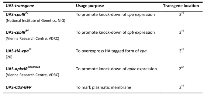
1.2 Fly Stocks: mutant allele and transgenic lines available for this study
In order to demonstrate that the genetic interaction between CP and aPKCCAXXDN (see Introduction Fig.7 and Fig.8) is due to a reduction of aPKC activity, I used a temperature sensitive allele of aPKC (apkcts) bearing a point mutation in a highly conserved residue of the kinase domain, which causes a temperature-dependent reduction of the kinase activity (Guilgur, L.G. and Martinho, R. et al., unpublished data). At permissive temperature (25°C), apkcts maternal mutants show a complete loss of epithelial integrity that leads to embryonic lethality (Guilgur, L.G. and Martinho, R. et al., unpublished data). This phenotype is similar to the one observed in maternal mutants for the apkc loss of function allele [79]. However, while zygotic mutants for the apkc null allele die as late as second instar larvae [15, 80], apkcts zygotic mutants are viable and morphologically normal at permissive temperature (25°C), but are lethal at restrictive temperature (30°C). At semi-permissive temperature (28°C), the viability is variable and adults show defects in abdominal dorsal closure (Guilgur, L.G. and Martinho, R. et al., unpublished data). In contrast to the apkc loss of function allele [15, 79, 80], the use of the apkcts allele (Martinho, R. et al., unpublished data) allowed me to modulate aPKC levels by switching the temperature and thereby gave me the opportunity to determine whether CP genetically interacts with aPKC in maintaining wing epithelial integrity.
18 The majority of fly stocks used in this work were available at the F.Janody’s Lab and some were kindly supplied by R.Martinho’s Lab. Oregon R flies were used as the wild-type stock. The Gal4 drivers and UAS-transgenes used in this study are reported in Table 1.
Table 1 - Gal4 drivers and UAS constructs used during the course of this work.
1.3 Fly Stocks generated
Stable stocks were generated using standard fly genetic methods and crossed with standard balancer stocks [83] or GFP marked balancer stock (CTG; Cyo, Twi-Gal4, UAS-2xEGFP) [84]. The fly stocks generated in this work are reported in Table 2.
Both apkcts(Martinho, R. et al., unpublished data) and cpa [85] mutant alleles are located in the right arm of the 2nd chromosome. Thus, to facilitate the process of generating a transgenic line with reduced levels of aPKC and CP, instead of the cpa Gal4 Drivers Spatial-temporal domain of activity in the wing disc
nubbin-Gal4 (nub-Gal4)
(a gift from G.Morata [81])
From 2nd instar larvae (48h) to end of larval stage in the prospective blade and distal hinge.
hedgehog‐Gal4 (hh‐Gal4)
(a gift from T.Tabata [82])
From end of embryogenesis to end of larval stage in the posterior compartments.
UAS-transgene Usage purpose Transgene location
UAS-cpaIR#2
(National Institute of Genetics, NIG)
To promote knock-down of cpa expression 3rd
UAS-cpbIRB4
(Vienna Research Centre, VDRC)
To promote knock-down of cpb expression 3rd
UAS-HA-cpaID
[20]
To overexpress HA tagged form of cpa 3rd
UAS-apkcIRKK100874
(Vienna Research Centre, VDRC)
To promote knock-down of apkc expression 2nd
19 mutant allele [85], I used a RNAi line against cpa, which is under UAS-Gal4 control (UAS-cpaIR#2) and is located in the 3rd chromosome. Although the RNAi technique do not completely abolish the expression of the target gene, I found that overexpressing UAS-cpaIR#2 in clones generated by the MARCM system recapitulates the cpa loss of function phenotype [20]: accumulation of actin filaments in all regions of the disc and cell extrusion and death specifically in the wing blade (see Results Fig.1E-H’ and Fig.2C-C’’ and E). Since the UAS-Gal4 system is more active at higher temperatures, cpa depletion can be modulated by switching the temperature.
In order to overexpress UAS-cpaIR#2 using nub-Gal4 driver in an apkcts mutant background, a recombinant line was produced between the apkcts mutant allele (w-; FRT42D,apkcts/CyO) and w-; nub-Gal4, both located on the second chromosome. To confirm the presence of nub-Gal4, recombinant flies (w-; nub-Gal4, FRT42D,apkcts/CyO) were crossed either with UAS-CD8-GFP or UAS-cpaIR#2 and selected either for GFP expression in nub-domain or for having cpa depleted wings (see Introduction Fig.8C). Since apkcts homozygous females are sterile, recombinant flies were first crossed with w-; FRT42D,apkcts/CyO, and F1 non CyO females were selected for absence of F2 progeny.
Table 2 - Fly stocks generated during the course of this work.
1.4 Genetic tools
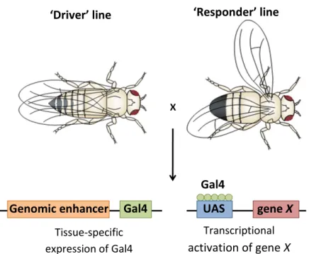
1.4.1 The UAS-Gal4 system
Expression of UAS constructs was performed at 25oC, unless mentioned otherwise, using the Gal4 system [86]. The Gal4 protein is an archetypal eukaryotic transcription factor isolated as an activator of genes responsible for galactose
Fly stocks generated w-; nub-Gal4,FRT42D,apkcts/CyO
w-; FRT42D,apkcts/CyO; UAS-cpaIR#2/TM6B
w-; FRT42D,apkcts/CTG
w-; FRT42D,apkcts/CTG; UAS-cpaIR#2/TM6B
20 metabolism in budding yeast [87]. The Gal4 gene has been widely cloned and used to genetically manipulate expression of genes in foreign organisms. For instance, in Drosophila, several well characterized enhancer sequences have been ‘trapped’ by the Gal4 coding gene to yield spatial-temporal specific drivers of gene expression [86]. Promoter sequences containing a consensus binding site for activating Gal factors, termed UAS sequences, have been introduced in a number of eukaryotic expression vectors, allowing for the production of transgenic constructs that are conditionally expressed upon the availability of Gal4. Thus, in Drosophila, one transgenic line, the ‘driver’, expresses Gal4 in a known temporal and spatial pattern and a second transgenic line, the ‘responder’, contains a UAS-transgene in a transcriptionally silent state due to the absence of Gal4. When ‘responder’ lines are mated to flies expressing the Gal4 driver, the resulting progeny (F1) express the transgene in a pattern that is identical to Gal4 driver expression pattern [88].
Fig. 1 - The UAS-Gal4 system in Drosophila. One of the parental stocks carries the Gal4 gene in close
proximity to a known enhancer of gene expression. This stock can be crossed to another stock containing a UAS-transgene (UAS-gene X), and the resulting progeny will express this transgene in the pattern of expression of the Gal4 factor, which is specified by its associated enhancer region. Adapted from St Johnston, D. (2002) [89].
Gal4 UAS Gal4
gene X Genomic enhancer
‘Driver’ line ‘Responder’ line
Tissue-specific expression of Gal4
Transcriptional
21
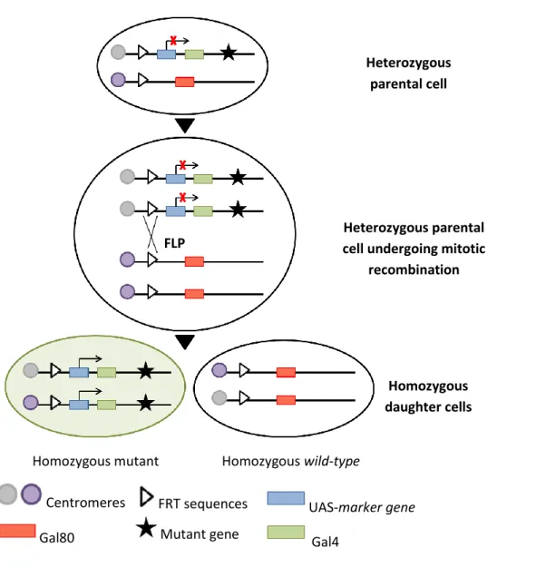
1.4.2 Generation of clones by mitotic recombination
The FLP/FRT system was used to generate mitotic clones to study recessive mutations that would cause embryonic lethality if present in the entire organism [89]. The Flipase (FLP) recombinase, a yeast λ integrase, directs site-specific mitotic recombination through recognition of Flipase Recombination Target (FRT) sites on homologous chromosome arms. Several P-element insertion stocks have been generated in Drosophila, introducing FRT sites close to the centromere of each chromosome as well as a transgene encoding FLP driven by a heat-shock inducible promoter (hsp70). In this sense, mitotic recombination between FRT sites in homologues chromatids occurs following a 1 hour treatment at 37oC of individuals at the stage of interest. As a result, in a heterozygous parental cell, segregation of recombinant chromosomes at mitosis results in the generation of two daughter cells: a homozygous clone for the mutation distal to FRT site and a ‘twin spot’ clone, which is genetically wild-type [90].
In this study, mosaic analysis with a repressible cell marker (MARCM) was used to generate apkcts mutant clones positively marked by GFP, as well as constitutively overexpress UAS-transgenes in marked clones. The MARCM technique is based on the combine use of Gal4 together with its repressor Gal80, providing a way of controlling the UAS-Gal4 system. When Gal80 is removed by FRT-mediated mitotic recombination, Gal4 expression is no longer suppressed and can drive the expression of a certain UAS-marker, like UAS-GFP or another UAS-transgene [90].
The fly stocks containing FLP and FRT sites, used during the course of this work to generate positively marked clones, are indicated in Table 2 and Table 3, and heat-shock was performed at 1st or at 2nd instar larvae in order to analyze whether clones induced at different developmental stages display differences in cell behaviour. Since apkcts allele has different outcomes depending on the temperature, after being heat-shocked, larvae were raised at either 25°C or 28°C or 30°C until late 3rd instar larvae.
22 Table 3 - FLP/FRT system stocks used during the course of this work.
Fig. 2 - Genetic basis of the mosaic analysis with MARCM system. After site-specific mitotic
recombination, a heterozygous parental cell can give rise to two daughter cells in which the chromosome arms distal to the recombination site become homozygous. Driven by a ubiquitous enhancer, Gal80 efficiently suppresses Gal4-dependent expression of a UAS-marker gene. If Gal80, but not Gal4 or UAS-marker gene, is inserted on the chromosome arm carrying the wild-type gene of interest, the daughter cell homozygous for the mutant gene no longer contains Gal80. Therefore, the marker gene can be specifically turned on by Ga4 in homozygous mutant cells. Adapted from Lee, T. & Luo, L. (1999) [91].
FLP/FRT stock Usage Purpose
y-,w-,hsFLP122, UAS-GFP; FRT42D, tub-Gal80; tub-Gal4/ TM6B
To generate clones positively marked by GFP
FLP
Heterozygous parental cell
Heterozygous parental cell undergoing mitotic
recombination
Homozygous daughter cells
Homozygous mutant Homozygous wild-type
Centromeres FRT sequences UAS-marker gene Gal4
23
2. Immunohistochemistry
2.1 Antibody staining
Third instar larvae were dissected in 0,1M phosphate buffer (pH 7,2) and fixed in 4% formaldehyde in PEM (0,1M PIPES pH 7,0; 2mM MgSO4; 1mM EGTA) for 30 minutes on ice. Larvae were then washed and permeabilized 3 times, for 20 minutes each, in PBS 0,2% Triton X-100 (PBT) and incubated with the primary antibodies diluted in PBT, supplemented with 10% donkey serum, overnight at 4oC. The larvae were then washed 3 times, for 20 minutes each, in PBT and incubated in the dark with secondary antibody diluted in PBT 10% donkey serum, for 2 hours at 4oC. Imaginal discs were washed again 3 times for 20 minutes with PBT before mounting between slides and cover-slides in Glycerol 60%. The primary antibodies used were: mouse anti-Arm, (N27A1, 1:10; Developmental Studies Hybridoma Bank (DSHB)); rabbit anti-activated Caspase3 (1:300; Cell Signalling Technology); rabbit anti-aPKC (1:2000; Santa Cruz); rat anti-E-Cad (CAD2, 1:25; DSHB); mouse anti-HA (1:5000; Covance). The secondary antibodies used were Donkey anti-mouse and anti-rabbit conjugated to TRITC or Cy5, (1:200; Jackson Immunoresearch).
2.2 Phalloidin staining
A Phalloidin conjugated dye was used to reveal F-actin. Third instar larvae were dissected, fixed and permeabilized as described in the previous section. Before mounting, tissues were incubated with TRITC conjugated Phalloidin, (1:200; Sigma), in PBT for 8-10 minutes and rinsed 3 times 5 minutes, before mounting in Glycerol 60%.
3. Image Acquisition and Analysis
Fluorescent images of wing imaginal discs were obtained with a Zeiss LSM 510 Meta confocal microscope using a 20x 0.80NA dry objective lens or a 40x 1.3NA oil objective lens. During confocal image acquisition, the detection parameters were
24 adjusted to avoid under- or overexposed pixels, and images were acquired though the full thickness of the wing disc either at 1µm for 20x lens or at 0.5 µm for 40x lens. Images of adult wings were obtained either with a Media Cybernetics EvolutionMP CCD camera attached to a Leica DM LB2 microscope using a 10x dry objective lenses for images at 0.48µm scale or with a DFC 420C CCD camera attached to a Leica MZ75 using 2x dry objective lenses for images at 1.55 µm. Images of adult flies were obtained on a DFC 420C CCD camera attached to a Leica MZ75 using 1.25x dry objective lenses. The ImageJ (NIH) and Imaris v6.4.2 (Bitplane) softwares were used for image processing and analysis, and for reconstructions of orthogonal XZ sections (in depth) and Z-projections.
For quantification of antibody staining, the average fluorescence intensity measured of three non-overlapping 350µm2 area regions in the anterior and posterior wing compartment of maximum Z-projection of the ten most-apical slices. In order to qualitatively evaluate colocalization between fluorophores, binary colocalization masks were obtained with ImageJ, using a cut off point of 60% relative to maximum pixel intensity. Using the same software, quantification of the colocalization was performed using the Mander’s Overlap coefficient (plugin available on ImageJ website), which indicates an overlap of the signals and thus represents the true degree of colocalization [92, 93]. All confocal images used for colocalization analysis were first median filtered (radius 2.0) to reduce background noise and to improve the efficiency of the colocalization analysis.
4. Classification of cell death observed in mosaic analysis
In order to determine whether reducing aPKC, using the apkcts allele, enhances the phenotype of cpa depleted clones, I qualitatively categorized each wing disc based on cell extrusion and anti-activated Caspase 3 staining observed within GFP positive cells : (A) no cell death – wing discs that do not display activated Caspase 3-expressing cells; (B) weak cell death – wing discs showing few activated Caspase 3 positive cells, but the majority of cells marked by GFP are still maintained within the epithelium; (C)
25 medium cell death – wing discs displaying additional Caspase-dependent cell death and most GFP positive cells are located basally; (D) strong cell death – all basal GFP labelled cells express activated Caspase 3.
Fig. 3 – Cell death classification of wing imaginal discs for different experimental conditions where levels of Cpa and/or aPKC were reduced. All panels show basal Z-projections of third wing discs with
clones positively labelled with GFP (green in A-D) and stained with TRITC-Phalloidin to reveal F-actin (red in A-D) and anti-activated Caspase 3, which identifies Caspase-dependent cell death (blue in A-D or grey in A’-D’). Dorsal is downwards.
5. Statistical analysis
A two-sample unpaired Student’s test two-tail was used to test for differences between wild-type wings and apkcts homozygous adult wings. A two-sample paired Student’s test one-tail was used to test for differences either in aPKC or E-Cad or Arm staining between wild-type cells (anterior wing compartment) and cpb depleted cells (posterior wing compartment). All samples followed a normal distribution (Shapiro-Wilk’s normality test, p>0.1) and variances were equal (Levene’s test for equality of variances, p>0.05). Statistical tests were computed with the STATISTICA v9 (StatSoft) programme. A A’ B D’ C’ B’ D C C3 C3 C3 C3 GFP; Phall; C3 GFP; Phall; C3 GFP; Phall; C3 GFP; Phall; C3
26
IV. RESULTS
1. Capping Protein cooperates with aPKC
In order to demonstrate that the genetic interaction between CP and aPKCCAXXDN is due to a reduction of aPKC activity, I investigated whether decreasing aPKC levels using mutant alleles enhances the CP loss of function phenotype. To do so, I used a temperature sensitive allele of aPKC (apkcts) bearing a point mutation in a highly conserved residue of the protein kinase domain, which causes a temperature-dependent reduction of aPKC kinase activity (Guilgur, L.G. and Martinho, R. et al., unpublished data). In contrast to apkc loss of function alleles [15, 79, 80], the use of the apkcts allele (Martinho, R. et al., unpublished data) allowed me to modulate aPKC levels by switching the temperature and thereby gave me the opportunity to determine whether CP interacts genetically with aPKC in maintaining wing epithelial integrity.
1.1 apkcts induces extrusion and death of Cpa-deleted cells in mosaic clones induced in the wing blade
First, I analyzed whether the cpa depletion phenotype was enhanced when cells were also homozygote mutant for the apkcts allele. In this sense, I made use of the MARCM system to induce clones homozygous mutant for apkcts that overexpress UAS-cpaIR#2. Heat-shock was performed in 1st or at 2nd instar larvae in order to analyze whether clones induced at different developmental stages display differences in cell behaviour. Since apkcts allele has different outcomes depending on the temperature, after being heat-shocked, larvae were raised at 25°C, 28°C or 30°C until late 3rd instar larvae. Discs were stained with anti-activated Caspase 3, which identify Caspase-dependent cell death and with TRITC-Phalloidin, to reveal F-actin. This allowed me to determine whether decreasing aPKC levels modulate cell death and F-actin accumulation of cpa depleted cells. It was difficult to obtain either cpaIR#2 or
27
APICAL BASAL APICAL BASAL
25°C
28°C
a
pkc
ts -/-clo
nes
U
AS
cpaR
I
#2clo
n
es
ap
kc
ts -/-; U
AS
cp
aR
I
#2clo
ne
s
C3 C3 C3 C3 C3 C3 A A’ B B’ C C’ D D’ E E’ F F’ G G’ H H’ J J’ K K’ I I’ L L’ Phall Phall Phall Phall Phall Phall GFP; Phall; C3GFP; Phall; C3 GFP; Phall; C3 GFP; Phall; C3
GFP; Phall; C3 GFP; Phall; C3 GFP; Phall; C3 GFP; Phall; C3