aPKC Phosphorylation of HDAC6 Results in
Increased Deacetylation Activity
Yifeng Du1, Michael L. Seibenhener1, Jin Yan2, Jianxiong Jiang3, Michael C. Wooten1*
1Department of Biological Sciences, Cellular and Molecular Biosciences Program, Auburn University, AL, 36849, United States of America,2Graduate Center for Toxicology, Markey Cancer Center, University of Kentucky College of Medicine, Lexington, KY, 40536, United States of America,3James L. Winkle College of Pharmacy, University of Cincinnati, Cincinnati, OH, 45267, United States of America
Abstract
The Class II histone deacetylase, HDAC6, has been shown to be involved in cell motility, aggresome formation and mitochondria transport. HDAC6 deacetylase activity regulatesα -tubulin acetylation levels and thus plays a critical role in these processes. In turn, HDAC6 activity can be regulated by interaction with various proteins including multiple kinases. Ki-nase mediated phosphorylation of HDAC6 can lead to either increased or reduced activity. Our previous research has shown that sequestosome1/p62 (SQSTM1/p62) interacts with HDAC6 and regulates its activity. As SQSTM1/p62 is a scaffolding protein known to interact directly with the zeta isoform of Protein Kinase C (PKCζ), we sought to examine if HDAC6 could be a substrate for PKCζphosphorylation and if so, how its activity might be regulated. Our data demonstrate that HDAC6 is not only present in a protein complex with PKCζbut can also be phosphorylated by PKCζ. We also show that specific phosphorylation of HDAC6 by PKCζincreases HDAC6 deacetylase activity resulting in reduced acetylated tu-bulin levels. Our findings provide novel insight into the molecular mechanism by which HDAC6, PKCζand SQSTM1/p62 function together in protein aggregate clearance. These results also highlight a new research direction which may prove fruitful for understanding the underlying cause of several neurodegenerative diseases.
Introduction
The class II histone deacetylase 6 (HDAC6) has been found to be associated with diverse cellu-lar processes. The deacetylation of multiple targets such as tubulin, hsp90, cortactin, and his-tone by HDAC6 is well documented [1,2]. The best characterized of these interactions is with theα-tubulin subunit of microtubules. HDAC6’s control of the acetylation state of these sub-units is an essential component of cell migration and motility [3,4], aggresome clearance [5], mitochondria transport [6] and dynein associated retrograde transport along microtubules [7]. While it is known that deacetylation ofα-tubulin influences the functional aspects of these processes, it is only recently that the exact modes of HDAC6 regulation are being elucidated. HDAC6 is predominantly localized to the cytoplasm where interaction with direct or indirect a11111
OPEN ACCESS
Citation:Du Y, Seibenhener ML, Yan J, Jiang J, Wooten MC (2015) aPKC Phosphorylation of HDAC6 Results in Increased Deacetylation Activity. PLoS ONE 10(4): e0123191. doi:10.1371/journal. pone.0123191
Academic Editor:Colin Johnson, Oregon State University, UNITED STATES
Received:November 21, 2014
Accepted:March 2, 2015
Published:April 10, 2015
Copyright:© 2015 Du et al. This is an open access article distributed under the terms of theCreative Commons Attribution License, which permits unrestricted use, distribution, and reproduction in any medium, provided the original author and source are credited.
Data Availability Statement:All relevant data are within the paper.
Funding:This work was supported by NIH-2RO1NS033661 (MCW). The funders had no role in study design, data collection and analysis, decision to publish, or preparation of the manuscript.
binding partners results in regulation of HDAC6’s activity [8,9]. For example, direct binding to tau, a microtubule associated stabilizing protein, directly inhibits HDAC6 deacetylase activi-ty leading to impairment of autophagy [10].
In addition to protein-protein interactions affecting its activity, HDAC6 has also been shown to be regulated by post-translational modifications such as phosphorylation. A number of different kinases have been implicated as phosphorylation agents. An EGFR mediated phos-phorylation pathway leads to reduced deacetylase activity of HDAC6 resulting in negatively regulated EGFR endocytosis and degradation [11]. Conversely, HDAC6 phosphorylation me-diated by G protein-coupled receptor kinase 2 (GRK2) and casein kinase 2 (CK2) regulate cell motility and aggresome formation by increasing the deacetylase activity of HDAC6 [4,5]. HDAC6 contains two deacetylase catalytic domains, DD1 and DD2. Interestingly, studies have indicated that phosphorylation sites potentially exist both within the two catalytic domains and outside of them [11–13], suggesting the possibility for indirect regulation of HDAC6’s catalytic activity through allosteric conformation changes [3–6].
Given the multifaceted role played by HDAC6 in cellular function, understanding kinase-mediated regulation of HDAC6 is important. PKCs can serve as important cytoskeleton regula-tors involved in cell polarization, directional sensing, and cell motility [14]. The classical PKC isoform PKCαrecruits and activates HDAC6 by phosphorylation. This interaction modulates HDAC6’s deacetylation ofβ-catenin, enhancing its nuclear translocation and promoter bind-ing [15,16]. Atypical PKC (aPKC) is essential for the regulation of cell polarization, cell motili-ty and migration of macrophages [14]. Recent investigations suggest that the aPKC-aurora A-NDEL1 pathway is crucial for the regulation of microtubule dynamics [17]. Inhibition of aPKC prevents the activation of HDAC6 and stabilizes primary cilia [18]. In addition, two cy-tosolic aPKC isoforms have been shown to phosphorylate AurA, which then targets HDAC6, stimulating tubulin deacetylation in primary cilia [19]. However, to date, there is little evidence indicating any direct association between aPKC and HDAC6. Recently our laboratory reported that the multimeric scaffolding protein SQSTM1/p62 binds to HDAC6, negatively regulating its deacetylase activity and affecting microtubule network equilibrium [20]. SQSTM1/p62 was originally isolated and characterized as an aPKC binding protein [21] with roles defined in ubiquitin binding [22] and cytoplasmic aggregate formation [23,24]. Thus we reasoned that the SQSTM1/p62 binding partner PKCzcould regulate HDAC6 deacetylase activity by direct phosphorylation of the HDAC6 protein. Here we show that HDAC6 is a substrate for PKCzphosphorylation and that the deacetylase activity of HDAC6 is induced by PKCz specific phosphorylation.
Materials and Methods
Cell Culture and Transfection
Human embryonic kidney (HEK) 293 cells and Mouse Embryonic Fibroblast (MEF) from the American Type Culture Collection were grown as described previously [25]. Transfections were achieved using jetPRIME DNA Transfection Reagent (Polyplus Transfection, VWR Inter-national) following the manufacturer’s directions. Cells were grown in DMEM supplemented with 10% Heat-inactivated Fetal Calf Serum (Atlanta Biologicals, Atlanta, GA) and antibiotic/ antimyotic solution (Sigma-Aldrich, St. Louis, MO) at 37°C with high humidity and 5% CO2environment.
Antibodies and Reagents
Antibodies forα-tubulin and acetyl-tubulin were also from Sigma-Aldrich (St. Louis, MO). Texas Red, Cy5 and Oregon Green fluorescent secondary antibodies were from Life Technolo-gies (Carlsbad, CA). aPKC pseudosubstrate inhibitor and Tubucin were purchased from EMD Biosciences (San Diego, CA). Purifed PKCzwas from EMD Biosciences.
Immunoprecipitation and Western Blot Analysis
HEK cells were lysed on tissue culture plates on ice with Triton Lysis Buffer (TLB, 50mM Tris pH 7.5, 150mM NaCl, 10mM NaF, 1mM Na3VO4, 0.5% Triton X-100, 10μg/ml leupeptin, 10μg/ml aprotinin and 1mM PMSF). Protein was determined by Bradford Assay and equal amount of whole cell lysate (500μg) was incubated with 3μg of appropriate primary antibody for 3 hours at 4°C followed by the addition of agarose beads (50μl) specific for the primary an-tibody. Immunoprecipitation was allowed to rotate overnight at 4°C. The immunoprecipitates were then washed five times with TLB. Proteins were released from agarose beads by boiling 2 minutes in SDS-PAGE sample buffer and then separated by 10% SDS-PAGE followed by trans-fer to nitrocellulose membrane and Western blot analysis with the corresponding antibodies. ECL solution (GE Healthcare, Piscataway, NJ) was used to visualize proteins.
Immunoprecipitation Activity Assay
FLAG-tagged HDAC6 was expressed in HEK cells and immunoprecipitated using anti-FLAG antibody as described above in Activity Lysis Buffer (150 mM NaCl, 50 mM Tris, 0.1% TX-100, 2 mM EDTA, 1 mM EGTA, 1 mM PMSF, 25μg/ml leupeptin, 25μg/ml aproti-nin, 1 mM Na3VO4, 2.5 mg/ml PnPP). Washing of the immunoprecipitate was with Activity
Wash Buffer (35mM Tris, 150mM NaCl, 15mM MgCl2, 1mM MnCl2, 0.5mM EGTA, 0.1%
TX100, 25μg/ml leupeptin, 25μg/ml aprotinin). Following the final wash, 0.5μg active re-combinant aPKCzenzyme (EMD Biosciences, San Diego, CA) was added in Activity Assay Buffer (35mM Tris, 15mM MgCl2, 1mM MnCl2, 0.5mM EGTA, 1mM Na3VO4) to the
pel-leted agarose beads along with 5μCi [γ-32P]-ATP (Perkin-Elmer, Akron, OH) and 10μM cold ATP. Phosphorylation reactions were allowed to react for 30 minutes at 30°C with constant agitation. Reactions were stopped by the addition of equal volume 2X Laemmli Sample Buffer and labeled immunoprecipitates were resolved on 10% SDS-PAGE gels. Gels were then fixed in 45% methanol/10% Acetic acid prior to drying and exposure to x-ray film.
Immunofluorescence
Statistical Analysis
For each experimental condition, a minimum of 25 individual cells were photographed and in-tensity values measured using NIS Elements Software AR 3.22.13 [Build 730] (Nikon). Means, standard errors, andt-tests were calculated manually [26].T-test statistics were calculated under the assumption of unequal group variances with one-tailedp-values.P-values less than 0.05 were considered significant.
Results and Discussion
Protein interactions are essential for cellular functions. Many of these interactions are part of signaling pathways that regulate protein activities. Phosphorylation is an important signaling mechanism resulting in modulation of a protein’s activity within a signaling cascade. We have previously shown that the activity of the histone deacetylase protein HDAC6 can be modulated by the scaffolding protein SQSTM1/p62 [20]. SQSTM1/p62, in complex with atypical protein kinase C (aPKC), has also been implicated as an important regulatory module controlling the activation of NF-κB by phosphorylation in stroma cells of cancer patients [27]. The aPKC, PKCz, can positively affect NF-κB at the transcriptional level by specific phosphorylation of its p65 subunit [28] while also displaying a negative regulatory role in IL-6 production in an
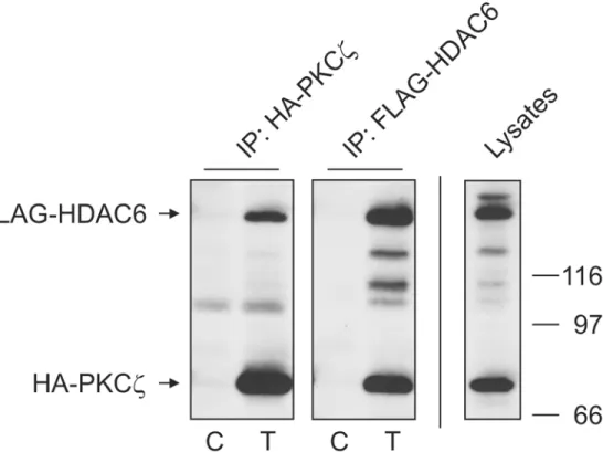
NF-κB independent manner [29,30]. It is well recognized that HDAC6 activity is regulated by phosphorylation. This regulation in turn has direct impact on cell motility [3,31] and clearance of cellular aggresomes [5]. Based on these reports and our previous work, we sought to deter-mine if an aPKC, specifically PKCz, could phosphorylate HDAC6 and regulate its deacetylase activity. Using tagged protein constructs, we exogenously expressed FLAG-HDAC6 and HA-PKCzin HEK cells and examined their interaction by immunoprecipitation (Fig 1). By im-munoprecipitating either tagged protein, we were able to visualize the other as part of the cap-tured immune complex. While not proving direct interaction, these results do indicate that the two proteins can exist as part of the same complex. This raises the possibility that HDAC6 could be regulated by the kinase activity of PKCz.
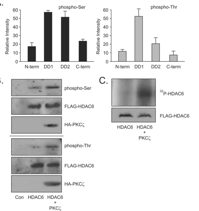
when PKCzwas overexpressed above basal levels (Fig 2B) indicating intracellular modification of the phosphorylation state of HDAC6. This increase in phosphorylation of FLAG-HDAC6 was further confirmed in an immune-complex kinase assay where FLAG-HDAC6 was immunoprecipitated and used as the substrate for purified PKCzγ-32P-ATP. An increase in
32
P- ATP associating with HDAC6 is clearly evident when PKCzwas added to the kinase assay (Fig 2C). Taken together, this data confirmed that HDAC6 was indeed a substrate for PKCz phosphorylation. It also suggests that PKCzphosphorylation sites are predominantly in the conserved DD1 and DD2 catalytic regions of HDAC6. Having shown that PKCzdoes interact in a complex with HDAC6 resulting in specific phosphorylation of HDAC6, we next wished to address if this phosphorylation event had a regulatory effect on the deacetylation activity of HDAC6.
α-Tubulin is a well characterized intracellular substrate of HDAC6 deacetylase activity. Acetylation ofα-tubulin at Lys40 is one characteristic of stable microtubules [33,34] while mistimed deacetylation of microtubules has been linked to neurodegenerative diseases [35]. Usingα-tubulin as a test substrate we have previously shown that the aPKC interacting protein SQSTM1/p62 plays a role in regulation of HDAC6 activity [20]. Thus, we reasoned that acety-lation ofα-tubulin would provide a readout of the phosphorylation effects on HDAC6 by PKCz. Using this model, we examined what effect inhibition of either HDAC6 or PKCzactivity had on acetylated tubulin levels. Non-transfected mouse embryonic fibroblasts (MEF) cells were treated with either the HDAC6 deacetylase activity specific inhibitor tubacin or myristoy-lated aPKC pseudosubstrate inhibitor to see effects of inhibition on endogenous protein
Fig 1. Atypical PKCζexists in a complex with HDAC6.HA-PKCζand FLAG-HDAC6 were transfected into HEK cells and reciprocal immunoprecipitations performed using antibodies for each tag as described in Materials and Methods. Control lanes (C) were transfected with empty plasmid while sample lanes (T) were transfected with both HA-PKCζand FLAG-HDAC6. 40μg of whole cell lystate (Lysate) was loaded to show construct expression in transfected cells. Co-precipitating bands were detected by SDS-PAGE and Western blot with tag-specific antibodies. Results are indicative of 3 independent experiments.
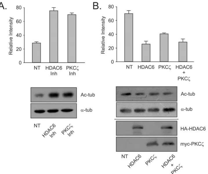
activity. The increase in acetylated tubulin in cells treated with aPKC inhibitor was comparable to that seen when cells were treated with HDAC6 specific inhibitor. Thus, preventing aPKC specific phosphorylation of HDAC6 had a similar effect on HDAC6 deacetylase activity as seen with specific HDAC6 inhibition (Fig 3A). Combined these results indicate that the kinase ac-tivity of PKCzis capable of upregulating the deacetylase activity of HDAC6.
Fig 2. Atypical PKCζphosphorylates HDAC6.(A) GST-deletion constructs of HDAC6 were expressed in HEK cells and captured by GST-pulldown assay. Purified PKCζenzyme was used to catalyze anin-vitrophosphorylation assay using the GST-deletion constructs as substrate. Phosphorylation of each construct was assessed using SDS-PAGE and phospho-specific antibodies for phospho-Serine and phospho-Threonine. Relative changes in
phosphorylation were measured by densitometry and analyzed using UN-SCAN-IT 6.1 Software (Orem, UT). (B) FLAG-HDAC6 was expressed in HEK cells either with or without HA-PKCζexpression.In vivophosphorylation increases were detected using phospho-specific antibodies. (C) FLAG-HDAC6 was expressed in HEK cells and immunoprecipitated with anti-FLAG antibody. The immunoprecipitate was used as a substrate in an immune-complex kinase assay with purified PKCζenzyme as the catalyst. Changes in phosphorylation were detected by incorporation of32P-ATP and autoradiography. All results
are indicative of 3 independent experiments.
With the data now indicating that inhibition of aPKC promotes acetylation ofα-tubulin due to HDAC6 inactivation, the next step was to demonstrate a reverse effect. We predicted that in-creasing either PKCzor HDAC6 would produce lowered tubulin acetylation levels. Increasing the amount of HDAC6 in the cytoplasm of fibroblasts has previously been shown to decrease acetylation ofα-tubulin [20]. When HDAC6 was exogenously overexpressed in HEK cells, the expected drop in acetylated tubulin was observed (Fig 3B). Overexpression of PKCzproduced a similar but less drastic decrease in acetylated tubulin. When PKCzwas co-expressed with HDAC6, the levels of acetylated tubulin present in the cells showed a drop comparable to the lev-els seen with HDAC6 alone (Fig 3B). This outcome was most likely due to the large amount of HDAC6 exogenously expressed in the cells producing a titer that resulted in hiding any extra ac-tivation effect shown by co-expression of PKCz. With tubulin acetylation as a specific readout of HDAC6 activity, we see that increasing levels of PKCzresults in upregulation of the activation
Fig 3. Phosphorylation by atypical PKCζupregulates the deacetylase activity of HDAC6.(A) MEF cells were treated with HDAC6 inhibitor (tubacin—
10μM for 4 hours) or aPKC inhibitor (myristoylated pseudosubstrate—20μM for 16 hours) and HDAC6 endogenous activity was assessed using changes in
acetylated-tubulin levels. A total of 40μg whole cell lysate was loaded in each lane. (B) HA-HDAC6 and myc-PKCζwere expressed separately or in concert in HEK cells and acetylated tubulin levels assessed by SDS-PAGE and Western blotting. Expression blots for lysates were loaded with 40μg lysate protein. Results are indicative of 3 independent experiments.
state of HDAC6 causing a corresponding reduction in acetylation levels ofα-tubulin. This sug-gests that HDAC6 phosphorylation by PKCzdoes result in an increase in deacetylase activity.
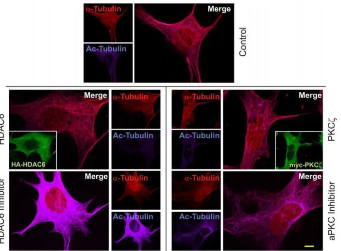
The impact of inhibition or overexpression of PKCzand HDAC6 on acetylation of tubulin was further visualized by immunofluorescence. MEF cells were transfected with tagged HDAC6 or treated with tubacin [12]. Bothα-tubulin and acetyl-tubulin were identified using specific antibodies and visualized with fluorescent tagged secondary antibodies. All images were produced using the same settings on a Nikon A1/T1 confocal microscope so that compar-isons in intensity could be analyzed between different cells. The intensity of the fluorescence from theα-tubulin and acetylated tubulin images were measured using NIS-Elements AR soft-ware (Nikon). Intensity values forα-tubulin were normalized across all samples and subtracted from the acetylated tubulin values of the corresponding panels. This allowed the remaining acetylated tubulin fluorescent intensity to be used as a measure of the activity of HDAC6. Con-trol non-transfected and untreated cells were first analyzed for basal levels ofα-tubulin acetyla-tion (483.55 +/- 23.38). Overexpression of HDAC6 (seen in inset,Fig 4) resulted in low relative
Fig 4. Atypical PKCζregulates HDAC6 deacetylase activity.Parental MEF cells were transfected to overexpress either HA-HDAC6 or myc-PKCζor were treated with HDAC6 inhibitor tubacin (10μM for 4 hours) or aPKC inhibitor (myristoylated pseudosubstrate—20μM for 16 hours). Cells were subsequently
fixed with 4% paraformaldehyde prior to immunofluorescence staining usingα-tubulin antibodies (1:100) detected with Cy5 secondary antibody (1:400) and acetyl-tubulin antibody (1:100) detected with Texas Red secondary antibody (1:400). Texas Red labeled acetyl tubulin was pseudocolored to 400nm using Nikon NIS Elements AR software to visually separate it from Cy5. Inset panels for HA-HDAC6 and myc-PKCζ(HA and myc antibodies used at 1:100) overexpression were detected with Oregon Green secondary antibody (1:400). Images from a minimum of 25 cells each were used for statistical analysis. Scale bar is 10μm.
levels of acetylated tubulin in the cell due to increased deacetylase activity upon the substrate (360.67 +/- 14.22;t(51) = 4.012,p= 0.0004; a representative panel is shown inFig 4). However, when MEF cells are treated with HDAC6 inhibitor, acetylated tubulin fluorescence intensity levels (1987.77 +/- 196.19;t(48) = -9.384,p<0.0001) were strikingly higher being approximate-ly 6-fold greater when compared to the HDAC6 overexpressing cells. Similarapproximate-ly, overexpression of PKCzresulted in low acetylated tubulin intensity values (377.26 +/- 59.49;t(52) = 2.519, p= 0.011) approximately equal to HDAC6 overexpression. Inhibition of PKCzactivity by pseudosubstrate treatment showed greater intensity of acetylated tubulin (624.91 +/- 35.58;t (48) = -4.708,p= 0.0001) with approximately a 2-fold increase over exogenous PKCz expres-sion. This was less than was seen with HDAC6 inhibition. However, it is entirely plausible that PKCzis not the only kinase that activates HDAC6. Thus, even when PKCzphosphorylation of HDAC6 is inhibited, HDAC6 deacetylase activity can still function, though obviously at a re-duced level. These results do however confirm that PKCzlevels in the cell or the activity state of PKCzcan regulate HDAC6 activity.
Multiple functions of HDAC6 have been well documented in the past decade. It is known to be involved in microtubule dynamics and cellular organization associated with neurodegenera-tive diseases and cancer [35,36]. Many kinases have been demonstrated to phosphorylate HDAC6 and regulate its activity. In the light of the results reported herein, we conclude that the aPKC isoform, PKCz, can phosphorylate HDAC6 and upregulate its deacetylase activity. This finding provides a clue into the molecular mechanism linking HDAC6 activity within normal cells to potential roles in neurodegenerative disease and cancer. Our results indicate that further investigations into the viability of using HDAC6 as a target for treatment of neuro-logical diseases may be warranted.
Author Contributions
Conceived and designed the experiments: YD MLS JJ MCW. Performed the experiments: YD MLS JY. Analyzed the data: YD MLS JY JJ MCW. Contributed reagents/materials/analysis tools: MLS MCW. Wrote the paper: YD MLS MCW.
References
1. Hubbert C, Guardiola A, Shao R, Kawaguchi Y, Ito A, Nixon A, et al. (2002) HDAC6 is a microtubule-as-sociated deacetylase. Nature 417: 455–458. PMID:12024216
2. Zhang X, Yuan Z, Zhang Y, Yong S, Salas-Burgos A, Koomen J, et al. (2007) HDAC6 modulates cell motility by altering the acetylation level of cortactin. Mol Cell 27:197–213. PMID:17643370
3. Williams KA, Zhang M, Xiang S, Hu C, Wu J-Y, Zhang S, et al. (2013) Extracellular signal-regulated ki-nase (ERK) phosphorylates histone deacetylase 6 (HDAC6) at serine 1035 to stimulate cell migration. J Biol Chem 288: 33156–33170. doi:10.1074/jbc.M113.472506PMID:24089523
4. Lafarga V, Aymerich I, Tapia O, Mayer F Jr, Penela P (2012) A novel GRK2/HDAC6 interaction modu-lates cell spreading and motility. EMBO J 31: 856–869. doi:10.1038/emboj.2011.466PMID:22193721 5. Watabe M, Nakaki T (2011) Protein kinase CK2 regulates the formation and clearance of aggresomes
in response to stress. J Cell Science 124: 1519–1532. doi:10.1242/jcs.081778PMID:21486957 6. Chen S, Owens GC, Makarenkova H, Edelman DB (2010) HDAC6 regulates mitochondrial transport in
hippocampal neurons. PLoS One 5:e10848. doi:10.1371/journal.pone.0010848PMID:20520769
7. Calderilla-Barbosa L, Seibenhener ML, Du Y, Diaz-Meco M-T, Moscat J, Yan J, et al. (2014) Interaction of SQSTM1 with the motor protein dynein—SQSTM1 is required for normal dynein function and
traffick-ing. J Cell Science 127: 4052–4063. doi:10.1242/jcs.152363PMID:25015291
8. Gao YS, Hubbert CC, Lu J, Lee YS, Lee JY, Yao TP. (2007) Histone deacetylase 6 regulates growth factor-induced actin remodeling and endocytosis. Mol Cell Biol 27: 8637–8647. PMID:17938201 9. Zhou J, Vos CC, Gjyrezi A, Yoshida M, Khuri FR, Tamanoi F, et al. (2009) The protein
farnesyltransfer-ase regulates HDAC6 activity in a microtubule-dependent manner. J Biol Chem 284: 9648–9655. doi:
10. Perez M, Santa-Maria I, Gomez de Barreda E, Zhu X, Cuadros R, Cabrero JR, et al. (2009) Tau—an
in-hibitor of deacetylase HDAC6 function. J Neurochem 109: 1756–1766. doi:10.1111/j.1471-4159.
2009.06102.xPMID:19457097
11. Deribe YL, Wild P, Chandrashaker A, Curak J, Schmidt MH, Kalaidzidis Y, et al. (2009) Regulation of epidermal growth factor receptor trafficking by lysine deacetylase HDAC6. Sci Signal 2: ra84. doi:10. 1126/scisignal.2000576PMID:20029029
12. Haggarty SJ, Koeller KM, Wong JC, Grozinger CM, Schreiber SL (2003) Domain-selective small-mole-cule inhibitor of histone deacetylase 6 (HDAC6)-mediated tubulin deacetylation. Proc Natl Acad Sci USA 100: 4389–4394. PMID:12677000
13. Zou H, Wu Y, Navre M, Sang BC (2006) Characterization of the two catalytic domains in histone deace-tylase 6. Biochem Biophys Res Commun, 34: 45–50.
14. Xiao H, Liu M (2013) Atypical protein kinase C in cell motility. Cell Mol Life Sci 70:3057–3066. doi:10.
1007/s00018-012-1192-1PMID:23096778
15. Chattopadhyay S, Fensterl V, Zhang Y, Veleeparambil M, Wetzel JL, Sen GC (2013) Inhibition of viral pathogenesis and promotion of the septic shock response to bacterial infection by IRF-3 are regulated by the acetylation and phosphorylation of its coactivators. MBio 4: e00636–12. doi:10.1128/mBio.
00636-12PMID:23532979
16. Zhu J, Coyne CB, Sarkar SN (2011) PKC alpha regulates Sendai virus-mediated interferon induction through HDAC6 andβ-catenin. EMBO J 30: 4838–49. doi:10.1038/emboj.2011.351PMID:21952047 17. Mori D, Yamada M, Mimori-Kiyosue Y, Shirai Y, Suzuki A, Ohno S, et al. (2009) An essential role of the aPKC-Aurora A-NDEL1 pathway in neurite elongation by modulation of microtubule dynamics. Nat Cell Biol 11:1057–1068. doi:10.1038/ncb1919PMID:19668197
18. He Q, Wang G, Wakade S, Dasgupta S, Dinkins M, Kong JN, et al. (2014) Primary cilia in stem cells and neural progenitors are regulated by neutral sphingomyelinase 2 and ceramide. Mol Biol Cell 25:1715–1729. doi:10.1091/mbc.E13-12-0730PMID:24694597
19. Pugacheva EN, Jablonski SA, Hartman TR, Henske EP, Golemis EA (2007) HEF1-dependent Aurora A activation induces disassembly of the primary cilium. Cell 129:1351–1363. PMID:17604723 20. Yan J, Seibenhener ML, Calderilla-Barbosa L, Diaz-Meco M-T, Moscat J, Jiang J, et al. (2013)
SQSTM1/p62 interacts with HDAC6 and regulates deacetylase activity. PLoS ONE 8: e76016. doi:10. 1371/journal.pone.0076016PMID:24086678
21. Sanchez P, De Carcer G, Sandoval IV, Moscat J, Diaz-Meco MT (1998) Localization of atypical protein kinase C isoforms into lysosome-targeted endosomes through interaction with p62. Mol Cell Biol 18:3069–80. PMID:9566925
22. Vadlamudi RK, Joung I, Strominger JL, Shin J (1996) p62, a phosphotyrosine-independent ligand of the SH2 domain of p56lck, belongs to a new class of ubiquitin-binding proteins. J Biol Chem 271:20235–20237. PMID:8702753
23. Zatloukal K, Stumptner C, Fuchsbichler A, Heid H, Schnoelzer M, Kenner L, et al. (2002) p62 Is a com-mon component of cytoplasmic inclusions in protein aggregation diseases. Am J Pathol 160:255–263.
PMID:11786419
24. Kuusisto E, Salminen A, Alafuzoff I (2002) Early accumulation of p62 in neurofibrillary tangles in Alzhei-mer's disease: possible role in tangle formation. Neuropathol Appl Neurobiol 28:228–237. PMID:
12060347
25. Wooten MW, Geetha T, Seibenhener ML, Babu JR, Diaz-Meco MT, Moscat J. (2005) The p62 scaffold regulates nerve growth factor-induced NF-kappaB activation by influencing TRAF6 polyubiquitination. J Biol Chem 280: 35625–35629. PMID:16079148
26. Sokal RR, Rohlf FJ (2012) Biometry: WH Freeman New York. 935p.
27. Hiruma Y, Honjo T, Jelinek DF, Windle JJ, Shin J, Roodman GD, et al. Increased signaling through p62 in the marrow microenvironment increases myeloma cell growth and osteoclast formation. Blood 2009; 113: 4894–4902. doi:10.1182/blood-2008-08-173948PMID:19282458
28. Duran A, Diaz-Meco MT, Moscat J (2003) Essential role of RelA Ser311 phosphorylation by zetaPKC in NF-kappaB transcriptional activation. EMBO J 22: 3910–3918. PMID:12881425
29. Leitges M, Sanz L, Martin P, Duran A, Braun U, Garcia JF, et al. (2001) Targeted disruption of the zetaPKC gene results in the impairment of the NF-kappaB pathway. Mol Cell 8: 771–780. PMID:11684013 30. Galvez AS, Duran A, Linares JF, Pathrose P, Castilla EA, Abu-Baker S, et al. (2009) Protein kinase
Czeta represses the interleukin-6 promoter and impairs tumorigenesis in vivo. Mol Cell Biol 29: 104–115. doi:10.1128/MCB.01294-08PMID:18955501
32. Zhang Y, Gilquin B, Khochbin S, Matthias P (2006) Two catalytic domains are required for protein dea-cetylation. J Biol Chem 281: 2401–2404. PMID:16272578
33. Piperno G, LeDizet M, Chang XJ (1987) Microtubules containing acetylated alpha-tubulin in mammali-an cells in culture. J Cell Biol 104: 289–302. PMID:2879846
34. LeDizet M, Piperno G (1987) Identification of an acetylation site of Chlamydomonas alpha-tubulin. Proc Natl Acad Sci U S A 84: 5720–5724. PMID:2441392
35. Li G, Jiang H, Chang M, Xie H, Hu L (2011) HDAC6 alpha-tubulin deacetylase: a potential therapeutic target in neurodegenerative diseases. J Neurol Sci 304: 1–8. doi:10.1016/j.jns.2011.02.017PMID:
21377170