SILICA NANOPARTICLES IN ORAL PEPTIDE DELIVERY FOR DIABETES
MELLITUS CONTROL AND TREATMENT
TESE APRESENTADA PARA OBTENÇÃO DO GRAU DE DOUTOR EM CIÊNCIAS QUÍMICAS E BIOLÓGICAS
Tatiana Andreani
Orientadora: Professora Doutora Eliana B. Souto
Coorientadora: Professora Doutora Amélia M. Lopes Dias da Silva
Composição do Júri:
___________________________________
___________________________________
___________________________________
___________________________________
___________________________________
___________________________________
___________________________________
Vila Real, 2014i
Silica nanoparticles in oral peptide delivery for Diabetes Mellitus control and treatment
Thesis submitted to the Universidade de Trás-os-Montes e Alto Douro in fulfillment of the requirements for the degree of Philosophy Doctor, in the Field of Chemical and Biological Sciences, under the guidance of Professors Eliana B. Souto and Amélia M. Silva
iii
Acknowledgements
I express my deep sense of gratitude and reverence to God for every moment experienced in Portugal and especially for the opportunity of learning during the whole PhD.
I am extremely grateful to my dear parents who support me over the distance and always believe in me. Thanks also to my sisters (Juli and Fabi), so loving and that always gave me motivation to follow my dreams - without all of you, none of this would have been possible.
Special thanks to Marco, my eternal love, for his supreme sacrifice, unwavering support and affection and for giving me optimism to never giving up.
I am grateful to my dear friend Joana Fangueiro for her friendship and for spending countless hours with me writing scientific papers and thesis and mainly for helping me in several down moments in my life.
I similarly extend heartfelt thanks to my friend and collaborators at Universidade Estadual Paulista (UNESP). Thank you – Professor Maria Palmira, Ana Luiza, Charlene, Leonardo and Esteban for receiving me and for acclimating me to the laboratory. Thank you for constant help and valuable advices. Without them would be difficult to carry out this work.
I would like to thank to my colleagues from UTAD and Fernando Pessoa University (UFP), Vânia, Ana Sofia, Bruno and Slavomira for their moral support, help and encouragement.
I would like to thank to my friends at Department of Geology – Mizé, Rui and Paula for our fun lunches at the canteen.
Thanks to technicians and professors at Department of Biology and Environment (UTAD) and at UFP for their help and support during the period of my project.
I would like to thank Lisete Fernandes from Microscopy Unit for her help with TEM images and X-Ray analysis. I also would like to thanks to Professor Verónica Bermudez and Mariana Almeida for their constant help in DSC studies and for interesting discussio ns about sol-gel technology.
Big thanks to Professor Maria Luiza García from Barcelona University, for receiving me in her laboratory and for her invaluable help in my investigation when I really needed it.
iv
encouragement and unending enthusiasm about our work. Their friendship and support made my PhD experience exciting and fruitful, and for this I will be forever grateful.
Finally, I would like to acknowledge the Fundação para Ciência e Tecnologia (FCT, Portugal) for financial support, grating me a PhD scholarship (SFRH/BD/60640/2009). FCT is also acknowledged under the reference PTDC/SAU-FAR/113100/2009 and PEst-C/AGR/UI4033/2011.
v
Resumo
O presente trabalho visou o desenvolvimento de nanopartículas de sílica (SiNP, do inglês “silica nanoparticles”) com a finalidade de explorar o potencial destes sistemas para a administração oral de péptidos, usando a insulina como fármaco modelo.
As SiNP foram produzidas a partir da hidrólise e condensação do silicato de tetraetila (TEOS) pelo método sol-gel e otimizadas usando um desenho fatorial 22. O tamanho médio (Z-Ave) e o índice de polidispersão (PI) das nanopartículas foram influenciados pela concentração de TEOS e pela velocidade de agitação durante a síntese das mesmas. Foram obtidas nanopartículas com 0,43 mol.L-1 de TEOS e uma velocidade de homogeneização de 5000 rpm, revelando um Z-Ave de 256,6 nm e um PI de 0,218. Trealose, manitol e sorbitol foram utilizados como crioprotetores a fim de melhorar a estabilidade das nanopartículas durante a liofilização. Calorimetria diferencial de varrimento (DSC) e raio-X revelaram que as SiNP na presença dos crioprotetores apresentaram uma forma cristalina. No entanto, a trealose foi a mais adequada para a manutenção da integridade das nanopartículas após a sua reconstituição em água, uma vez que este dissacarídeo oferece maior resistência das camadas em torno das nanopartículas, a qual promove maior interação com os grupos SiOH presentes na superfície da sílica.
O revestimento das SiNP com quitosano, alginato de sódio, PEG 6000 e PEG 20000 foi realizado para desenvolver uma formulação destinada à administração oral de insulina por meio da combinação das vantagens das SiNP e das propriedades mucoadesivas desses polímeros. A associação da insulina às nanopartículas foi realizada por adsorção após a produção das mesmas. No caso das nanopartículas revestidas, a insulina foi previamente dissolvida na solução polimérica e posteriormente adicionada às SiNP.
Estudos de DSC revelaram que os picos endotérmicos e exotérmicos dos polímeros puros foram deslocados para temperaturas mais elevadas nas SiNP revestidas, provavelmente devido à interação de ligação de hidrogénio entre os grupos de SiOH das SiNP e os grupos funcionais específicos do PEG (OH), do quitosano (NH) ou do alginato de sódio (COOH). Esta alteração resultou em maior estabilidade das nanopartículas. A difração de raio-X mostrou que as SiNP revestidas exibiram uma estrutura menos ordenada comparado com os polímeros puros. A associação da insulina às nanoparticulas resultou numa estrutura mais cristalina, provavelmente devido à solubilização da insulina nas soluções poliméricas
vi
revestimento das nanopartículas, além das bandas de absorção de amida que são características essenciais dos espectros de proteínas. Análises por dicroísmo circular (CD) demonstraram que a estrutura da insulina foi ligeiramente afetada pelo revestimento durante a síntese das nanopartículas.
Termogramas obtidos por nano DSC mostraram que as SiNP não revestidas podem promover melhor interação com os grupos polares dos liposomas. Além disso, as SiNP e o PEG 6000 foram mais eficazes em proteger a insulina contra a desnaturação térmica.
A mucoadesividade das nanopartículas foi avaliada in vitro pela interação com mucina a pH gástrico e intestinal. SiNP revestidas com quitosano ou alginato apresentaram melhor capacidade de adsorver a mucina, em comparação com as SiNP não revestidas e as SiNP revestidas com PEG (SiNP-PEG 6000).
O revestimento das nanopartículas resultou numa libertação da insulina mais rápida tanto a pH 2,0 como a pH 6,8, comparando com as nanopartículas não revestidas, devido à baixa interação entre a insulina e os grupos SiOH da superfície da sílica. O perfil de libertação de insulina foi afetado apenas pelas SiNP não revestidas e pelas SiNP-PEG 6000 em relação à solução de insulina. Embora as SiNP não revestidas e as SiNP-PEG 6000 tenham provocado redução da libertação de insulina a pH gástrico, 60% da insulina foi libertada após 2 horas de incubação.
Os estudos in vitro de permeação da insulina foram realizados por meio do saco intestinal invertido de ratos, comparando as nanopartículas não revestidas e SiNP-PEG 6000 e SiNP-PEG 20000. No entanto, a presença do PEG não alterou significativamente o comportamento da permeação da insulina na mucosa intestinal de ratos.
As nanopartículas mostraram baixa toxicidade nas linhagens celulares testadas (HepG2 e Caco-2) nas concentrações de 50-500 μg/mL. A incorporação da insulina nas nanopartículas não afetou a viabilidade celular. Doses mais elevadas de SiNP-PEG 6000 apresentaram toxicidade nas linhas celulares selecionadas. Imagens obtidas por microscopia óptica revelaram a presença de vacúolos no interior das células Caco-2, indicando uma possível endocitose dessas nanopartículas.
As SiNP podem ser consideradas como sistemas promissores para a dministração oral da insulina aplicando uma tecnologia simples, de baixo-custo e sem danos ao ambiente.
ix
Abstract
The present work has been focused on the synthesis of silica nanoparticles (SiNP) intended to oral peptide delivery for Diabetes mellitus control and treatment, using insulin as model drug.
SiNP were synthesized from the hydrolysis and condensation of tetraethyl orthosilicate (TEOS) by sol-gel technology. Nanoparticles were optimized using a 22factorial design approach. The mean particle size (Z-Ave) and polydispersity index (PI) were influenced by TEOS concentration and by stirring speed during nanoparticle synthesis. Optimized SiNP were obtained using 0.43 mol.L-1of TEOS and the homogenization speed of 5000 rpm, depicting a Z-Ave of 256.6 nm and a PI of 0.218.
Trehalose, mannitol and sorbitol were used as cryoprotectants to improve SiNP stabilization during lyophilization. Differential scanning calorimetry (DSC) and X-ray studies demonstrated that SiNP in the presence of different cryoprotectans showed a crystalline behavior. However, trehalose was more suitable in maintaining particle integrity after reconstitution of lyophilized nanoparticles in water, due to the higher spatial resistance of its layers around the nanoparticles, leading to stronger interaction with SiOH groups in comparison to mannitol or sorbitol.
After formulation optimization, the next step was to coat SiNP with chitosan, sodium alginate, PEG 6000 and PEG 20000 to develop a system for oral insulin administration by combining the SiNP advantages and the mucoadhesive properties of hydrophilic polymers. Insulin was associated to different nanoparticles by adsorption after nanoparticles production. In the case of coated nanoparticles, insulin was previously dissolved in the polymer solution and then added to SiNP.
Interaction between insulin and nanoparticles was assessed by DSC, X-ray and Fourier-transform infrared (FTIR) studies. DSC showed that endothermic and exothermic peaks of pure polymers were shifted to higher temperatures in all coated SiNP, probably due to the interaction by hydrogen bounds between SiNP SiOH groups and specific functional groups of PEG (OH), chitosan (NH) or sodium alginate (COOH), resulting in more stable nanoparticles. X-ray diffraction showed that coated SiNP displayed less ordered structure compared with pure polymers. The association of insulin to nanoparticles resulted in more crystalline structure due to the insulin solubilization into the polymers solutions leading to the
x spectra.
Insulin secondary structure was assessed by Circular Dichroism (CD) after protein dissolution into polymer solutions during nanoparticle synthesis, showing that insulin structure was slightly affected by coating.
Nano DSC was used to evaluate the interaction between nanoparticles and the biomembrane models (liposomes), and the thermal insulin stability dissolved in the different polymers. The interaction between nanoparticles and liposomes showed that uncoated SiNP could promote higher interaction with the polar head groups of liposomes. Also, uncoated SiNP were more effective in protecting insulin from thermal denaturation.
The nanoparticles mucoadhesive properties were assessed by in vitro interaction with mucin at gastric and intestinal pH. SiNP coated with chitosan or alginate showed better ability in adsorbing to mucin in comparison to uncoated SiNP and PEG-coated SiNP.
It was observed that nanoparticles coated with mucoadhesive polymers resulted in faster insulin release at pH 2.0 and 6.8 in comparison to uncoated nanoparticles due to the low interaction between insulin and SiOH present onto the silica surface. Insulin release profile was only significantly affected by uncoated SiNP and PEG 6000-coated SiNP in comparison to insulin solution. Although, uncoated and PEG-coated SiNP reduced insulin release at gastric pH, 60% of insulin was released at pH 2.0 after 2 h.
In vitro insulin permeation studies were conducted through everted rat intestine
comparing uncoated and PEG-coated SiNP. However, the presence of PEG onto the silica surface did not significantly change the permeation behavior of insulin through the intestinal mucosa.
In general, all nanoparticles showed to be biocompatible, revealing low toxicity in different human cancer cell lines (HepG2 and Caco-2) at tested concentrations (50-500 μg/mL). The presence of insulin did not affect the cell viability significantly. Higher toxicities were observed for PEG 6000-coated SiNP at high concentrations. Microscope images revealed the formation of vacuoles in the Caco-2 cell body, indicating a possible endocytosis of these nanoparticles.
The present work allows the conclusion that coating of silica nanoparticles with mucoadhesive polymers influences their physicochemical properties, insulin release, as well as the cell viability. These studies and findings show the feasibility of applying silica
xi
xiii
Thesis Contents
Acknowledgements ... iii
Resumo ...v
Abstract ... ix
Thesis Contents ... xiii
Figure Captions ...xix
Table Captions……….…..……...xxv
List of Abbreviations………..……….xxix
Chapter I...1
General Introduction ………...1
1. A critical review on development of silica matrices and their biomedical applications ....3
1.1. Sol-gel technology ...3
1.1.1. Silica gel ...4
1.1.1.1. Silica gel and drug release ...4
1.1.1.2. Silica gel for biological species immobilization ...5
1.1.2. Silica nanoparticles ...9
1.1.2.1. Stöber Method ...9
1.1.2.2. Microemulsion technique (polymerization) ... 10
1.1.2.3. Application of silica nanoparticles in biomedicine ... 12
1.1.2.3.1. Cell Imaging ... 12
1.1.2.3.2. Biomolecular and cell separation ... 12
1.1.2.3.3. Controlled drug release ... 14
1.1.2.3.4. Gene therapy ... 17
1.1.3. Multifunctional silica nanoparticles: A new approach in theragnostic nanomedicine………18
1.2. Diabetes mellitus ... 19
1.2.1.1. Incretin hormones ... 23
1.2.1.1.1. Glucagon-like-peptide 1 ... 23
1.2.1.2. Insulin ... 31
1.2.1.2.1. Novel advances for oral insulin delivery ... 32
1.2.1.2.2. Application of nanotechnology for oral insulin delivery ... 35
1.2.1.2.2.1. Liposomes ... 35
1.2.1.2.2.2. Solid lipid nanoparticles ... 35
1.2.1.2.2.3. Polymeric nanoparticles ... 36
xiv
1.5. References ... 41
Chapter II ... 57
Design of silica nanoparticles ... 57
2.1. Introduction... 59
2.2. Materials and methods... 61
2.2.1. Materials ... 61
2.2.2. Experimental factorial design ... 61
2.2.3. Synthesis of silica nanoparticles ... 62
2.2.4. Preparation of freeze dried nanoparticles ... 62
2.2.5. Size and size distribution ... 62
2.2.6. Transmission electron microscope (TEM) analysis ... 63
2.3. Results and discussions ... 64
2.3.1. Effect of TEOS concentration and different HSH speeds ... 64
2.3.2. Analysis of lyophilized nanoparticles ... 69
2.3.3. TEM analysis ... 69
2.5. References ... 71
Chapter III ... 73
Effect of cryoprotectants on reconstitution of silica nanoparticles ... 73
3.1. Introduction... 75
3.2. Materials and methods... 77
3.2.1. Materials ... 77
3.2.3. Preparation of freeze-dried nanoparticles ... 77
3.2.4. Size and size distribution ... 78
3.2.5. Atomic force microscopy (AFM) studies ... 78
3.2.6. Thermal analysis ... 79
3.2.7. X-ray diffraction (XRD) analysis... 79
3.3. Results and discussions ... 80
3.3.1. Size and size distribution ... 80
3.3.2. AFM studies ... 82 3.3.3. DSC analysis ... 83 3.3.4. XRD analysis ... 85 3.4. Conclusions... 88 3.5. References ... 89 Chapter IV ... 91
xv
4.2. Materials and methods... 96
4.2.1. Materials ... 96
4.2.2. Synthesis of nanoparticles ... 96
4.2.3. Particle size and zeta potential analysis ... 97
4.2.4. Association efficacy ... 97
4.2.5. Differential scanning calorimetry (DSC) analysis ... 97
4.2.5. Fourier transform infrared (FTIR) analysis ... 98
4.2.6. X-ray diffraction (XRD) analysis... 98
4.3. Results and discussions ... 99
4.3.1. Size and zeta potential analysis ... 99
4.3.2. Association Efficacy (AE) ... 100
4.3.3. DSC analysis ... 101 4.3.4. X-ray analysis ... 105 4.3.5. FTIR analysis ... 106 4.4. Conclusions... 108 4.5. References ... 109 Chapter V ... 113
Coated silica nanoparticles for mucosal adhesion ... 113
5.2. Materials and methods... 117
5.2.1. Materials ... 117
5.2.2. Synthesis of nanoparticles ... 117
5.2.3. Size and zeta potential analysis ... 118
5.2.4. Association efficacy (AE)... 118
5.2.5. Analysis of insulin secondary structure ... 118
5.2.6. Thermal analysis of insulin ... 119
5.2.7. Interaction studies between nanoparticles and biomembrane models ... 119
5.2.8. In vitro interaction between mucin and different nanoparticles ... 119
5.3. Results and discussions ... 120
5.3.1. Size and zeta potential analysis ... 120
5.3.2. Association Efficacy (AE) of insulin ... 120
5.3.3. Analysis of insulin secondary structure ... 121
5.3.4. Thermal analysis of insulin ... 123
5.3.5. Interaction studies between nanoparticles and biomembrane models ... 125
xvi
Chapter VI ... 135
In vitro insulin release and permeation from nanoparticles ... 135
6.1. Introduction... 137
6.2. Materials and methods... 139
6.2.1. Materials ... 139
6.2.2. Synthesis of nanoparticles ... 139
6.2.3. Apparatus and chromatography system... 140
6.2.4. Insulin assay by high performance liquid chromatography (HPLC) ... 140
6.2.5. In vitro insulin release from nanoparticles ... 140
6.2.6. Release profile analysis ... 141
6.2.7. Circular dichroism (CD) ... 141
6.2.8. Permeation studies through everted rat intestine ... 142
6.2.9. Morphological studies ... 142
6.2.10. Statistical analysis ... 142
6.3. Results and discussions ... 143
6.3.1. In vitro insulin release and modeling ... 143
6.3.2. Circular dichroism (CD) ... 148
6.3.3. Permeation studies through everted rat intestine ... 150
6.3.4. Morphological studies ... 151
6.4. Conclusions... 152
6.5. References ... 153
Chapter VII ... 155
Comparative cytotoxicity response of nanoparticles ... 155
7.1. Introduction... 157
7.2. Materials and methods... 159
7.2.1. Materials ... 159
7.2.3. Synthesis of nanoparticles ... 159
7.2.4. Cell cultures and maintenance ... 160
7.2.5. In vitro cytotoxicity assay ... 160
7.2.6. Evaluation of alterations on cell morphology ... 161
7.2.7. Statistical analysis ... 161
7.3. Results and discussions ... 162
7.4. Conclusions... 169
xvii
8.1. Conclusion remarks of current study ... 175
Chapter IX ... 179
Supplements ... 179
Supplement 1: Bradford method for insulin quantification ... 181
Supplement 2: HPLC validation for insulin assay ... 183
Chapter X ... 189
Publication list ... 189
xix
Figure Captions
Chapter I- General Introduction……….1
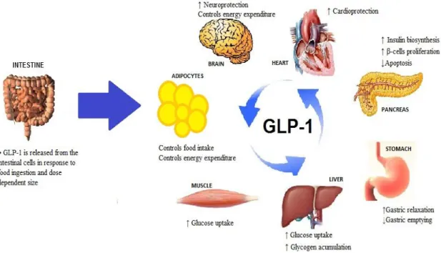
Figure 1.1. Immobilization procedures for cell attachment in sol-gel matrices. The hydrolysis and condensation of silica precursors is carried out at low pH, using HCl as catalytic agent leading to the sol formation. The pH of sol is neutralized and then the cells are added to sol, causing rapid gelation and cell entrapment within a silica-gel structure ...9 Figure 1.2. Schematic procedure of silica growth in W/O microemulsion technique. (1) Sol-gel solution composed by silica precursor, water and ammonium. (2) Surfactant in organic solvent. (3) Emulsification between the organic phase and the aqueous phase in order to prepare the microemulsion. (4) Compartmentalization of sol-gel within water droplets ... 11 Figure 1.3. Structure and mechanism of GMNC. GMNC is constituted by magnetic nanoparticles, silica coating, avidin-biotin linkage and a molecular beacon. Molecular beacon consists in a loop of nucleic acid with a fluorophore and a quencher group attached at opposite ends. When the beacon is a closed loop mode, the quencher and the fluorophore are held together which causes the quenching of the fluorescent emission of the dye. If the target molecules (DNA or RNA) are complementary to the strand in loop, molecular beacon hybridizes resulting in an opened loop shape and consequently the separation of quencher and fluorophore leads to the fluorescence detection. ... 14 Figure 1.4. GLP-1 release and its main action on several target organ. GLP-1 is released from the intestinal A-cells in response to food ingestion and dose-dependent on meal size. Through the activation of GLP-1 receptors, it slows gastric emptying, induces gastric relaxation and satiety. It induces insulin biosynthesis and secretion, pancreatic beta cell proliferation and reduces cytotoxic-induced apoptosis. It exerts protection of the cardiovascular and neuronal system. GLP-1 acts on the adipocytes and brain centers to control food intake and energy expenditure. It increases glucose uptake by muscle and liver cells. ... 24
xx
intact GLP-1, producing inactive fragments of GLP-1; b) DDP-IV inhibitors inactivate the enzyme and c) active GLP-1 bind to incretin receptors of pancreatic β-cell, increasing the insulin release, the proliferation of β-cell and decreasing of glucagon secretion ... 26
Chapter II- Design of silica nanoparticles………57
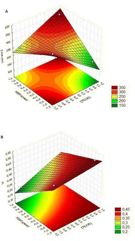
Figure 2.1. Pareto chart of the Z-Ave (A) and PI (B) for silica nanoparticles...66 Figure 2.2. Surface response chart of the effect of TEOS concentration and HSH speed on Z-Ave (A) and PI (B) of SiNP. ... 68 Figure 2.3. TEM images of optimized SiNP synthesized by HSH after freeze-drying process in the absence of cryoprotectants agents. ... 69
Chapter III- Effect of cryoprotectants on reconstitution of silica nanoparticles………..73
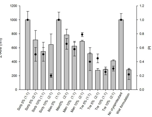
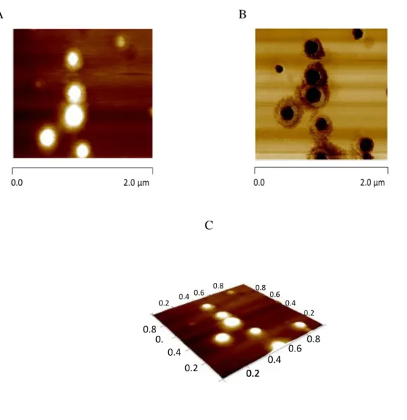
Figure 3.1. Z-Ave (nm) ± SD and PI ± SD (●) of SiNP in the presence of different cryoprotectants. The % (w/v) as well as the ratio cryoprotectants:nanoparticles (v/v) are indicated for each cryoprotectant agents. Sorb: sorbitol; Man: mannitol; Tre:trehalose……..82 Figure 3.2. Morphological characterization of SiNP in the presence of trehalose 10 % (1:1, v/v) Morphological characterization of SiNP in the presence of trehalose 10 % (1:1, v/v). AFM height image (A), phase image (B) and 3D image (C)... 83 Figure 3.3. DSC patterns of the freeze-dried SiNP in the presence of different cryoprotectant agents (10 %, 1:1). The superposition of the 3 DSC curves of SiNP and different cryoprotectants (B). ... 84 Figure 3.4. X-ray diffraction patterns of the cryoprotectant agents alone and of their mixture with silica nanoparticles after freeze-drying process. ... 86
xxi
SA……...103 Figure 4.2. Thermograms of (a) chitosan, (b) SiNP-CH and (c) Ins-SiNP CH………...103 Figure 4.3. Thermograms of (a) PEG 6000, (b) SiNP-PEG 6000 and (c) Ins- SiNP-PEG
6000……….104 Figure 4.4. Thermograms of (a) PEG 20000, (b) SiNP-PEG 20000 and (c) Ins- SiNP-PEG 20000………...104 Figure 4.5. XRD patterns of (a) Ins-SiNP-SA, (b) SiNP-SA and (c) sodium alginate………....105 Figure 4.6. XRD patterns of (a) Ins-SiNP-CH, (b) SiNP-CH and (c) chitosan………..105 Figure 4.7. XRD patterns of (a) Ins-SiNP-PEG, (b) SiNP-PEG 6000 and (c) PEG 6000…..105 Figure 4.8. XRD patterns of (a) Ins-SiNP-PEG 20000, (b) SiNP-PEG 20000 and (c) PEG
20000………...105 Figure 4.9. FTIR spectra of (a) Ins-SiNP-CH, (b) Ins-SiNP-SA, (c) Ins-SiNP-PEG 20000, (d) Ins-SiNP-PEG 6000 and (e) Ins-SiNP………107
Chapter V- Coated silica nanoparticles for mucosal adhesion……….113
Figure 5.1. Far-UV CD spectra of native insulin in PBS (black line), insulin associated to PEG 6000 (black dotted line) and insulin associated to PEG 20000 (grey line)………121 Figure 5.2. Far-UV CD spectra of native insulin in HCl/KCl buffer (black line), insulin associated to sodium alginate (black dotted line) and insulin associated to chitosan (grey line) ... 122 Figure 5.3. DSC transition profiles of insulin associated to PEG 6000 (a), to SA (b), to CH (c), to SiNP (d) and human insulin solution (e). ... 124
xxii
(curve c) SiNP-AS (curve d) and in the absence of nanoparticles (curve e). ... 126 Figure 5.5. DSC curves of DPPC liposomes in contact with SiNP in different scans (a) 1 scan, (b) 2 scan, (c) 3 scan, (d) 4 scan and in the absence of nanoparticles (e). ... 127 Figure 5.6. Influence of mucin on the nanoparticle ZP. Different concentrations of mucin in
g/mL: 0 (white bars), 100 (light-grey bars), 250 (dark-grey bars) and 500 (black filled bars),and several coating agents, as denoted in the graph, at two different pH environment, pH 2.0 (A) and pH 6.8 (B). Measurements were performed after 30 min incubation (mean ± SD, n=3)……….129
Chapter VI- In vitro insulin release and permeation from nanoparticles………...135
Figure 6.1. Cumulative release profiles of insulin from different nanoparticles. Experiments were conducted in HCl/KCl buffer at pH 2.0 (A) and in PBS at pH 6.8 (B) (n = 6 ± SD)…144 Figure 6.2. Far-UV CD spectrum of native insulin (black line), insulin released from SiNP-PEG 6000 (black dotted line) and SiNP-SiNP-PEG 20000 (grey line) at pH 2.0 (A) and pH 6.8 (B) buffers. ... 149 Figure 6.3. The influence of SiNP and PEG-coated SiNP on insulin permeation from everted gut sacs incubated in TC-199 buffer over 60 min (n = 6 ± SD). ... 150 Figure 6.4. Morphological characterization of SiNP (left) and Ins-SiNP coated with PEG 6000 (right). ... 151
Chapter VII- Comparative cytotoxicity response of nanoparticles………..155 Figure 7.1. Viability of Caco-2 (leftmost columns) and HepG2 (rightmost columns) cells after 48 h exposure to 50, 100, 200 and 500 μg/mL of uncoated SiNP, unloaded (light grey bars) and insulin-loaded (dark grey bars). Cell viability is expressed as % of control (untreated cells). For each cell line, three independent experiments (each with 8 replicates) were carried out………163
xxiii
alginate (b). Cell viability is expressed as % of control (untreated cells). For each cell line, three independent experiments (each with 8 replicates) were carried out. ... 165 Figure 7.3. Viability of Caco-2 (leftmost columns) and HepG2 (rightmost columns) cells after 48 h exposure to 50, 100, 200 and 500 μg/mL of SiNP coated with PEG 6000 (a) and SiNP coated with PEG 20000 (b). Cell viability is expressed as % of control (untreated cells). For each cell line, three independent experiments (each with 8 replicates) were carried out. ... 167 Figure 7.4. Appearance of Caco-2 cells observed under optical inverted microscope. (A) control cells and (B) cells incubated with SiNP-PEG 6000 at 500 µg/mL (amplification 400x) ... 168
Chapter IX- Supplements……….179
Figure S2.1. Representative chromatogram of insulin at 2 μg/mL in HCl/KCl buffer (pH 2.0)………..184 Figure S2.2. Representative chromatogram of insulin at 2 μg/mL in PBS (pH 6.8). ... 184 Figure S2.3. Representative chromatogram of insulin at 2 μg/mL in TC-199 buffer (pH 7.2) ... 184 Figure S2.4. Representative chromatogram of HCl/KCl buffer (pH 2.0). ... 185 Figure S2.5. Representative chromatogram of PBS (pH 6.8). ... 185 Figure S2.6. Representative chromatogram of TC-199 without lactose buffer (pH 7.2). ... 185
xxv
Table Captions
Chapter I- General Introduction……….1
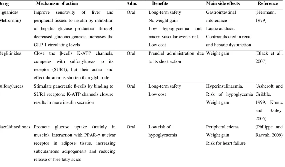
Table 1.1. Relevant achievements in biosensing from silica sol-gel matrices………7 Table 1.2. Encapsulation of cells into silica matrices……….8 Table 1.3. Examples of drugs incorporated in silica nanoparticles and their properties……..16 Table 1.4. Anti-hyperglycemic drugs available, respective mechanism of action, administratio administration route (Adm) and main side effects………21 Table 1.5. Drugs on the market and under development for GLP-1 therapies, the GLP-1 receptors agonist and DDP-IV inhibitors………..28 Table 1.6. Oral systems for insulin delivery that are currently being investigated in different clinical phases………...34
Chapter II- Design of silica nanoparticles………57
Table 2.1. Initial 22 factorial design, providing the lower (−1), upper (+1) and central point (0) level values for each variable. ... 61 Table 2.2. Influence of TEOS (mol.L-1) and HSH speed (rpm) on the formation of silica nanoparticles. ... 64 Table 2.3. Analysis of the Z-Ave by ANOVA statistical test. ... 65 Table 2.4. Analysis of PI by ANOVA statistical test. ... 67
xxvi
Table 3.1. Controlled parameters applied for the synthesis of nanoparticles...77 Table 3.2. DSC parameters for cryoprotectant agents and their mixture with SiNP………...85
Chapter IV- Coating of silica baboparticles with mucoadhesive polymers………...91 Table 4.1. Physicochemical properties of different insulin-associated nanoparticles………..99 Table 4.2. DSC parameters of the polymers and unloaded and loaded-nanoparticle produced by sol-gel technology... 102
Chapter V- Coated silica nanoparticles for mucosal adhesion………...113
Table 5.1. Z-Ave, PI and ZP and EA of different insulin-associated nanoparticles………....……...120 Table 5.2. Observed parameters of secondary structure of insulin dissolved in PBS, PEG 6000 or PEG 20000. ... 122 Table 5.3. Observed parameters of secondary structure of insulin dissolved in HCl/KCl buffer, chitosan and sodium alginate. ... 123 Table 5.4. Calorimetric parameters of human insulin and insulin associated to SiNP, SA, CH and PEG 6000. ... 125 Table 5.5. Calorimetric parameters of DPPC liposomes in the presence of different nanoparticles. ... 127 Table 5.6. Calorimetric parameters of DPPC liposomes in the presence of SiNP at different scans... 128
xxvii
Table 6.1. MRT and half-life for insulin solution, Ins-SiNP and Ins-SiNP-PEG 6000 at gastric pH………145 Table 6.2. MRT and half-life for insulin solution, Ins-SiNP and Ins-SiNP-PEG 6000 at intestinal pH………145 Table 6.3. Mathematic modeling for insulin release at gastric and intestinal conditions………....147 Table 6.4. Observed parameters of secondary insulin solution structure and of insulin release from SiNP-PEG 6000 and SiNP-PEG 20000 at pH 2.0………..148 Table 6.5. Observed parameters of secondary insulin solution structure and of insulin release from SiNP-PEG 6000 and SiNP-PEG 20000 at pH 6.8………..148
Supplements………...……179
Table S2.1. Linear regression equations obtained from validation process. ... 183 Table S2.2. Results of precision test for insulin from standard concentrations in HCl/KCl (pH 2.0), PBS (pH 6.8) and in TC-199 without lactose (pH 7.2) buffers (n = 3). ... 186 Table S2.3. Validation parameters for insulin quantification in HCl/KCl buffer (pH 2.0). ... 187 Table S2.4. Validation parameters for insulin quantification in PBS (pH 6.8). ... 187 Table S2.5. Validation parameters for insulin quantification in TC-199 buffer (pH 7.2). ... 188
xxix
List of Abbreviations
Adm Administration
AE Association efficacy
AFM Atomic force microscopy
AIC Akaike information criteria
APC Allophycocyanin
APDMOS 3-aoryloxypropyl) dimethoxymethylsilane
APS Adaptor molecules containing PH and SH2 domains
ATP Adenosine triphosphate
AUC Area under curve
BMI Body mass index
BSA Bovine serum albumin
CBLC Cas-Br-M (murine) ecotropic retroviral transforming sequence c
CH Chitosan
CD Circular dichroism
CTAB Cetyltrimethyl-ammonium bromide
DGS Diglyceroxysilane
DLS Dynamic light scattering
DNA Deoxyribonucleic acid
DPP-IV Dipeptidyl peptidase-IV
DPPC Dipalmitoylphosphatidylcholine
DRG Dorsal root ganglia
DSC Differential scanning calorimeter
EDTA Diaminoethanetetraacetic acid
FDA Food and Drug Administration
FITC Fluorescein isothiocyanate
FITR Fourier-transform infrared
FP Farmacopéia portuguesa
xxx
GIP Glucose-dependent insulinotropic polypeptide
GIT Gastrointestinal tract
GLP-1 Glucagon-like peptide 1
GMNC Genomagnetic nanocapturers
GPTMS Glycidoxypropyl trimethoxysilane
h Hour(s)
HAS Human serum albumin
HbHNL Hydroxynitrile lyase
HIM2 Hexyl-insulin monoconjugate
HIV Human immunodeficiency virus
HP55 Hydroxypropyl methylcellulose phthalate HPLC High performance liquid chromatography HPMCP Hydroxypropyl methylcellulose phthalate
HRP Horseradish perosidase
HSH High shear homogenization
HVD-I Hepatic-directed vesicle insulin
ICH International Conference on Harmonization Ins-CH Insulin dissolved in chitosan solution
Ins-HCl/KCl Insulin dissolved in HCl/KCl buffer
Ins-PBS Insulin dissolved in Phosphate-buffered saline Ins-PEG 6000 Insulin dissolved in PEG 6000 solution
Ins-PEG 20000 Insulin dissolved in PEG 20000 solution Ins-SA Insulin dissolved in sodium alginate solution Ins-SiNP Insulin associated to silica nanoparticles
Ins-SiNP-CH Insulin associated to silica nanoparticles coated with chitosan Ins-SiNP-PEG
6000
Insulin associated to silica nanoparticles coated with PEG 6000
Ins-SiNP-PEG 20000
Insulin associated to silica nanoparticles coated with PEG 20000
Ins-SiNP-SA Insulin associated to silica nanoparticles coated with sodium alginate IRS Insulin receptor substrate
xxxi
µDSC Micro differential scanning calorimetry
MPS Mononuclear phagocyte system
MRI Magnetic resonance image
MSCs Mesenchymal stem cells
MRT Mean release time
MSiNP Mesoporous silica nanoparticles
MWNT Multi-walled carbon nanotubes
nDSC Nano differential scanning calorimetry
NIR Near infra-red
PACA Poly(alkyl cyanocrylate)
PBS Phosphate-buffered saline
PC Phosphatidylcholine
PCL Poly(ε-caprolactone)
PCS Photon correlation spectroscopy
PDGF Platelet Derived Growth Factor
pDNA Plasmid DNA
PEG Poly(ethylene glycol)
PEI Polyethlyenimine
γ-PGA Poly(γ-glutamic acid)
PH Pleckstrin homology
PI Polydispersity index
PLA Poly(lactide acid)
PLE Poly(lactic acid-co-ethylene oxide)
PLGA MS Poly (D, L-lactic-co-glycolic acid) microspheres
PLGA-PEG-PLGA
Poly [(DL-lactide-co-glycolide)-b-ethylene glycol-b-(DL-lactide-coglycolide)]
PPAR-γ Peroxisome proliferator-activated receptor-gamma
QD Quantum dots
RES Reticulo-endothelial system
RITC Rodhamine B isothiocyanate
xxxii
SD Standard deviation
SH Shc
SiNP Silica nanoparticles
SiNP-Sorb Silica nanoparticles lyophilized with sorbitol SiNP-Man Silica nanoparticles lyophilized with mannitol SiNP-Tre Silica nanoparticles lyophilized with trehalose SiNP-CH Silica nanoparticles coated with chitosan SiNP-PEG 6000 Silica nanoparticles coated with PEG 6000 SiNP-PEG 20000 Silica nanoparticles coated with PEG 20000 SiNP-SA Silica nanoparticles coated with sodium alginate
SiRNA Small interfering RNA
SLN Solid lipid nanoparticles
SNAC Sodium N-(8-[2-hydroxybenzoyl]-amino) caprylic acid)
S/O/O Solid-in-oil-in-oil
SUR1 Sulfonylurea receptor 1
T2DM Type 2 diabetes mellitus
TEOS Tetraethyl ortosilicate
Tg Glass transition temperature
TEM Transmission electron microscope
TFA Trifluoroacetic acid
TL Tomato lectin
Tm Transition temperature
TMC Trimethyl chitosan
TMOS Tetramethyl ortosilicate
UEA1 Ulex europaeus agglutinin 1
UI Under investigation
UV Ultraviolet
VEGF Vascular endothelium growth factor
XRD X-ray diffraction
Z-Ave Mean particle size
xxxiii
Chapter I
General Introduction
3
1. A critical review on development of silica matrices and their biomedical
applications
1.1. Sol-gel technology
The sol-gel technology, for a long time, has been used for synthesizing a variety of silica materials, either crystalline or amorphous of controlled porosity, as well as fibers (Turinske et al., 2013), films (Ghach et al., 2013), glasses (Ferrer et al., 2002) and nanoparticles (Deng et al., 2005) due to its ability to form products at mild conditions. This technology involves hydrolysis and condensation of metal alkoxysilanes precursors (Si(OR)4),
such as tetraethyl orthosilicate (TEOS), tetramethyl orthosilicate (TMOS) or inorganic salts, such as sodium silicate (Na2SiO3) under acidic or alkaline conditions. The hydrolysis reaction
of TEOS can be expressed by the following formulae:
Si(OC2H5)4 + H2O → Si(OC2H5)3OH + C2H5OH
The hydrolysis of TEOS molecules forms silanol groups (SiOH). The condensation between the silanol groups or between silanol groups and alkoxysilanes precursor can lead to particles with siloxane bonds (Si-O-Si). Examples of condensation reactions are given bellow:
Water condensation 2Si–(OH)4 → 2 SiO2 + 4 H2O
Alcohol condensation
Si-(OH)4 + Si-(OR)4 → SiO2 + C2H5OH
Due to the low partial positive charge (δ) of silicon, the introduction of catalytic agents for nucleophilic attack is required. The catalyst most used in sol-gel process is mineral acids or ammonium, but neutral salts and transition metal alkoxides have also been employed.
4
1.1.1. Silica gel
1.1.1.1. Silica gel and drug release
Sol-gel technology provides several alternatives to synthesize various kinds of silica gel, which structure depends on many parameters, such as type and concentration of precursors, water to precursor ratio, nature and concentration of catalyst agent, solvent, temperature, as well as aging and dry processes (Iller, 1979; Jafarzadeh et al., 2009; Rahman et al., 2004; Sato et al., 1990).
The silica gels obtained by sol-gel process are usually classified according to the dispersion medium used in the gel preparation. Hydrogel consists of hydrophilic polymer networks with three dimension configuration enabling the imbibing of high amounts of water or biological fluids (Gehrke and Lee, 1990; Peppas and Mikos, 1986). In contrast, an alcogel is a colloidal gel which alcohol is the dispersion medium. After gel preparation, gel aging and drying can lead to the production of a xerogel or an aerogel. A xerogel is based on a solid from gel by drying at ambient pressure. This kind of drying usually results in shrinkage of the network, and further expelling of the liquid from the pores. The shrinkage mechanism is generally attributed to the new bond formation through condensation reaction.
Alternatively, when solvent removal occurs under supercritical conditions, the surface tension between liquid and vapor can be avoided and an aerogel is produced without porous collapse (Mohanan et al., 2005). Aerogels consist of pearl-necklace–like network of particles which 99 % of their bulk volume is empty (Pierre and Pajonk, 2002).
Although sol-gel based silica xerogels and aerogels have been exhaustively investigated as drug delivery carriers (Caputo et al., 2012; Costache et al., 2013), the release mechanisms of drugs from the gels have not been elucidated. Data demonstrate that the control release can be based on simple diffusion or a combination of the diffusion and erosion process (Ritger and Peppas, 1987; Wu et al., 2005). These processes are affected by several aspects, such as, molecular size and chemical characteristics of the drug, the matrix size, pH, interactions between the drug and the matrix and the texture of the gel (Prokopowicz, 2009). The hydrolysis and condensation reactions can be influenced by water/alkoxide increasing the surface area that can control and sustain the release of encapsulated compounds (Tan et al., 1996).
5
Recently, an interesting pH responsive sol-gel hybrid hydro-xerogel for drug release of doxorubicin was reported by Angelopoulou and co-workers (Angelopoulou et al., 2012). For this purpose, the authors used a Si/Ca or Mg based-xerogel with a dextran hydrogel. The hybrid-xerogel showed good apatite deposition properties. For doxorubicin, the release was more pronounced under low pH, reaching ~70 % of drug released after 180 h. In contrast, at neutral pH, only 20 % of doxorubicin was released.
1.1.1.2. Silica gel for biological species immobilization
Enzyme-based electrode has been widely studied for developing sensitive and selective biosensors. The traditional method of enzyme immobilization consists in non-covalent or non-covalent attachment (Guibault, 1984). However, biosensor development for enzyme immobilization may be limited due to the lack of simple and generic protocols. Thus, a simple and low cost method to immobilize and stabilize enzymes is required.
Sol-gel technology has been used for encapsulating a large number of proteins and cells. The produced sol-gel glasses can protect the biomolecules from denaturation, maintaining their activities, as well as can permit the growth and proliferation of entrapped cells. However, several factors could affect the bio species encapsulation in sol-gel matrices (Carturan et al., 2004; Kuncová et al., 2004). The hydrolysis and condensation of silicon alkoxides lead to the release of alcohol molecules as byproduct, being a potential obstacle for enzyme and cell encapsulation. Many approaches have been adopted to avoid the presence of alcohol. One way, is the alcohol removal via evaporation under vacuum. The alcohol-free route has been used for encapsulating horseradish perosidase (HRP), hydroxynitrile lyase (HbHNL) or allophycocyanin (APC) (Macmillan et al., 2009). However, the removal of alcohol is not complete, since its release can proceed for prolonged periods. Thus, aqueous sol-gel route using sodium silicate as precursors has been developed to avoid the generation of alcohol. The encapsulation is carried out at room-temperature and neutral pH, minimizing the degradation of biomolecules. HRP and glucose-6-phosphate dehydrogenase (G6PDH) were successfully encapsulated in this aqueous route processing (Bhatia et al., 2000). The use of biocompatible alcohols such as polyol-based silanes can also avoid denaturation by alcohol. The stability of entrapped enzymes is improved, since glycerol is produced under hydrolysis reaction. Recently, the use of diglyceroxysilane (DGS) as precursor and poly(ethylene glycol) (PEG) as stabilizer agent has been employed for HRP immobilization in
6
monodisperse spherical silica nanoparticles. The large PEG molecular weights lead to better steric stabilization and more monodisperse particles (Voss et al., 2007).
Glucose biosensor analysis was developed by combining immobilized glucose oxidase in silica gel with an oxygen sensing film using sol-gel technology (Chang et al., 2010). Even in the presence of high concentrations of ascorbic acid or chloride ion, no interference was detected and the activity of the enzyme was preserved after 2 months of storage. In addition, the evaluation of glucose concentrations was conducted using a 96-well plate and a fluorescence plate reader that permit an optimization of the tests in a laboratory scale.
Table 1.1 summarizes examples of various enzymes entrapped in silica sol-gel matrices for biosensor application.
Another exciting research area is the development of sol-gel based matrices for tissue derived cell growth aiming at cell therapy and at living organism immobilization. However, active cell immobilization still remains a challenge. Cells are more sensitive than biomolecules, requiring material with high biocompatibility, as well as appropriate immobilization methods. In addition, nutrients, oxygen and degradation products should be easily diffused by the material porous (Meunier et al., 2010). Hence, sol-gel matrices can also act as template allowing the growth and proliferation of the host cells and can be remodeled in a living tissue, as well as promote successful immobilization of numerous microorganisms as shown in Table 1.2.
7
Table 1.1. Recent achievements in biosensing from silica sol-gel matrices.
Sensor preparation Transducer Detection range Sensitivity (μA/mM) Response time (s) Km (mM) Reference
Glucose oxidase in chitosan-SiO2 gel
dropped on Pt/MWNT nanoparticles
Amperometric 1 μM to 23mM 5.89 5 14.4 (Zou et al., 2008)
L-lactate oxidase in silica-sol-gel film on Pt/MWNT nanoparticles
Amperometric 0.2 to 2.0 mM 6.36 5 - (Huang et al.,
2008) Cholesterol oxidase + cholesterol
esterase in chitosan-SiO2/MWCNT bionanocomposite film Differential pulse voltammetry 0.15 to 7.68 mM 3.8 10 0.052 (Solanki et al., 2009)
Glucose oxidase in silica sol–gel film onto Prussian Blue modified electrode
Amperometric 0 to 4.75 mM - 12 6.7 (Li et al., 2004)
HRP in sol-gel chitosan –APDMOS film Amperometric 5.0×10⁻⁹ to 1.0×10⁻⁷ M
- 2 1.3×10⁻³ (Wang et al., 2003)
Acetylcholinesterase/choline oxidase in gold nanoparticles and MWCNTs by silica sol-gel process
Amperometric 0.005 to 0.4 mM 3.395 15 - (Hou et al., 2012)
8
Table 1.2. Encapsulation of cells into silica matrices. Cell and/or
factors
Silica source
Application Comments Reference
3T3 mouse fibroblasts and CRL-2595 epithelial cells TEOS Wound healing
Survive of cells after
encapsulation; cell death due to the lack of cell attachment
(Nieto et al., 2009)
Myoblasts and VEGF
TMOS Diaphragm
repair
VEGF control release; nontoxicity; inflammatory response by high VEGF concentration (Conconi et al., 2009) Rat (DRG) neurons TMOS/ Polyurea
Neural repair Attachment and growth of DRG neurons on the surface
(Sabri et al., 2010)
SaOs-2 osteosarcoma
GPTMS/γ -PGA
Bone defect Hybrid with good mechanical properties; Support and growth of cell line without toxicity
(Poologasundaram pillai et al., 2012) hMSCs TEOS- PEG-RGD Neural, muscular and bone repair
Nanocomposite thixotropic gel for 3D cell culture. High
expression and differentiation of cells. Differences in cell
morphology with stiffness changes
(Pek et al., 2010)
Abbreviations: VEGF, vascular endothelium growth factor; DRG, dorsal root ganglia; GPTMS, glycidoxypropyl trimethoxysilane; γ-PGA, Poly(γ-glutamic acid); MSCs, mesenchymal stem cells.
However, the presence of acidic or alkaline catalysis in the sol-gel technology and the addition or generation of alcohol used to dissolve the silica precursor in the reaction media are highly harmful, affecting, thus, the cell viability (Carturan et al., 2004; Kuncová et al., 2004). To overcome these limitations, several techniques have been employed in sol-gel process, including a sequence of events, such as, low pH hydrolysis, alcohol removal and previous neutral condensation-gelation before the cell entrapment in colloidal silica (Baca et al., 2007; Conroy et al., 2000). The typical procedure to entrapped cells in sol-gel matrices is shown in Figure 1.1.
9
Figure 1.1. Immobilization procedures for cell attachment in sol-gel matrices. The hydrolysis and
condensation of silica precursors are carried out at low pH, using HCl as catalytic agent leading to the sol formation. The pH of sol is neutralized and then the cells are added to sol, causing rapid gelation and cell entrapment within a silica-gel structure (Andreani et al., 2012a).
1.1.2. Silica nanoparticles
Silica nanoparticles can be produced mainly by two methods: Stöber method and microemulsion technique.
1.1.2.1. Stöber Method
Many processes can be based on sol-gel reactions to produce silica nanoparticles. One of the most popular is the so-called Stöber method which allows synthesizing monodisperse silica spheres from aqueous alcohol solutions in the presence of ammonium as catalyst agent (Stober et al., 1968). Although some works have performed the production of nanoparticles under acidic conditions (Takahashi et al., 2000), Stöber process has been widely applied for the preparation of colloidal monodispersed silica spheres between 50 and 2000 nm size. The formation of nanometer silica particles highly depends on the reaction parameters. By optimizing the concentration of TEOS, ammonium, water and alcohol, Park and co-workers (Park et al., 2002) synthesized ultra-fine silica particles with a mean size of 13.7 ± 4.5 nm. The effect of these factors on the resulting particle size and distribution of silica particles has
10
been extensively studied. The particle size can increase with the increase of TEOS and ammonium concentrations. This phenomenon is attributed to the increase of primary particles at the induction period leading to secondary particles and consequently aggregation. The presence of ammonium increases the hydrolysis and condensation rate of TEOS, resulting in the increase of silica nanoparticles (Rahman et al., 2007). However, some works have demonstrated that with increasing the ammonium concentration smaller particles were produced (Bagwe et al., 2004).
The main advantage of Stöber method is the ability to form homogenous spherical silica particles in comparison to the systems catalyzed under acidic conditions which usually result in gel structures (Prokopowicz, 2009).
1.1.2.2. Microemulsion technique (polymerization)
Silica nanoparticles with controlled size can be produced using the compartmentalization of sol-gel solution into nano droplets that work as nano-reactors. To synthesize particles with dimensions comparable to these nano-reactors, two approaches have been employed, such as spray-drying and emulsion polymerization.
Spray-drying process consists in a production of silica microspheres by atomization of pre-hydrolyzed sol-gel solution into a heated reactor, evaporating the fluid within the droplets. The droplets size is usually controlled by many parameters, including sol-gel viscosity and surface tension as well as by flow rate and atomizer characteristics (Barbe et al., 2004). Although this process can produce controlled release microspheres, several limitations can result from this method. Since the spray-dried process produced particles that are non-pores particles, the release rate is controlled by dissolution of the silica matrix in the releasing medium. This phenomenon can lead to a burst release in the sol-gel isoelectric point and when using less water-soluble drugs. The coagulation of the droplets inside the reactor, due to increasing the water to alkoxide ratio, can also occur (Kortesuo et al., 2001; Kortesuo et al., 2002). In addition, the particles synthesized by spray-drying process are limited to the micrometer range, and thus, they cannot be applied in intravenous administration. Finally, the right temperature used during the processing prevents its application for encapsulating biomolecules such as, enzymes and other proteins.
The combination of water-in-oil emulsion (W/O) with alkoxide hydrolysis contributes for a better assembling of these materials and retains the release control of sol-gel technology.
11
The technique is based on the immiscibility between non-polar organic solvent and polar sol-gel solution leading to a compartmentalization of sol-sol-gel within water droplets stabilized by a surfactant (Figure 1.2). The water droplet size is directly controlled by the emulsion parameters, including the used solvent, nature of surfactant and water/surfactant ratio (Rosen, 1989).
Figure 1.2. Schematic procedure of silica growth in W/O microemulsion technique. (1) Sol-gel
solution composed by silica precursor, water and ammonium. (2) Surfactant in organic solvent. (3) Emulsification between the organic phase and the aqueous phase in order to prepare the microemulsion. (4) Compartmentalization of sol-gel within water droplets (Andreani et al. 2012b).
In recent years, some sol-gel/emulsion methods have been adopted to synthesize silica particles (Guo et al., 2012; Hwang et al., 2005; Teng et al., 2010; Zhang et al., 2010). By combining sol-gel technology with emulsion polymerization, Barbé and co-workers (Barbé et al., 2008), demonstrated that silica micro- and nanoparticles can maintain the control of the release rate of encapsulated active agents compared with xerogel monoliths. However, this process is limited to small hydrophilic molecules, and the presence of solvents and surfactants may be harmful for biomolecules encapsulation. Therefore, this technique is always
12
associated to extensively centrifugation and washing cycles to remove the surfactant and solvent molecules before the contact of biological species.
1.1.2.3. Application of silica nanoparticles in biomedicine
1.1.2.3.1. Cell Imaging
Cellular imaging using fluorescent optical microscopy is an important technique in biomedical application to monitor several tissues, and thus, to recognize defect cells. Quantum Dots (QD), gold nanoparticles, organic dyes and phosphors have been extensively used for tumor detection (Aravind et al., 2012). However, the high production cost of gold nanoparticles and phosphors, as well as the decomposition, short lifetime and accumulation of heavy metal ions in the body, in the case of QD (Atabaev et al., 2012; Derfus et al., 2004b) can limited their use in life sciences. Very recently, Wang and co-workers (Wang et al., 2012) developed a new strategy for the synthesis of near infrared (NIR) PbS-QD functionalized with a silica-polymers dual layer, leading to an improvement of PbS-QD stability, as well as prevention of PbS-QD ionization in Pb2+, eliminating, thus the cytotoxicity events.
Silica nanoparticles can also act as carriers for dye molecules, being a great potential for bioimaging. Compared to a single dye molecule, dye-doped silica nanoparticles demonstrated a highly signal quality with greater sensitivity and photostability without cytotoxic effects (Derfus et al., 2004a; Li et al., 2013; Ow et al., 2004; Yang et al., 2013). Several organic dye molecules can be doped into silica nanoparticles. Recently, Accomasso and co-workers (Accomasso et al., 2012) reported the development of fluorescent silica nanoparticles using cyanine dyes prepared via microemulsion method. Cyanine-doped silica nanoparticles easily penetrate into stem cells and do not affect the cell viability and growth which are essential properties for cell tracking agent.
1.1.2.3.2. Biomolecular and cell separation
Magnetic nanoparticles of iron oxide have been widely developed for many applications in, for example, drug and gene delivery (Parveen et al., 2012), magnetic resonance imaging (MRI) (Amiri et al., 2013), magnetic hyperthermia for tumor cells (Sadhukha et al., 2013), biolabelling and biomolecular separation (Herr et al., 2006; Smith et al., 2007). Magnetic separation has the ability to separate biological molecules and cells due
13
to the external magnetic field exerted in magnetic nanoparticles. However, these nanoparticles tend to aggregate and can lead to the potential toxicity to the biological systems. To circumvent such issues, silica-coated magnetic nanoparticles have been successfully used in bio-separation of nucleic acids, cells and peptides.
In 2003, Zhao and co-workers developed the first bio-separation in a functionalized silica surface (Zhao et al., 2003). The authors created genomagnetic nanocapturers (GMNC) for collection and detection of DNA/mRNA molecules by hybridization events followed by magnetic separation. GMNC is constructed by bioconjugation of DNA probe molecules at magnetic nanoparticles surface via avidin-biotin linkage (Figure 1.3).
Silica-coated magnetic nanoparticles have been also developed to detect the presence of DNA sequences related to human immunodeficiency virus (HIV) (Liang et al., 2007). This strategy is based on the hybridization reaction between Ag/SiO2 core-shell
nanoparticles-based RAMAN tags functionalized with oligonucleotides (detection probe) and the amino groups functionalized silica-coated magnetic nanoparticles (captureprobe). The silica coating demonstrated signal amplification of hybridization assay, allowing better DNA detection sequences by RAMAN spectroscopy.
14
Figure 1.3. Structure and mechanism of GMNC. GMNC is constituted by magnetic nanoparticles,
silica coating, avidin-biotin linkage and a molecular beacon. Molecular beacon consists in a loop of nucleic acid with a fluorophore and a quencher group attached at opposite ends. When the beacon is a closed loop mode, the quencher and the fluorophore are held together which causes the quenching of the fluorescent emission of the dye. If the target molecules (DNA or RNA) are complementary to the strand in loop, molecular beacon hybridizes resulting in an opened loop shape and consequently the separation of quencher and fluorophore leads to the fluorescence detection.
1.1.2.3.3. Controlled drug release
After the discovery of mesoporous materials for drug delivery, ordered mesoporous silica nanoparticles (MSiNP) have attracted considerable attention on biomedical application. Mobil Crystalline Materials (MCM-41, MCM-48) and Santa Barbara Amorphous (SBA-15) are examples of mesoporous silica that consist in a honeycomb-like porous structure with mesopores (2-10 nm) suitable for encapsulate large amounts of drug (Slowing et al., 2008).
MCM-41 are the most type of MSiNP used for controlled drug release. Generally, a cationic surfactant, such as cetyltrimethyl-ammonium bromide (CTAB) is employed as template in the synthesis of MCM-41. When CTAB concentration is above the critical micelle concentration (CMC), the surfactant can self-aggregate into micelles. Thus, the silica
15
precursor is hydrolyzed at the polar region of micelles, leading to the silica matrix around the micelle surface. The removal of CTAB results in MCM-41.
Unlike MCM-41, MCM-48 have bicontinuous channels, which can confer more suitable drug loading and release (Alfredsson and Anderson, 1996). However, the MCM-48 synthesis, under cationic or anionic surfactant as templates and under high temperatures, may produce particles in micrometer dimensions. Methods for circumventing this problem have been suggested, such as utilizing the modified Stöber method at room temperature employing pluronic F127 as surfactant to synthesize monodispersed spherical MCM-48 nanoparticles (Kim et al., 2010).
As alternative to the use of surfactants, amphiphilic triblock and diblock copolymers, under acidic media, have been extensively used as template for synthesizing silica mesoporous, such as SBA-15, which possesses a 2D hexagonal symmetry (p6mm) (Zhao et al., 1998a; Zhao et al., 1998b). In comparison to MCM-41, SBA-15 possesses larger sizes (~200 nm), thicker pore walls and wider pore sizes ranging of 5 to 30 nm.
Due to the easy of introducing of several organic groups onto MSiNP surface by covalent bounding or electrostatic interaction, various interesting hybrid structures have also been investigated. The preparation of single colloidal mesoporous silica nanoparticles coated with an intact supported lipid bilayer using three different kinds of lipids has been reported. The colloidal mesoporous silica nanoparticles were synthesized by sol-gel method using TEOS as silica source and the surfactant CTAB as pore template. The resulting nanoparticles were then suspended in the lipid solution. The hybrid system loaded with the anticancer drug colchicine is readily taken up by cells and lead to the depolymerization of microtubules with remarkably enhanced efficiency as compared to the same dose of drug in solution. The potential use of the hybrid system was demonstrated by delivery of colchicine into HuH7 liver cancer cells. The diffuse release of the drug inside the living cells inhibits, after 120 min, the microtubule polymerization and hence induces cell death, increasing drug efficiency. Also, this novel drug delivery system shows the advantage of providing a stable colloidal suspension in aqueous media, carrying large amounts of drug inside the silica mesoporous (Cauda et al., 2010). Table 1.3 summarizes the several drugs incorporated in silica nanoparticles.
16
Table 1.3. Examples of drugs incorporated in silica nanoparticles and their properties.
Incorporated drugs
Properties Reference
Camptothecin Suppression of tumor growth in mice with low toxicity effects
(Lu et al., 2010)
Camptothecin Surface functionalization of SiNP with THSC demonstrated high colloidal stability in aqueous medium and internalized by cancer cells leading to the suppression of tumor growth in mice
(Botella et al., 2011)
Rifampin/ Isoniazid
MSiNP coated with PEI or cyclodextrin for rifampin and isoniazid delivery, respectively, showed high internalization efficacy in M. tuberculosis-infected macrophages
(Clemens et al., 2012)
Paclitaxel MSiNP functionalized-PEI for siRNA attachment with enhancing paclitaxel release to pancreatic cancer cells with reduced cytotoxicity
(Xia et al., 2009)
Doxorubicin Delivering of siRNA simultaneously with doxorubicin into multidrug-resistance human ovarian cancer cells suppress the non-pump resistance and improve the anticancer action of doxorubicin
(Chen et al., 2009)
Ibuprofen MSiNP-coated chitosan polymer for pH-responsive delivery of ibuprofen. Under low pH, the amino groups on chitosan are protonated and, thus, ibuprofen can be released. In contrast, at physiological
conditions, a chitosan insoluble gel is formed, avoiding the release of ibuprofen
(Popat et al., 2012)
Insulin Insulin release was dependent on porous size. The large porous led to faster release of peptide
(Tozuka et al., 2010)
Abbreviations: SiRNA, small interfering RNA; PEI, polyethlyenimine; SiNP, silica nanoparticles.
Recently, several studies have used PEG coating on the surface of various nanoparticulate systems to make them water-soluble and/or biocompatible (Du et al., 2009; Hervé et al., 2008). PEG is an important polymer used for drug delivery due to its water solubility and its ability to prevent opsonization, consequently increasing circulation lifetime of the drugs or vehicles (Andrade et al., 1996). In order to study the potential application as
17
intravenous drug delivery vehicle, PEGylated silica spheres have been successfully synthesized. Immobilization of bovine serum albumin was carried out to evaluate the protein adsorption after PEGylation. The addition of PEG onto silica surface can avoid the reticulo-endothelial systems (RES) compared to bare silica. The influence of PEG’s chain length was also analyzed, demonstrating that PEG concentrations above 300 g/mol maintain constant the reduction of protein adsorption (Yagüe et al., 2008).
1.1.2.3.4. Gene therapy
Gene therapy has gained more attention as a promising strategy for the treatment of several disorders, such as cancer and infectious diseases. The most challenge in gene delivery processes is the optimization of gene carriers that confers not only gene transfection efficiency but also low toxicity.
The successful plasmid DNA (pDNA) gene delivery was achieved in vivo, using rat Achilles tendon and MSiNP (Suwalski et al., 2010). The release of pDNA entrapped in MSiNP was sustained for at least 2 weeks with gradual decrease of the luciferase activity. Plasmid encoded Platelet Derived Growth Factor (PDGF) gene in MSiNP demonstrated high gene transfection in injured tendon, resulting in a strong organization of collagen fibers with no inflammation or necrosis signs (Suwalski et al., 2010).
He and co-workers (He et al., 2011) reported the development of a hybrid for non-viral gene delivery. The hybrid nanoparticles (PEI-SiNP) complexed-DNA showed higher internalization in COS-7 cells in comparison to PEI-DNA complexes. Also, PEI-SiNP demonstrated serum-resistant properties with low cytotoxicity.
Another example involves in vitro and in vivo SiRNA delivery (using siRNA targeting to VEGF) by an expansion of amine functionalized-MSiNP porous with an average of 23 nm (Na et al., 2012). The expansion of silica pores allowed high loading capacity of siRNA, as well as maintained the chemical stability of the nucleic acid upon exposures to nucleases. Fluorescence microscopy images indicated the suppression of blood vessel by downregulation VEGF, indicating the high efficiency in silencing of the targeted gene.