www.scielo.br/aabc
Molecular basis of mammalian cell invasion
by
Trypanosoma cruzi
NOBUKO YOSHIDA
Departamento de Microbiologia, Imunologia e Parasitologia, Escola Paulista de Medicina Universidade Federal de São Paulo, R. Botucatu, 862 – 04023-062 São Paulo, SP, Brasil
Manuscript received on February 25, 2005; accepted for publication on February 28, 2005; presented byLUCIAMENDONÇAPREVIATO
ABSTRACT
Establishment of infection byTrypanosoma cruzi, the agent of Chagas´ disease, depends on a series of events involving interactions of diverse parasite molecules with host components. Here we focus on the mechanisms of target cell invasion by metacyclic trypomastigotes (MT) and mammalian tissue culture trypomastigotes (TCT). During MT or TCT internalization, signal transduction pathways are activated both in the parasite and the target cell, leading to Ca2+mobilization. For cell adhesion, MT engage surface glycoproteins, such as gp82 and gp35/50, which are Ca2+signal-inducing molecules. InT. cruziisolates that enter host cells in gp82-mediated manner, parasite protein tyrosine kinase as well as phospholipase C are activated, and Ca2+ is released from IP3-sensitive stores, whereas inT. cruziisolates that attach to target cells mainly through gp35/50, the signaling pathway involving adenylate cyclase appears to be stimulated, with Ca2+release from acidocalciosomes. In addition,T. cruziisolate-dependent inhibitory signals, mediated by MT-specific gp90, may be triggered both in the host cell and the parasite. The repertoire of TCT molecules implicated in cell invasion includes surface glycoproteins of gp85 family, with members containing binding sites for laminin and cytokeratin 18, enzymes such as cruzipain, trans-sialidase, and an oligopeptidase B that generates a Ca2+-agonist from a precursor molecule.
Key words:Trypanosoma cruzi, trypomastigotes, cell invasion, signal transduction, Ca2+mobilization.
OVERVIEW
Trypanosoma cruziis transmitted by insect vectors
when these blood-sucking triatomines deposit on the skin their feces containing MT, the infective forms of the parasite. Through the ocular mucosa or lesions in the skin, the parasites find their way to invade host cells. Metacyclic forms may enter mammalian hosts also by oral route. According to Coura et al. (2002), more than half of the acute cases of Chagas disease recorded between 1968 and 2000 in Brazilian Ama-zon can be attributed to microepidemics of orally
E-mail: nyoshida@ecb.epm.br
in which the parasites go through multiple rounds of cell invasion and intracellular replication, their pro-liferation is controlled by the host immune response. Mammalian cell invasion byT. cruzihas been
extensively studied in vitro by using MT cultured in liquid media and TCT, as counterparts of insect-borne and bloodstream parasites respectively. Dif-ferentT. cruzi isolates and a variety of cell types,
mostly cells that are not professional phagocytes, have been used (Table I). The picture emerging from these studies is that T. cruzi penetration into host
cells is a multi-step process involving various para-site and host cell molecules that, in a concerted series of events, leads to intracellular Ca2+ mobilization
in both cells (Docampo et al. 1996, Burleigh and Andrews 1998, Yoshida 2003). Attachment of try-pomastigotes is receptor-mediated and is restricted to cell surface domains (Schenkman et al. 1991a). For instance, parasites bind to and enter HeLa cells at their edges and invade polarized MDCK cells through their basolateral domains (Mortara 1991, Schenkman et al. 1988a). To invade mammalian cells, MT and TCT engage distinct sets of surface molecules that differentially interact with host com-ponents. The MT-specific glycoprotein gp82 binds to gastric mucin (Neira et al. 2003) whereas mem-bers of the TCT gp85 family have been shown to bind components of extracelullar matrix, such as fi-bronectin and laminin (Ouaissi et al. 1986a, Gior-dano et al. 1994). Although MT gp82 and TCT gp85 display differential adhesive properties and in-teract with different receptors on target cells, they are related molecules. They have considerable se-quence identity (40-60%) and are included in theT. cruzigp85/sialidase superfamily (Araya et al. 1994,
Colli and Alves 1999). Mucin-like glycoproteins constitute another group of molecules implicated in host cell invasion that are differentially expressed in MT and TCT. Mucins from MT are protease-resistant gp35/50 molecules (Mortara et al. 1992, Schenkman et al. 1993a), whereas TCT mucins are larger molecules, migrating in SDS-PAGE as dif-fuse bands between 70 and 200 kDa (Almeida et al. 1994, Schenkman et al. 1991b).
MT SURFACE MOLECULES THAT MEDIATE BIDIRECTIONAL SIGNAL TRANSDUCTION
AND HOST CELL INVASION
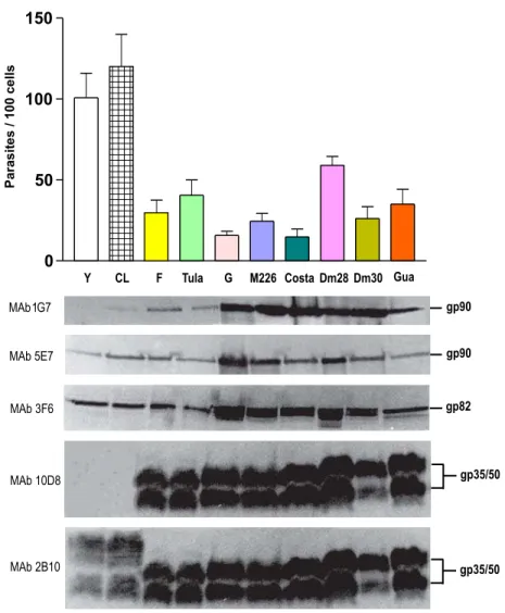
Studies with MT of 10 T. cruzi isolates, derived
from different sources in distinct geographical re-gions, have revealed two groups of parasites that differentially express surface glycoproteins and dis-play differential ability to invade mammalian cells in vitro (Fig. 1). The two groups are highly diver-gent. By molecular phylogenetic analysis, Briones et al. (1999) have found that the distance between the twoT. cruzilineages is larger than the distances
amongLeishmaniaspp.
MT of highly invasive isolates are deficient in gp90 and gp35/50, identified respectively by mon-oclonal antibodies (MAbs) 1G7 and 10D8, but ex-press the variant forms of gp90 and gp35/50, identi-fied by MAbs 5E7 and 2B10. MT of poorly invasive isolates express a gp90 molecule that is detectable by MAbs 1G7 and 5E7, and gp35/50 molecules rec-ognized by both MAbs 10D8 and 2B10. Expression of gp82, which reacts with MAb 3F6, is ubiquitous among these isolates (Fig. 1).
Gp35/50, gp82 and gp90 bind to as yet unde-fined host cell receptors. The interaction of any of these molecules with its receptor triggers bidirec-tional signaling cascades. Whether the MT-target cell interaction results in productive infection de-pends on which surface molecule is predominantly engaged. To attach to and enter host cells, MT of highly invasive CL isolate, for instance, engage gp82 (Ramirez et al. 1993), which efficiently trig-gers Ca2+signaling in MT and host cells (Ruiz et al.
1998), whereas MT of poorly invasive G isolate ap-pear to rely mainly on gp35/50 for their internaliza-tion (Yoshida et al. 1989). Gp35/50 molecules are not as effective as gp82 in promoting invasion, due probably to their poor Ca2+signal-inducing activity
(Dorta et al. 1995). If the interaction is mediated by gp90, which is devoid of Ca2+signaling activity,
productive infection is precluded.
Extensive studies with MT ofT. cruziisolates
TABLE I
Mammalian cells and T. cruzi isolates used in invasion assays.
T. cruziisolate or clone
CL, CL-14, Costalimai, Dm28, Dm28c, Dm30, F, MD, G, Guafitas, M226, MD, 569, 588, Silvio X-10/4, RA, Tulahun, Y
Mammalian cell type
Chinese hamster ovary (CHO) cell
Human carcinoma-derived epithelial HeLa cell Human umbilical vein endothelial cell (HUVEC) L6E9myoblast
LLC-MK2cell
Madin-Darby canine kidney (MDCK) cell Mouse 3T3 fibroblast,
My1Lu mink lung cell
Normal rat kidney (NRK) fibroblast Primary canine cardiac myocyte
Vero cell derived from African green monkey fibroblast
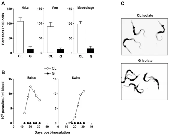
and poorly invasive parasites, have shown that their infectivity is not related to the target cell type. Re-gardless of the mammalian cell type, the number of internalized parasites of CL isolate is always several fold higher than that of G isolate (Fig. 2A). These in vitro findings closely correlate to the observations in vivo. Regardless of the mouse strain or the route of parasite administration, MT of CL isolate produce high parasitemias, in contrast to MT of G isolate that invariably produce subpatent infection (Fig. 2B). Presumably the mechanisms of host cell entry acting in vitro prevail in vivo. Invasion of epithelial HeLa cells by MT may be equivalent to the invasion of gas-tric mucosal epithelium, upon oral infection. Like-wise, MT entry into mouse peritoneal macrophages, whether the parasites are seeded onto cultured cells or are inoculated intraperitoneally, may be equiva-lent.
One interesting observation is that MT of CL and G isolates are morphologically different (Fig. 2C). As compared to CL isolate, parasites of G isolate are shorter and the kinetoplast is more proximal to the posterior end. Isolate-dependent morphological variations are also observed in blood
trypomastigotes and the different morphologies ap-parently denote physiological differences (Brener 1973). Slender forms appear to penetrate host cells better than the stout forms (Brener 1969). In someT. cruziisolates, slender trypomastigotes are prevalent
during the first days of infection, whereas in other isolates stout forms predominate during the entire infection in mice (Brener 1973).
GP82,THECA2+SIGNAL-INDUCINGMOLECULE THATPROMOTESCELLINVASION OFHIGHLY INVASIVET. cruziISOLATES
The role of the metacyclic stage-specific surface molecule gp82 in mammalian cell invasion was first determined by inhibition of MT internalization us-ing MAb 3F6 or the purified native glycoprotein (Ramirez et al. 1993). Subsequent studies indi-cated that gp82 promotes MT entry into host cells by inducing the activation of signaling cascades and Ca2+ mobilization in both cells (Ruiz et al. 1998,
Yoshida et al. 2000). Recent studies withT. cruzi
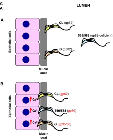
Fig. 1 – Mammalian cell invasion by MT of differentT. cruziisolates and profile of surface molecules. In the upper panel is shown the infectivity of MT of the indicated isolates, expressed as the number of internalized parasites per 100 cells, upon incubation with HeLa cells for 3h at 37◦C. The values correspond to the means± SD of five experiments performed in duplicates, in which at least 500 Giemsa-stained cells were counted. Shown in the lower panel are the profiles of surface molecules identified by monoclonal antibodies directed to gp90, gp82 and gp35/50.
in the surface profile of clone CL-14 MT was the deficient expression of gp82 (Atayde et al. 2004).
Gp82 is a glycoprotein containing N-linked oligosaccharides (Ramirez et al. 1993) that is an-chored to MT plasma membrane by glycosylphos-phatidylinositol (GPI) moiety (Cardoso de Almeida and Heise 1993). The identity of amino acid se-quences of gp82 deduced from cDNA clones of G and CL metacyclic forms is 97.9%, and 100%
Fig. 2 – Differential in vitro and in vivo infectivity ofT. cruziisolates CL and G. A) MT infectivity upon incubation of parasites for 3h with different cell types (HeLa, Vero or mouse peritoneal macrophages). The values, expressed as the number of internalized parasites per 100 cells, correspond to the means±SD of 10 (HeLa), 8 (Vero) or 5 (macrophage) experiments performed in duplicates, in which at least 500 Giemsa-stained cells were counted. B) Course of infection upon inoculation of 4×10⁵MT by oral route into Balb/c mice, or by intraperitoneal route into outbred Swiss mice. Each data point corresponds to the mean parasitemia of 6 animals. C) Purified MT of CL and G isolates stained with Giemsa. Note that MT of CL isolate are longer and the kinetoplast is located more distal to the posterior end, as compared to G isolate.
The central domain of MT gp82 shares 60-65% identity with the C-terminal region of TCT glyco-proteins of gp85 family. However, within the se-quences containing the cell binding site and the epi-tope for MAb 3F6, there are significant differences between MT and TCT molecules, and these include substitutions of acidic amino acids for uncharged or positively charged residues, substitutions of un-charged residues for lysine or arginine, and substi-tutions of residues with polar side chains for those with nonpolar side chains and vice-versa.
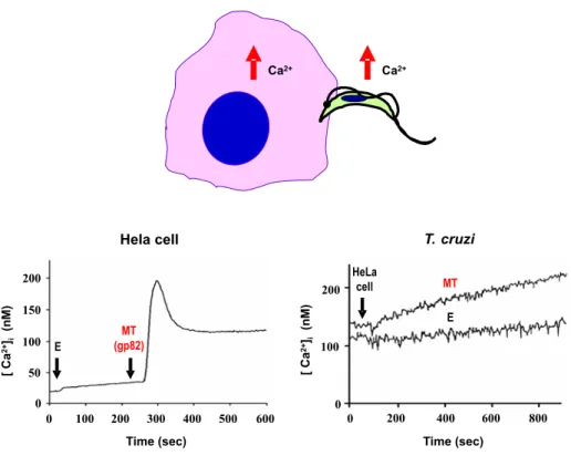
Binding of gp82 triggers in host cells a tran-sient increase in intracellular Ca2+ concentration,
in the same manner as the soluble extracts of MT
(Fig. 4). Non infective epimastigotes do not induce Ca2+ signaling (Tardieux et al. 1994, Dorta et al.
1995) unless they are transfected with T. cruzi
ex-pression vector carrying gp82 cDNA (Manque et al. 2003). Gp82 triggers Ca2+response in mammalian
cells susceptible toT. cruziinfection, such as HeLa
and Vero cells, but not in T. cruzi-resistant human
leukemic K562 cells.
The kinetics of Ca2+mobilization in MT is
re-Fig. 3 – Schematic representation ofT. cruzigp82. The sequence deduced from a cDNA clone derived from MT of G isolate is represented, showing the epitope for MAb 3F6 and the host cell binding site, which were mapped using synthetic peptides and truncated recombinant constructs of gp82.
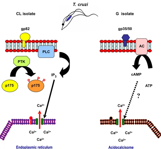
Fig. 5 – Activation of different signal transduction pathways in MT of phylogenetically distinctT. cruziisolates during host cell invasion. In the highly invasive CL isolate, the recognition of the surface molecule gp82 by its receptor leads to PTK activation, phosphorylation of p175, activation of PLC and IP3generation, culminating with IP3-mediated Ca2+release from internal stores, probably endoplasmic reticulum. The components of the G isolate signaling cascade, triggered at the cell surface by gp35/50, are mostly unknown; cAMP generated by adenylyl cyclase may be involved, and acidocalcisomes appear to be the source of Ca2+required for parasite internalization.
sults in protein tyrosine kinase (PTK) activation and phosphorylation of p175, a protein that is un-detectable in noninfective epimastigotes (Favoreto et al. 1998). Activation of PTK and Ca2+response
are associated events, being both inhibited by gp82, as well as by genistein, a specific inhibitor of PTK (Yoshida et al. 2000). The signaling cascade in-duced by gp82 includes the participation of phos-pholipase C (PLC), that generates inositol 1,4,5-triphosphate (IP3), as inferred from experiments in
which the parasite infectivity was impaired by PLC inhibitor U73122, as well as by drugs, such as hep-arin, a competitive IP3-receptor blocker, and caffein,
which affects Ca2+release from IP
3-sensitive stores
(Yoshida et al. 2000). Our hypothesis based on these findings is that in MT ofT. cruziisolates that
engage gp82 to enter target cells, such as the CL isolate, a signaling cascade is initiated at the para-site cell surface by gp82 and proceeds downstream through a sequence involving PTK, PLC and IP3, leading to Ca2+mobilization, possibly from
endo-plasmic reticulum (Fig. 5).
been reported. A neuron survival factor neurturin, for instance, signals through multicomplex recep-tors that consist of receptor tyrosine kinase and a member of a GPI-linked family of receptors that de-termine ligand specificity (Buj-Bello et al. 1997). In T cells, PTKs can be found in complexes im-munoprecipitated with antibodies directed to GPI-anchored proteins (Stefanova et al. 1991, Shenoy-Scaria et al. 1992, Thomas and Samelson 1992). An alternative possibility is the interaction of GPI-anchored proteins and kinases mediated by trans-membrane linker proteins, as suggested by Brown (1993). Also awaiting elucidation is the question concerning the connection between PTK or p175 and PLC.
GP82,AMEDIATOR OFINVASION OFGASTRIC MUCOSALEPITHELIUM
Recently, the involvement of gp82 in oralT. cruzi
infection was inferred from experiments in which the infectivity of CL isolate MT was greatly reduced upon treatment with anti-gp82 MAb 3F6 (Neira et al. 2003). In addition to playing a critical role in promoting invasion of gastric mucosal epithelium, the ability of gp82 to bind to gastric mucin may be important for the establishment of infection by oral route.Shigella dysenteriae, for instance, whose
pathogenic potential is correlated with its ability to invade and multiply within the cells of colonic epithelium, preferentially adheres to colonic mucin (Sudha et al. 2001).
The role of gp82 in establishing efficient T. cruziinfection by oral route has been reinforced by
studies using gp82-deficient isolates, such as 569 and 588 (Cortez et al. 2003). When administered orally into mice, MT of these gp82-deficientT. cruzi
isolates produce patent parasitemia, but to greatly reduced levels when compared to isolate CL. Al-though the in vivo infectivity of isolates 569 and 588 is lower than that of the CL isolate, the abil-ity to enter host cells in vitro is similar. MT of isolates 569 and 588 can efficiently invade cells in vitro because they express gp30, a surface glyco-protein detectable by MAb 3F6 and displaying Ca2+
signal-inducing activity. Gp30-mediated HeLa cell entry of isolates 569 and 588, which is inhibited by MAb 3F6 as well as by native gp30, is dependent on the parasite signal transduction involving PTK activation and Ca2+mobilization from
thapsigargin-sensitive stores, similarly to the gp82-mediated sig-naling. Notwithstanding the functional similarity between gp82 and gp30, MT of gp82-deficient lates are less infective by oral route than the CL iso-late, and this is probably due to the low efficiency of gp30 in binding to gastric mucin. Accordingly, in the presence of high concentration of gastric mucin, to mimic the in vivo condition, the rate of HeLa cell invasion of gp82-deficient MT, but not of CL iso-late, was significantly reduced (Cortez et al. 2003). These findings indicate that the host cell invasion by MT can be mediated either by gp82 or gp30, but efficient mucosal infection depends on the expres-sion of gp82, which promotes the adheexpres-sion to gastric mucin, the first step towards the penetration into the underlying epithelial cells.
GP35/50,THECA2+SIGNAL-INDUCINGMOLECULE THATPROMOTESCELLINVASION OFPOORLY INVASIVET. cruziISOLATES
The involvement of mucin-like gp35/50 molecules in mammalian cell invasion was determined by inhibition of MT internalization using MAb 10D8 or the purified native glycoprotein (Yoshida et al. 1989, Ruiz et al. 1993). Like gp82, gp35/50 bind to target cells in a receptor-mediated manner and triggers bidirectional Ca2+response, but to a lower
degree than gp82 (Ruiz et al. 1998).
Gp35/50 mucins are expressed in metacyclic forms and epimastigotes of allT. cruziisolates
mucins and, depending on theT. cruziisolate, may
contain galactofuranose residues in addition to ga-lactopyranose (Previato et al. 1994, Acosta-Serrano et al. 1995, Salto et al. 2000). MAb 10D8, that recognize gp35/50 mucins in MT of poorly invasive isolates, such as G and Tulahuen, and partially neu-tralize their infectivity (Yoshida et al. 1989), reacts with epitopes containing galactofuranose, whereas MAb 2B10 appears to react with galactopyranose-containing epitopes present in all isolates.
T. cruzi mucins are the main acceptors of
sialic acid in trans-sialidase (TS)-mediated reaction (Schenkman and Eichinger 1993). TS is an enzyme that specifically transfers (α2-3)-linked sialic acid
from extrinsic host-derived macromolecules to O-linked oligosaccharides ofT. cruzimucin-like
gly-coproteins (Schenkman et al. 1991b, Schenkman and Eichinger 1993). The enzyme, that is up to 30 times more active in TCT than in MT (Acosta-Serrano et al. 2001), was first identified in TCT and bloodstream trypomastigotes as a developmentally regulated sialidase that releases sialic acid from hu-man erythrocytes and plasma glycoprotein (Pereira 1983). UsingT. cruziepimastigotes, Previato et al.
(1985) found that sialic acid from exogenous sialy-lated glycoconjugates is incorporated into parasite macromolecules. TS preferentially tranfers sialyl residues to available galactose acceptors and acts as a sialidase in the absence of appropriate amounts of suitable acceptors (Schenkman and Eichinger 1993). In MT, sialyl residues are transferred ex-clusively into gp35/50 mucins (Schenkman et al. 1993a).
Sialyl residues of gp35/50 mucins are not re-quired for MT invasion, they may rather impair the interaction with target cells. Treatment of MT of G isolate with bacterial neuraminidase, for instance, removes sialic acid from gp35/50 and increases the parasite infectivity (Yoshida et al. 1997). Resialy-lation of gp35/50, by incubation of parasites with
T. cruziTS and sialyl lactose, restores the reactivity
with lectin or monoclonal antibody specific for sialic acid and, accordingly, reduces the rate of MT entry into target cells to levels similar to those before
de-sialylation. Compatible with this finding, the capac-ity to bind to host cells and to trigger Ca2+response
was found to be higher in desialylated gp35/50 as compared to its sialylated counterpart (Yoshida et al. 1997). Infectivity of CL isolate MT is not affected by neuraminidase treatment because these parasites rely on gp82, rather than on gp35/50, for their internalization.
In MT that preferentially engage gp35/50 to invade host cells, as is the case of G isolate, the signaling cascade triggered in the parasite is distinct from that induced by gp82 (Fig. 5). PTK and PLC are not implicated, instead cyclic AMP (cAMP) may play a role, as deduced from the increased parasite infectivity upon treatment with adenylyl cyclase ac-tivator forskolin (Neira et al. 2002). The Ca2+
re-quired for cell invasion appears to be released from acidocalcisomes, the vacuoles containing a Ca2+/H+
exchange system (Docampo et al. 1995), provided that treatment of MT with a combination of iono-mycin plus NH4Cl or nigericin, that releases Ca2+
from these acidic compartments, significantly di-minishes target cell entry (Neira et al. 2002). How gp35/50, an GPI-anchored molecule, relays the ex-ternal signal to the parasite interior, and what are the components required for that process are questions that remain unanswered.
GP90,THEMT-SPECIFICDOWNREGULATOR OF HOSTCELLINVASION
The property of gp90 as a negative regulator of tar-get cell invasion was demonstrated by experiments using antisense oligonucleotides targeted to gp90 gene sequences. Treatment of MT of G isolate with antisense oligonucleotides reduced the expression of gp90 and increased the parasite ability to enter host cells, whereas their sense counterparts or the mismatched sequences had no effect (Málaga and Yoshida 2001).
is not mobilized in the parasite upon binding of anti-gp90 MAb 1G7, in contrast to what happens upon interaction with monoclonal antibodies directed to gp82 or gp35/50 (Ruiz et al. 1998). In MT, the in-teraction of gp90 with its receptor may trigger an inhibitory pathway, similarly to cells of the immune system where, in addition to activation signals, sig-naling cascades acting as negative regulators can be induced (Veillette et al. 2002, Vivier et al. 2004). Like NK cell surface inhibitory receptors that an-tagonize activation pathways using protein tyrosine phosphatases (Vivier et al. 2004), gp90 mediates the activation of MT tyrosine phosphatase that counter-acts the action of PTK by dephosphorlylating p175 (Manque et al. 2003).
Like gp82 and gp35/50, gp90 is also anchored to the plasma membrane via GPI (Schenkman et al. 1988b). The type of association of these GPI-anchored molecules with other components of the plasma membrane is unknown. One possibility is the interaction through their extracellular domains. Another possibility is the association through GPI lipid moiety, and in this case the nature of the lipid may influence which plasma membrane molecule is recruited. Of note in this regard is that the lipid portion of gp35/50 GPI from noninfective epimas-tigotes is composed essentially of 1-O
-hexadecyl-2-O-hexadecanoyl-PI and of 1-O-hexadecyl-2-O
-octadecanoyl-PI, whereas that of metacyclic stage gp35/50 is mainly ceramide-PI (Acosta-Serrano et al. 1995).
MECHANISMS OF MT INVASION OF GASTRIC MUCOSAL EPITHELIUM UPON ORAL INFECTION
MT, but not blood trypomastigotes, have uniquely specialized functions for mucosal invasion and effi-ciently enter gastric mucosal epithelium (Hoft 1996, Hoft et al. 1996). This is in accord with the no-tion that metacyclic and bloodstream trypomastig-otes are morphologically similar but are physiologi-cally distinct (Tyler and Engman 2001). Hoft (1996) suggested that MT express stage-specific surface molecules required for adhesion to mucosal
epithe-lial surface receptors and/or for penetration of mucin coat. This hypothesis has been supported by results from experiments of oral infection in mice and in vitro cell invasion assays mimicking the in vivo con-ditions (Neira et al. 2003, Cortez et al. 2003). Based on these data, the following sequence of events can be visualized (Fig. 6). When MT reach the stom-ach, they resist destruction because they are pro-tected by protease-resistant gp35/50 mucins, which are abundant on the parasite surface. Pepsin diges-tion leaves intact the gp82 domain containing both the target cell and the gastric mucin-binding sites. The parasites bind to gastric mucin through gp82 (Fig. 6A) and traverse the mucus to reach the under-lying epithelial cells. It has as yet to be demon-strated, but it is possible that T. cruzi has
muci-nase activity, likeEntamoeba histolytica, which
ex-presses cyteine proteinases that disrupt the poly-meric structure of colonic mucin (Moncada et al. 2003), or Trichomonas vaginalis, which invades
vaginal mucous layer by secreting mucinase (Lehker and Sweeney 1999). Preliminary experiments in-dicate that T. cruzi secrets an enzyme that acts on
gastric mucin.
Once the mucous barrier is overcome, MT of
T. cruziisolates such as CL efficiently invade gastric
epithelial cells by engaging gp82 (Fig. 6B) and trig-gering bidirectional Ca2+response. MT of G isolate
may reach the epithelial cells as effectively as the CL isolate MT, but their entry may be impaired by gp90. Some parasites manage to be internalized by engaging gp35/50 (Fig. 6B).
On the other hand, MT of T. cruzi isolates
Fig. 6 – Model of molecular interaction ofT. cruzimetacyclic forms with gastric mucin and gastric mucosal epithelium upon oral infection of mice. In the stomach, MT of different isolates resist destruction by pepsin and acidic pH because they are protected by protease-resistant gp35/50 mucins. A) MT of CL and G isolates bind to gastric mucin via gp82, as the first step to traverse the mucin layer and reach the underlying epithelial cells. Most parasites of gp82-deficient isolate 569 or 588 do not interact with the mucin coat, but a small number of parasites do so, in gp30 mediated manner. B) Once the epithelial cells are reached, MT of CL isolate attach to and efficiently invade them in gp82-dependent manner, whereas MT of gp82-deficient isolate 569 or 588 rely on gp30 for cell invasion. MT of G isolate enter epithelial cells poorly, due to the preferential adhesion through gp35/50, which has lower Ca2+signal-inducing activity than gp82.
TCT MOLECULES IMPLICATED IN HOST CELL INVASION
Diverse TCT molecules have been implicated in host cell invasion. These include surface and/or se-creted components such as Tc-85 containing bind-ing sites for laminin and cytokeatin 18 (Giordano et al. 1994, Magdesian et al. 2001), mucins and TS (Schenkman et al. 1991b), cystein proteinases (Meirelles et al. 1992) and members of the prolyl oligopeptidase family of serine proteases (Burleigh et al. 1997, Grellier et al. 2001).
TC-85 FAMILY AND OTHERTCT COMPONENTS WITHAFFINITY FOREXTRACELLULARMATRIX
mo-tif (VTVXNVFLYNR) of the gp85/TS superfamily, at the C-terminal domain, and does not contain the laminin-binding site (Magdesian et al. 2001). In the sequence deduced from laminin-binding Tc-85 cDNA, there is a stretch of 122 amino acid residues downstream to this conserved motif (Giordano et al. 1999), whereas in MT gp82 the same motif localizes closer to the C- terminus, being followed by 38 residues, of which 14 correspond to the hy-drophobic GPI-anchor sequence (Araya et al. 1994). It is possible that this subterminal localization of conserved motif in MT gp82 precludes its interac-tion with cytokeratin C18.
The involvement of fibronectin in target cell invasion by TCT was deduced from experiments in which the peptide RGDS, corresponding to fi-bronectin cell attachment site, was found to bind to the parasite surface and to inhibit its internalization (Ouaissi et al. 1986b). By using affinity chromatog-raphy, the TCT ligand for fibronectin was purified and identified as an 85 kDa protein that inter-acts with cells bearing fibronectin molecules, such as human monocytes and neutrophils as well as 3T3 fibroblasts (Ouaissi et al. 1986a).
Another TCT surface molecule with affinity for extracellular matrix components is penetrin, a 60 kDa protein that selectively binds to heparin, heparin sulfate and collagen, and promotes fibrob-last adhesion and penetration (Ortega-Barria and Pereira 1991). An intriguing observation is that the recombinant penetrin, expressed inEscherichia coli
and localized on its surface, induced the bacterial attachment to and penetration into Vero cells in a proteoglycan- and collagen-inhibitable manner (Ortega-Barria and Pereira 1991). Assays to probe the role of host cell heparin and heparan sulfate glycosaminoglycans in T. cruzi invasion showed
that proteoglycan-deficient mutants of Chinese hamster ovary (CHO) cells are poor targets for TCT penetration (Herrera et al. 1994). Penetrin has not been further characterized and its structure remains undefined.
A member of the prolyl oligopeptidase (POP) family of serine proteases, with specificity for
hu-man collagen types I and IV, has been identified in cell-free extracts of trypomastigotes, amastigotes and epimastigotes (Santana et al. 1997, Grellier et al. 2001). The 80 kDa enzyme, denominated POP Tc80, also hydrolyses fibronectin and appears to be implicated in host cell invasion. Selective and ir-reversible inhibitors of POP Tc80 were found to block TCT entry into nonphagocytic mammalian cells (Grellier et al. 2001, Bastos et al. 2005).
In T. cruzi infection in vivo, the ability of
diverse trypomastigote molecules in binding to laminin, fibronectin, collagen, heparin, heparan sul-fate, heparin, in addition to the hydrolytic activity of some of them, may be essential for the parasite tran-sit through the extracellular matrix towards target cells.
TSAND SIALIC ACID ACCEPTOR MOLECULES
The role of TS in mammalian cell invasion has as yet to be fully clarified. On the basis that polyclonal antibodies that block TS activity enhance invasion of host cells by TCT in vitro, Cavallesco and Pereira (1988) proposed that the enzyme negatively modu-latesT. cruziinfection. They also found that,
com-pared to the minor subset of trypomastigotes rec-ognized by anti-TS antibodies (TS+), the TS-
pop-ulation showed enhanced ability to enter host cells. By using anti-TS monoclonal antibodies and various cell types and parasite isolates, Prioli et al. (1990) confirmed the previous results, reinforcing the hy-pothesis that TS down regulatesT. cruziinfection.
Subsequently, however, contradictory data were re-ported by the same group. Pereira et al. (1996) prepared pure TS+ and TS- populations and tested
them for host cell invasion. They found that TS+
trypomastigotes were highly invasive whereas TS
-parasites were extremely inefficient in invading ep-ithelial cells and fibroblasts. Furthermore, introduc-tion of small amounts of TS into suspensions of non-penetrating TS-trypomastigotes converted them to
se-creted (Souto-Padrón et al. 1990). The shed acute phase antigen (SAPA), present in T. cruzi-infected
patients and identified as TS, is the secreted form of enzyme that generates strong immune response (Pollevick et al. 1991). Whether the secreted form of TS and the enzyme expressed on TCT surface act simultaneously during interaction with target cells, or the action of one of them predominates depend-ing on the circumstances, is not clear. Accorddepend-ing to Ming et al. (1993), TS may function as a counter-receptor for parasite binding toα2,3-sialyl receptors
on host cells as a prelude to TCT invasion. Alterna-tively, after binding of TCT to target cells through another molecule, secreted TS may transfer sialic acid from the mammalian cell membrane to the parasite mucins. These transference reactions could disrupt the binding of sialoadhesins, allow-ing the parasites to detach and find a new bindallow-ing site in order to proceed towards their internalization (Schenkman and Eichinger 1993).
In TCT, the sialic acid is incorporated by TS mainly into mucins migrating in SDS-PAGE as a broad band of 70-200 kDa (Schenkman et al. 1991a, b). Mucins and sialyl residues apparently are not pri-mary requirements for TCT invasion (Schenkman et al. 1993b), although monoclonal antibodies to sialic acid-containing epitopes inhibit parasite adhesion to host cells (Schenkman et al. 1991b). On the other hand, the involvement of target cell sialic acid inT. cruziinternalization was reported by different
groups, using sialic acid-deficient CHO (Lec2) cells. Trypomastigotes entered Lec2 cells to a much lower extent than parental CHO cells, but sialylation by TS restored parasite adhesion and invasion (Ciavaglia et al. 1993, Ming et al. 1993, Schenkman et al. 1993b).
CRUZIPAIN, THE MAJOR T. cruzi CYSTEINE PRO -TEINASE
Murta et al. (1990) identifiedT. cruziantigen gp57/
51 as a cysteine proteinase which is active across pH range 5-7.5. This enzyme, named cruzipain, is expressed in all developmental forms of differ-ent T. cruziisolates (Murta et al. 1990, Paiva et
al. 1998). By using peptidyl diazomethane deriva-tives, a class of irreversible inhibitors of cysteine proteinase, the involvement of cruzipain enzyme in host cell invasion and intracellular development was inferred (Meirelles et al. 1992). The role of cruzi-pain in vivo was also determined. Engel et al. (1998) cured experimental T. cruzi infection by treating
mice with peptide-fluoromethyl ketones, inhibitors that inactivate cruzipain and arrest intracellular replication as well as intercellular transmission in vitro (Harth et al. 1993).
The participation of cruzipain in host cell inva-sion by TCT is associated with its ability to generate bradykinin, according to Scharfstein et al. (2000), who investigated the involvement of B2 type of
bradykinin receptor (B2R) using human umbilical
vein endothelial cells (HUVECs) or CHO cells over-expressing B2R (CHO-B2R). They found that
cap-topril, an inhibitor of bradykinin degradation by kininase II, potentiated TCT entry into HUVECs and CHO-B2R, but not into mock-transfected CHO
cells, whereas the B2R antagonist HOE 140 or
mon-oclonal antibody to bradykinin blocked these ef-fects. Purified cruzipain enhanced parasite inva-sion and triggered Ca2+ mobilization in CHO-B
2R
in a manner inhibitable by HOE 140 or cruzipain inhibitor E-64, indicating that the enzyme plays a role in generating the kinin agonist from cell-bound kininogen. This kinin-mediated signal transduction route is not ubiquitous, its activation depending on the cell type and the parasite isolate used (Scharf-stein et al. 2000).
It has also been shown that MGTA, an inhibitor of kininase I, selectively decreases TCT infectivity for B1R-expressing cells and that addition of B1R or
B2R antagonists to host cells coexpressing these
re-ceptors inhibit parasite infectivity to a similar extent (Todorov et al. 2002). Because the combined appli-cation of both antagonists had no additive effect on both cardiomyocytes and HUVECs, the authors de-duced that B1R and B2R operate interdependently to
meet the intracellular Ca2+concentration threshold
required for efficient TCT internalization.
place within the secluded spaces formed by juxta-position of parasite and the target cell, inasmuch as membrane-permeable but not soluble cruzipain inhibitors block parasite invasion of cells that natu-rally overexpress kinin receptors (Scharfstein et al. 2000, Todorov et al. 2002). Cruzipain appears to be modulated by both the host and T. cruzi
com-ponents. On the premise that kininogen molecules may be displayed on cell surfaces by binding to glycosaminoglycans, Lima et al. (2002) examined whether the ability of cruzipain to release kinins from high molecular weight kininogen is mod-ulated by heparin sulfate. In the presence of hep-arin sulfate, they found an enhancement of 6-fold in cruzipain activity towards synthetic substrates and of up to 35-fold by direct measurement of bradykinin. On the other hand, a tight-binding cys-teine proteinase inhibitor, chagasin, was identified in T. cruzi (Monteiro et al. 2001). It is localized
in the flagellar pocket and cytoplasmic vesicles of TCT, and its expression is inversely correlated with that of cruzipain.
T. cruziOLIGOPEPTIDASEB
A soluble factor of unknown structure, secreted by TCT, is so far the sole component of this T. cruzi
developmental form reported to directly trigger Ca2+response in host cells. This soluble factor is
generated by the action of a 120 kDa alkaline pep-tidase on precursors present only in infective try-pomastigotes (Burleigh and Andrews 1995). The purified peptidase is devoid of Ca2+ signaling
ac-tivity on its own and is also present in noninfective epimastigotes (Burleigh and Andrews 1995).
By cloning and sequencing of the correspond-ing cDNA, the TCT peptidase was found to be a cytosolic enzyme closely related to members of the prolyl oligopeptidase family of serine endopepti-dases, and was denominatedT. cruzioligopeptidase
B (Burleigh et al. 1997). The oligopeptidase B null mutant trypomastigotes are defective in mo-bilizing Ca2+ from thapsigargin-sensitive stores in
mammalian cells, and in establishing infection in vitro and in vivo (Caler et al. 1998). Based on
experimental evidences, it has been proposed that the Ca2+ agonist generated by oligopeptidase B is
exported from the parasite, binds to a receptor on the surface of target cells, activating phospholipase C and generating IP3, which binds to its receptor
on the membrane of the endoplasmic reticulum and promotes Ca2+release.
OTHERT. cruziMOLECULES
Several other T. cruzimolecules have been
impli-cated in host cell invasion. Surface antigens with metalloprotease activity, which are homologous to
Leishmania gp63, were identified in MT and TCT
and affinity-purified antibodies to these antigens in-hibited host cell invasion by ∼50% (Cuevas et al.
2003).
Moro et al. (1995) characterized a secreted
T. cruzi protein with peptidyl-prolyl cis-trans
iso-merase activity, susceptible to inhibition by the im-munosuppressant FK506 and related drugs, and showed that the addition of the recombinant protein to simian epithelial or HeLa cells en-hances parasite invasion. The monomeric protein has a peptidyl-prolyl cis-trans isomerase core, en-compassing the characteristic rotamase hydropho-bic active site, and its mechanism of action may be the triggering of host cell signal, with or without the contribution of rotamase activity (Pereira et al. 2002).
A 67 kDa lectin-like glycoprotein, which binds to desialylated human erythrocyte membranes in a galactose-dependent way and recognizes receptors in mouse cardiac tissue and human cardiac aortic endothelium, has been described and the cell in-vasion inhibitory effect of anti-gp67 antibodies re-ported (Silber et al. 2002).
SIGNAL TRANSDUCTION IN HOST CELL DURING
T. cruzi INVASION
CA2+ SIGNALING ANDLYSOSOME RECRUITMENT IN TARGETCELLS
cytosolic-free Ca2+transients in normal rat kidney (NRK)
fi-broblasts, and that parasite entry is inhibited by depletion of host cell cytosolic-free Ca2+or
pre-treatment with Ca2+ channel blockers. In addition
to NRK cells, soluble fraction of TCT induced Ca2+
response in a variety of cell types, such as hamster CHO and Dede, dog MDCK, monkey CV-1, human A7 (Burleigh and Andrews 1995), L6E9myoblasts
(Moreno et al. 1994) and isolated primary canine cardiac myocytes (Barr et al. 1996). MT were also found to trigger Ca2+ signaling in diverse cell
types which included human epithelial HeLa cells and macrophages (Dorta et al. 1995, Wilkowsky et al. 1996). IP3, generated upon PLC activation, mediated intracellular Ca2+ mobilization triggered
by TCT soluble factor (Rodriguez et al. 1995). According to Andrews (1995), host cell Ca2+
response induces the recruitment of lysosomes to the site ofT. cruzipenetration. At that site,
lysoso-mal markers are immediately incorporated into par-asitophorous vacuole without accumulation of poly-merized actin around the recently internalized par-asites, and invasion is facilitated by disruption of microfilaments (Tardieux et al. 1992). Lysosome redistribution and TCT invasion of NRK or L6E9
cells is inhibited upon treatment with microtubule-binding drugs nocodazole, colchicine, vinblastine and taxol, or after microinjection with antibodies to kinesin, indicating that lysosome transport is me-diated by microtubule/kinesin (Rodriguez et al. 1996). Recently, Jaiswal et al. (2002) reported that lysosomes that fused were predominantly pre-docked at the plasma membrane, Ca2+ being
primarily responsible for fusion and not recruit-ment of lysosomes to the cell surface. By fusing with the plasma membrane, lysosomes would con-tribute to formation of the parasitophorous vacuole (Andrews 1995).
Elevation in intracellular free Ca2+
concentra-tion triggered lysosome fusion and exocyto-sis, as deduced from the appearance on the plasma membrane of the lysosomal glycoprotein lgp120, and the release of the lysosomal enzyme beta-hexosaminidase or the lysosomally processed form
of cathepsin D (Rodriguez et al. 1997). Ca2+
-dependent exocytosis of lysosomes is cAMP-regulated and is enhanced by isopreternol, a
β-adrenergic agonist that activates adenylyl cyclase
through heterotrimeric G protein Gs (Rodriguez et
al. 1999). Sinaptotagmin VII, a ubiquitously ex-pressed sinaptotagmin isoform that is localized on the membrane of lysosomes in different cell types and regulates exocytosis of these organelles, ap-pears to mediateT. cruziinvasion. TCT entry was
impaired in CHO cells loaded with antibodies that recognize the Ca2+-binding domain of
sinaptotag-min VII and inhibit the Ca2+-triggered exocytosis of
lysosomes (Caler et al. 2001).
Targeted lysosome exocytosis may not be the predominant mechanism by which TCT gain access to non-professional phagocytic cells. It has been found that only a minimal fraction of invading TCT associate with host cell lysosomes whereas the ma-jority of parasites induce plasma membrane invagi-nation and the TCT-containing vacuoles gradually acquire lysosomal markers (Woolsey et al. 2003). The newly formingT. cruzicompartments first
inter-act with an early endosome compartment and sub-sequently with other late endosomes, before inter-action with lysosomes (Wilkowsky et al. 2002).
HOSTCELLACTINCYTOSKELETON
As a rapid and transient reorganization of host cell microfilaments is induced by TCT soluble factor and live trypomastigotes, probably as a direct con-sequence of increased intracellular Ca2+
concentra-tion, it has been proposed that this disassembly of the cortical actin cytoskeleton plays a role inT. cruzi
fibrob-last, L-6 skeletal muscle myoblast and resident peri-toneal macrophages (Rosestolato et al. 2002), in ad-dition to heart muscle cells (Barbosa and Meirelles 1995). As regards MT invasion, it was significantly inhibited by treatment of HeLa cells with cytocha-lasin B or latrunculin B (Osuna et al. 1993), but unaffected by cytochalasin D (Schenkman and Mortara 1992). It is not clear why the results from different groups differ so widely, provided that ap-parently the experimental conditions and the drug concentration used are similar.
Recent data implicate the actin cytoskeleton in the intracellular retention of parasites. Woolsey and Burleigh (2004) demonstrated that cytochalasin D treatment of host cells inhibits early lysosome as-sociation with invading TCT by uncoupling the cell penetration step from lysosome recruitment and/or fusion, and prolonged disruption of actin microfila-ments results in significant loss of internalized par-asites from infected cells. That a significant frac-tion of the internalized parasite is not retained in-side host cells for a productive infection was con-firmed by Andrade and Andrews (2004), by block-ing lysosome-mediated TCT invasion through phos-phoinositide 3-kinase inhibition.
An interesting observation, that reinforces the role of the cortical actin cytoskeleton disassembly in T. cruziinvasion, has been made in human
pla-centa syncytiotrophoblasts. Using immunohisto-chemical techniques, Sartori et al. (2003a) ob-served the presence of actin in the syncytiotro-phoblasts throughout the brush border in placentae from non-chagasic women but, after culture with trypomastigotes, this labeling disappeared, indicat-ing that the parasite induced disassembly of the cortical actin cytoskeleton.
PHOSPHOINOSITIDE(PI)-3 KINASES, PROTEINKINASES ANDPHOSPHATASES
Among the mechanisms of T. cruzi invasion
are those dependent on lipid as well as protein ki-nases. Infection of macrophages with trypomastig-otes stimulates the formation of the lipid products of PI 3-kinases and treatment with wortmannin, an
inhibitor of PI 3-kinases, impaires parasite internal-ization (Todorov et al. 2000). Immunofluorescence microscopy, using antibodies against p85, the regu-latory subunit of PI 3-kinase, localized the enzyme at the site of parasite interaction with macrophages, which was rich in F-actin (Vieira et al. 2002). In ad-dition to phagocytic human macrophages and J774 murine cells, nonphagocytic Vero, L6E9 and 3T3
cells become less susceptible to T. cruziinfection
upon treatment with wortmannin (Wilkowsky et al. 2001). According to Woolsey et al. (2003), host cell PI 3-kinases activated by TCT early in the cell inva-sion process regulate lysosome-dependent parasite entry. Treatment ofT. cruziwith wortmannin also
inhibited parasite internalization (Wilkowsky et al. 2001), indicating that both parasite and target cell PI 3-kinase activities are implicated in cell invasion. The involvement of protein tyrosine kinase (PTK) inT. cruziinvasion of macrophages has been
reported by Vieira et al. (1994). By treating ei-ther TCT or macrophages with genistein, a spe-cific PTK inhibitor, these authors found a signifi-cant decrease in parasite endocytosis. Monoclonal anti-phosphotyrosine antibodies revealed an accu-mulation of tyrosine-phosphorylated residues at the site of parasite association with the macrophage sur-face, colocalizing with host cell F-actin-rich do-mains (Vieira et al. 2002). Activation of parasite PTK is required for MT and TCT entry into non-phagocytic cells (Favoreto et al. 1998) but, in con-trast to what is seen in macrophages, host cell PTK activity is not involved, as inferred from the lack of inhibition of TCT or MT internalization upon treatment of RNK or HeLa cells with genistein (Rodriguez et al. 1995, Favoreto et al. 1998).
Other protein kinases also participate in T. cruzientry into host cells. Wilkowsky et al. (2001)
(Villalta et al. 1999).
Activation of the host cell PTK is not required for T. cruzi invasion of nonphagocytic cells. On
the other hand, protein tyrosine phosphatases appear to be involved. Invasion of TCT induced tyrosine dephosphorylation of several proteins in L6E9
my-oblasts, and the parasite internalization was greatly reduced in the presence of protein tyrosine phos-phatase inhibitors and in the presence of excess phosphotyrosine, but not of phosphoserine or phos-phothreonine (Zhong et al. 1998). In human HEp2 tumor cells, infection with T. cruziresulted in the
alterations of their placental alkaline phosphatase activity as well as in a different pattern of actin or-ganization, compared to control cells, and the in-terference in the enzyme activity before infection decreased the invasion rate (Sartori et al. 2003b).
TARGETCELLSURFACEMOLECULESIMPLICATED INSIGNALTRANSDUCTION ANDTCT INVASION
In addition to Ca2+signaling through bradykinin
re-ceptor, the signaling route mediated by transform-ing growth factor-β (TGFβ) receptor may be
acti-vated during TCT invasion of target cells. In a series of experiments, Ming et al. (1995) showed the re-quirement of TGFβ pathway forT. cruziinvasion
of epithelial cells. They found that TCT attached to TGFβreceptor-deficient epithelial cell lines, but
were unable to penetrate. Susceptibility toT. cruzi
infection was restored by transfection with TGFβ
receptor genes, and treatment with TGFβ greatly
enhanced parasite internalization. As a TGFβ
-responsive reporter gene is induced in TGFβ
-sensitive cell lines by TCT, but not by noninvasive epimastigotes, Ming et al. (1995) postulated thatT. cruzi may directly trigger activation of the TGFβ
-signaling pathway required for invasion. The pu-tative TGFβ-like factor from TCT has never been
characterized.
In macrophages, the heterodimeric β1
inte-grins, which belong to a ubiquitous family of in-tegral membrane proteins that link the extracellular matrix to the cortical cytoskeleton, may be involved in signal transduction and T. cruzi
internaliza-tion. Fernandez et al. (1993) observed that, when added to human macrophages, monoclonal antibod-ies toβ1 subunit of VLA integrin family specifically
blocked T. cruzi uptake, without interfering with
the uptake of Leishmania pinfanoior Escherichia coli. As that inhibition correlated with the ability
to block fibronectin binding to macrophages, it is uncertain whether the parasite interacts with VLA directly or through the binding to fibronectin.
Galectin 3, which increases K-Ras activation and triggers a Ras signal (Elad-Sfadia et al. 2004), is another host cell component that may partici-pate inT. cruzi invasion. In experiments with
hu-man coronary artery smooth muscle cells, that ex-press galectin-3 on the surface and also secret it, Kleshchenko et al. (2004) found that exogenously added galectin-3 increases trypomastigote binding.
T. cruziadhered poorly to cells with reduced
expres-sion of galectin-3, but the adheexpres-sion property was restored by exogenous galectin-3.
MECHANISMS OF TARGET CELL INVASION BY BLOOD TRYPOMASTIGOTES
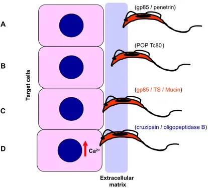
From the data of in vitro studies with TCT, the possible mechanisms that the bloodstream trypomastigotes may use to invade host cells can be envisaged (Fig. 7). Before reaching the target cells, in many instances the parasites have to over-come the barrier of extracellular matrix, in the same manner as MT encounter the mucin coat in the stom-ach. Through the surface molecules of gp85/TS su-perfamily, the parasites bind to fibronectin/laminin (Fig. 7A) and pave the way for the action of en-zymes, such as the serine protease POP Tc80 that hydrolyses collagen/fibronectin (Fig. 7B).
Upon encountering the target cells, trypomas-tigotes attach to them in a manner mediated by Tc-85, TS or mucin (Fig. 7C). This interaction induces the activation ofT. cruzioligopeptidase B that
gen-erates a Ca2+ signaling factor from a precursor
molecule. Triggering of host cell Ca2+
con-Fig. 7 – Model of molecular interaction ofT. cruzibloodstream trypomastigotes with target cells. The extracellular matrix may constitute in many instances a barrier trypomastigotes have to over-come to reach the host cells. A) TCT attach to the extracellular matrix using molecules such as gp85 and penetrin, which have affinity for laminin, fibronectin, collagen, heparin. B) To translocate through the matrix, the parasite may use POP Tc80, which has collagen/fibronectin hydrolyzing activity. C) Adhesion to and invasion of target cells can be mediated by gp85, TS and/or mucins. D) Binding of TCT to target cells induce the activation of oligopeptidase B that generates a Ca2+ agonist, as well as the secretion of cruzipain that indirectly triggers Ca2+response in host cells.
fines of parasite-target cell juxtaposition, and its ac-tion on the kininogen generates bradykinin, which also induces Ca2+ response by interacting with its
receptor (Fig. 7D). Trypomastigotes ofT. cruzi
iso-lates that trigger both signaling pathways may be more invasive. By testing differentT. cruziisolates,
Aparicio et al. (2004) have found a correlation be-tween the level of secreted cruzipain and the capac-ity of trypomastigotes to invade host cells. The low invasive capacity of G isolate, with low levels of cruzipain, may result from the lack of activation of cruzipain-mediated signaling pathway. In addition to the referred signaling routes, parasite-target cell interactions mediated by TGFβ receptors and β1
integrins may take place, further contributing to the process of parasite internalization.
CONCLUDING REMARKS
Activation of signal transduction pathways trig-gered in the parasite and the host cell, leading to intracellular Ca2+mobilization, Ca2+-induced
reor-ganization of the host cell actin cytoskeleton and lysosome recruitment, constitutes the general mechanism by which T. cruzi trypomastigotes
invade mammalian cells. With a plethora ofT. cruzi
molecules that have been identified and character-ized structural and functionally, plus the identifica-tion of target cell components involved, the whole process is beginning to be understood at the molecular level. The picture is complex. Not only MT and TCT engage different molecules to interact with host cells, but different T. cruziisolates may
sig-naling pathways. In addition, some molecular in-teractions may trigger inhibitory signals that down regulate trypomastigote invasion. Furthermore, as the in vivo infection is concerned, the interaction of parasites with host components before reaching the target cells has also to be considered. Great progress has been made towards understanding the mammalian cell invasion byT. cruzi, but a lot more
work has to be done before we can draw a more complete and detailed picture of that process.
ACKNOWLEDGMENTS
This work was supported by Fundação de Amparo à Pesquisa do Estado de São Paulo (FAPESP) and Conselho Nacional de Desenvolvimento Científico e Tecnológico (CNPq). I thank Dr. Sergio Schenk-man, Dr. José Daniel Lopes and Dr. Mauro Cortez for reading the manuscript.
RESUMO
O estabelecimento da infecção porTrypanosoma cruzi, o agente da doença de Chagas, depende de uma série de eventos envolvendo interações de diversas moléculas do parasita com componentes do hospedeiro. Focalizamos aqui os mecanismos de invasão celular por tripomastigo-tas metacíclicos (TM) e por tripomastigotripomastigo-tas de cultura de tecido (TCT). Durante a internalização de TM ou TCT, vias de transdução de sinal são ativadas tanto no para-sita como na célula alvo, acarretando a mobilização de Ca2+. Para adesão, TM utiliza as glicoproteínas de super-fície como a gp82 e gp35/50, que são moléculas indutoras de sinal de Ca2+. Em isolados de T. cruzique entram na célula hospedeira de maneira dependente de gp82, a proteína tirosina quinase assim como a fosfolipase C do parasita são ativadas, e Ca2+é liberado de reservatórios sensíveis a IP3, enquanto em isolados deT. cruzique se ligam às células alvo através de gp35/50, a via de sinali-zação envolvendo adenilil ciclase parece ser estimulada, com liberação de Ca2+de acidocalciossomos. Além disso, dependendo do isolado deT. cruzi, sinais inibitórios medi-ados por gp90 específica de TM podem ser desencademedi-ados tanto na célula hospedeira como no parasita. O repertório de moléculas de TCT implicadas na invasão celular inclui glicoproteínas de superfície da família gp85, com mem-bros contendo sitos de ligação à laminina e citoqueratina 18, enzimas como a cruzipaína, trans-sialidase, e uma
oligopeptidase B que gera um agonista de Ca2+ a par-tir de uma molécula precursora.
Palavras-chave:Trypanosoma cruzi, tripomastigotas,
in-vasão celular, transdução de sinal, mobilização de Ca2+.
REFERENCES
ACOSTA-SERRANOA, SCHENKMAN S, YOSHIDAN, MEHLER A, RICHARDSON JM AND FERGUSON MAJ. 1995. The lipid structure of the GPI-anchored mucin-like sialic acid acceptors of Trypanosoma cruzichanges during parasite differentiation from epimastigotes to infective metacyclic trypomastig-ote forms. J Biol Chem 270: 27244–27253. ACOSTA-SERRANO A, ALMEIDA IC, FREITAS
-JUNIOR LH, YOSHIDA N AND SCHENKMAN S. 2001. The mucin-like glycoprotein super-family of Trypanosoma cruzi: structure and biological roles. Mol Biochem Parasitol 114: 143–150.
ALMEIDAIC, FERGUSON MA, SCHENKMAN S AND TRAVASSOSLR. 1994. Lytic anti-α-galactosyl an-tibodies from patients with chronic Chagas’ disease recognize novel O-linked oligosaccharides on mucin-like glycosyl-phosphatidylinositol glycoproteins of Trypanosoma cruzi. Biochem J 304: 793–802. ALVESMJ, ABUING, KUWAJIMAVYANDCOLLIW.
1986. Partial inhibition of trypomastigote entry into cultured mammalian cells by monoclonal antibodies against a surface glycoprotein ofTrypanosoma cruzi. Mol Biochem Parasitol 21: 75–82.
ANDRADELOANDANDREWSNW. 2004. Lysosomal fusion is essential for the retention ofTrypanosoma cruziinside host cells. J Exp Med 200: 1135–1143. ANDREWS NW. 1995. Lysosome recruitment during host cell invasion byTrypanosoma cruzi. Trends Cell Biol 5: 133–137.
APARICIOIM, SCHARFSTEINJANDLIMAAP. 2004. A new cruzipain pathway of human cell invasion byTrypanosoma cruzirequires trypomastigote mem-branes. Infect Immun 72: 5892–5902.
ARAYAJE, CANOMI, YOSHIDANANDFRANCO DA SILVEIRAJ. 1994. Cloning and characterization of a gene for the stage-specific 82-kDa surface antigen of metacyclic trypomastigotes ofTrypanosoma cruzi. Mol Biochem Parasitol 65: 161–169.
basis of non virulence ofTrypanosoma cruziclone CL-14. Int J Parasitol 34: 851–860.
BARBOSA HSANDMEIRELLESMN. 1995. Evidence of participation of cytoskeleton of heart muscle cells during the invasion of Trypanosoma cruzi. Cell Struct Funct 20: 275–284.
BARRSC, HANW, ANDREWSNW, LOPEZJW, BALL BA, PANNABECKER TL AND GILMOUR-JR RF. 1996. A factor from Trypanosoma cruzi induces repetitive cytosolic free Ca2+ transients in isolated primary canine cardiac myocytes. Infect Immun 64: 1770–1777.
BASTOS IM ET AL. 2005. Molecular, functional and structural properties of the prolyl oligopeptidase of Trypanosoma cruzi(POP Tc80) that is required for parasite entry into mammalian cells. Biochem J 388: 29–38.
BRENER Z. 1969. The behavior of slender and stout forms ofTrypanosoma cruziin the blood-stream of normal and immune mice. Ann Trop Med Parasitol 63: 215–220.
BRENERZ. 1973. Biology ofTrypanosoma cruzi. Ann Rev Microbiol 27: 347–382.
BRIONES MRS, SOUTO RP, STOLF BS ANDZIN -GALEZB. 1999. The evolution of twoTrypanosoma cruzi subgroups inferred from rRNA genes can be correlated with the interchange of American mam-malian faunas in the Cenozoic and has implications to pathogenicity and host specificity. Mol Biochem Parasitol 104: 219–232.
BROWND. 1993. The tyrosine kinase connection: how GPI-anchored proteins activate T cells. Curr Opinion Immunol 5: 349–354.
BUJ-BELLO A, ADU J, PIÑON LGP, HORTON A, THOMPSONJ, ROSENTHALA, CHINCHETRUM, BUCHMANVLANDDAVIESAM. 1997. Neurturin responsiveness requires a GPI-linked receptor and the Ret receptor tyrosine kinase. Nature 387: 721– 724.
BURLEIGHBANDANDREWSNW. 1995. A 120-kDa al-kaline peptidase fromTrypanosoma cruziis involved in the generation of a novel Ca2+-signaling factor for mammalian cells. J Biol Chem 270: 5172–5180. BURLEIGH B AND ANDREWS NW. 1998. Signaling
and host cell invasion byTrypanosoma cruzi. Curr Opinion Microbiol 1: 461–465.
BURLEIGH B, CALER EV, WEBSTER P ANDAN -DREWSNW. 1997. A cytosolic serine endopeptidase fromTrypanosoma cruziis required for the genera-tion of Ca2+ signaling in mammalian cells. J Cell Biol 136: 609–620.
CALER EV, VAENA DE AVALOS S, HAYNES PA, ANDREWS NW AND BURLEIGH B. 1998. Oligo-peptidase B-dependent signaling mediates host cell invasion byTrypanosoma cruzi. EMBO J 17: 4975– 4986.
CALEREV, MORTYRE, BURLEIGHBANDANDREWS NW. 2000. Dual role of signaling pathways lead-ing to Ca2+and cyclic AMP elevation in host cell invasion byTrypanosoma cruzi. Infect Immun 68: 6602–6610.
CALER EV, CHAKRABARTI S, FOWLERKT, RAO S AND ANDREWS NW. 2001. The exocytosis-regulatory protein sinaptotagmin VII mediates cell invasion by Trypanosoma cruzi. J Exp Med 193: 1097–1104.
CARDOSO DEALMEIDAMLANDHEISEN. 1993. Pro-teins anchored via glycosylphosphatidylinositol and solubilizing phospholipases in Trypanosoma cruzi. Biol Res 26: 285–312.
CAVALLESCORANDPEREIRAMEA. 1988. Antibody toTrypanosoma cruzineuraminidase enhances infec-tion in vitro and identifies a subpopulainfec-tion of trypo-mastigotes. J Immunol 140: 617–625.
CIAVAGLIAMC, CARVALHOTUANDSOUZAW. 1993. Interaction ofTrypanosoma cruziwith cells with al-tered glycosylation patterns. Biochem Biophys Res Commun 193: 718–721.
COLLIWANDALVESMJM. 1999. Relevant glycocon-jugates on the surface ofTrypanosoma cruzi. Mem Inst Oswaldo Cruz 94 (Suppl. 1): 37–49.
CORTEZM, NEIRAI, FERREIRA D, LUQUETTI AO, RASSI A, ATAYDE VD AND YOSHIDA N. 2003. Infection by Trypanosoma cruzi metacyclic forms deficient in gp82 but expressing a related surface molecule gp30. Infect Immun 71: 6184–6191. COURA JR, JUNQUEIRA ACV, FERNANDES O, VA
-LENTE SAS AND MILES MA. 2002. Emerging Chagas disease in Amazonian Brazil. Trends Para-sitol 18: 171–176.
antigens with metalloprotease activity and a possible role in host cell infection. Infect Immun 71: 5739– 5749.
DINOIAJM, D’ORSO I, ASLUNDL, SANCHEZ DO AND FRASCHAC. 1998. TheTrypanosoma cruzi mucin family is transcribed from hundreds of genes having hypervariable regions. J Biol Chem 273: 10843–10850.
DOCAMPOR, SCOTTDA, VERCESIAEANDMORENO SNJ. 1995. Intracellular Ca2+ storage in acido-calcisomes ofTrypanosoma cruzi. Biochem J 310: 1005–1012.
DOCAMPOR, SCOTTDA, VERCESIAEANDMORENO SNJ. 1996. The role of Ca2+in the process of cell invasion by intracellular parasites. Parasitol Today 12: 61–65.
DORTA ML, FERREIRA AT, OSHIRO MEM AND YOSHIDAN. 1995. Ca2+ signal induced by Try-panosoma cruzi metacyclic trypomastigote surface molecules implicated in mammalian cell invasion. Mol Biochem Parasitol 73: 285–289.
ELAD-SFADIAG, HAKLAIR, BALANEANDKLOOG Y. 2004. Galectin-3 augments K-Ras activation and triggers a Ras signal that attenuates ERK but not phosphoinositide 3-kinase activity. J Biol Chem 279: 34922–34930.
ENGEL JC, DOYLE OS, HSICH I AND MCKERROW JH. 1998. Cysteine protease inhibitors cure an ex-perimentalTrypanosoma cruziinfection. J Exp Med 188: 725–734.
FAVORETO-JRS, DORTAMLANDYOSHIDAN. 1998. Trypanosoma cruzi175 kDa protein tyrosine phos-phorylation is associated with host cell invasion. Exp Parasitol 89: 188–194.
FERNANDEZ MA, MUNOZ-FERNANDEZ MA AND FRESNOM. 1993. Involvement of beta 1 integrins in the binding and entry ofTrypanosoma cruziinto human macrophages. Eur J Immunol 23: 552–557. GIORDANO R, CHAMMAS R, VEIGA SS, COLLI W
ANDALVESMJM. 1994. An acidic component of the heterogeneous Tc-85 protein family from the sur-face ofTrypanosoma cruziis a laminin binding gly-coprotein. Mol Biochem Parasitol 65: 85–94. GIORDANO R, FOUTS DL, TEWARI D, COLLI W,
MANNINGJEANDALVESMJM. 1999. Cloning of
a surface membrane glycoprotein specific for the in-fective form ofTrypanosoma cruzihaving adhesive properties to laminin. J Biol Chem 274: 3461–3468. GRELLIER P ET AL. 2001. Trypanosoma cruzi pro-lyl oligopeptidase Tc80 is involved in nonphagocytic mammalian cell invasion by trypomastigotes. J Biol Chem 276: 47078–47080.
HARTH G, ANDREWS N, MILLS AA, ENGEL JC, SMITH R AND MCKERROW JH. 1993. Peptide-fluoromethyl ketones arrest intracellular replication and intercellular transmission ofTrypanosoma cruzi. Mol Biochem Parasitol 58: 17–24.
HERRERA EM, MING M, ORTEGA-BARRIA E AND PEREIRA MEA. 1994. Mediation ofTrypanosoma cruzi invasion by heparan sulfate receptors on host cells and penetrin counter-receptors on the try-panosomes. Mol Biochem Parasitol 65: 73–83. HOFTDF. 1996. Differential mucosal infectivity of
dif-ferent life stages ofTrypanosoma cruzi. Am J Trop Med Hyg 55: 360–364.
HOFTDF, FARRARPL, KRATZ-OWENSKANDSHAF -FERD. 1996. Gastric invasion byTrypanosoma cruzi and induction of protective mucosal immune re-sponses. Inf Immun 64: 3800–3810.
JAISWALJK, ANDREWS NWANDSIMONSM. 2002. Membrane proximal lysosomes are the major vesi-cles responsible for calcium-dependent exocytosis in nonsecretory cells. J Cell Biol 159: 625–635. KLESHCHENKO YY, MOODY TN, FURTAK VA,
OCHIENG J, LIMA MF AND VILLALTA F. 2004. Human galectin-3 promotesTrypanosoma cruzi ad-hesion to human artery smooth muscle cells. Infect Immun 72: 6717–6721.
LEHKERMWANDSWEENEYD. 1999. Trichomonad invasion of the mucous layer requires adhesins, mucinases, and mortality. Sex Transm Infect 75: 231–238.
LIMA APCA, ALMEIDA PC, TERSARIOL ILS, SCHMITZV, SCHMAIERAH, JULIANOL, HIRATA IY, MÜLLER-ESTERL W, CHAGAS JR AND SCHARFSTEINJ. 2002. Heparan sulfate modulates kinin release byTrypanosoma cruzithrough the ac-tivity of cruzipain. J Biol Chem 277: 5875–5881. MAGDESIAN MH, GIORDANO R, JULIANO MA,
cruzi: identification of a parasite ligand and its host-cell receptor. J Biol Chem 276: 19382–19389. MÁLAGASAND YOSHIDAN. 2001. Targeted
reduc-tion in expression ofTrypanosoma cruzisurface gly-coprotein gp90 increases parasite infectivity. Infect Immun 69: 353–359.
MANQUEPM, EICHINGERD, JULIANOMA, JULIANO L, ARAYAJANDYOSHIDAN. 2000. Characteriza-tion of the cell adhesion site ofTrypanosoma cruzi metacyclic stage surface glycoprotein gp82. Infect Immun 68: 478–484.
MANQUE PM, NEIRA I, ATAYDE VD, CORDEROE, FERREIRAAT, FRANCO DASILVEIRAJ, RAMIREZ MANDYOSHIDAN. 2003. Cell adhesion and Ca2+ signaling activity in stably transfectedTrypanosoma cruziepimastigotes expressing the metacyclic stage-specific surface molecule gp82. Infect Immun 71: 1561–1565.
MEIRELLES MN, JULIANO L, CARMONA E, SILVA SG, COSTAEM, MURTAACANDSCHARFSTEIN J. 1992. Inhibitors of the major cysteinyl proteinase (gp57/51) impair host cell invasion and arrest the intracellular development of Trypanosoma cruziin vivo. Mol Biochem Parasitol 52: 175–184.
MINGM, CHUENKOVAM, ORTEGA-BARRIAEAND PEREIRAMEA. 1993. Mediation ofTrypanosoma cruzi invasion by sialic acid on the host cell and trans-sialidase on the trypanosome. Mol Biochem Parasitol 59: 243–252.
MINGM, EWENMEANDPEREIRAMEA. 1995. Try-panosome invasion of mammalian cells requires ac-tivation of the TGFβ signaling pathway. Cell 82: 287–296.
MONCADA D, KEELLER K AND CHADEE K. 2003. Entamoeba histolyticacysteine proteinases disrupt the polymeric structure of colonic mucin an alter its protective function. Infect Immun 71: 838–844. MONTEIRO ACS, ABRAHAMSON M, LIMA APCA,
VANNIER-SANTOS MA AND SCHARFSTEIN J. 2001. Identification, characterization and localiza-tion of chagasin, a tight-binding cysteine protease inhibitor inTrypanosoma cruzi. J Cell Science 114: 3933–3942.
MORENOSNJ, SILVAJ, VERCESIAEANDDOCAMPO R. 1994. Cytosolic-free calcium elevation in Trypa-nosoma cruzi is required for cell invasion. J Exp Med 180: 1535–1540.
MORO A, RUIZ-CABELLO F, FERNANDEZ-CANO A, STOCKRPANDGONZALEZA. 1995. Secretion by Trypanosoma cruzi of a peptidyl-prolyl cis-trans isomerase involved in cell infection. EMBO J 14: 2483–2490.
MORTARARA. 1991.Trypanosoma cruzi: amastigotes and trypomastigotes interact with different struc-tures on the surface of HeLa cells. Exp Parasitol 73: 1–14.
MORTARARA, SILVAS, ARAGUTHMF, BLANCOSA ANDYOSHIDAN. 1992. Polymorphism of the 35-and 50-kilodalton surface glycoconjugates of Try-panosoma cruzimetacyclic trypomastigotes. Infect Immun 60: 4673–4678.
MURTA AC, PERSECHINI PM, PADRON TS, SOUZA W, GUIMARÃES JÁ AND SCHARFSTEIN J. 1990. Structural and functional identification of gp57/51 antigen of Trypanosoma cruzi as a cysteine pro-teinase. Mol Biochem Parasitol 43: 27–38.
NEIRAI, FERREIRAATANDYOSHIDAN. 2002. Ac-tivation of distinct signal transduction pathways in Trypanosoma cruziisolates with differential capac-ity to invade host cells. Int J Parasitol 32: 405–414. NEIRA I, SILVA FA, CORTEZ M AND YOSHIDA N. 2003. Involvement ofTrypanosoma cruzimetacyclic trypomastigote surface molecule gp82 in adhesion to gastric mucin and invasion of epithelial cells. Infect Immun 71: 557–561.
ORTEGA-BARRIA E AND PEREIRA MEA. 1991. A novel Trypanosoma cruzi heparin binding protein promotes fibroblast adhesion and penetration on en-gineered bacteria and trypanosomes into mammalian cells. Cell 67: 411–421.
OSUNAA, RODRIGUEZ-CABEZASN, BOY M, CAS -TANYS S AND GAMARRO F. 1993. The invasion mechanism of the metacyclic forms ofTrypanosoma cruzi in nonphagocytic host cells. Biol Res 26: 19–26.
OUAISSIMA, CORNETTEJAND CAPRONA. 1986a. Identification and isolation of Trypanosoma cruzi trypomastigote cell surface protein with proper-ties expected of a fibronectin receptor. Mol Biochem Parasitol 19: 201–211.