Abstracts of the 10th International Conference on
Cachexia, Sarcopenia and Muscle Wasting, Rome, Italy,
8
–10 December 2017 (Part 1)
1-01
Body composition changes over three years in
older adults: a descriptive longitudinal analysis
Maria Teresa Tomás1,3,Alejandro Galán-Mercant2,3&
Beatriz Fernandes1,3
1Escola Superior de Tecnologia da Saúde de Lisboa, Portugal,2Universidade de Jaén,
Spain,32GHRG—Gerontology and Geriatric Health Research Group
Introduction: Many studies analyse body composition
changes in older adults. However, few studies analyse body composition in elderly people with functional measures. Studies using Double X-Ray analysis (DXA) or Bioimpedance analysis proved to be reliable but expensive or only possible in a laboratory environment.
The purpose of our study was to analyse changes in body composition over three years using anthropometric measures in a sample of elderly people in order to perceive functional changes.
Methods: Forty-three participants (12 men; 31 women) aged 60 years and over and independent in activities of daily life
were assessed using anthropometric measures in afirst
mo-ment and past three years. Weight, height, waist and hip cir-cumference were measured, and body mass index (BMI) and waist-to-hip ratio (WHR) were also calculated. Skeletal mus-cle mass (SMM) was also calculated using Al-Gindan et al. (2014) equations and normalized for height to found skeletal muscle index (SMI) in order to analyze cut-off points associ-ated with physical disability according to Janssen et al (2004). Results: A significant difference was found over three years in SMM (p = 0.007), SMI (p = 0.027), BMI (p = 0.041) and WHR (p = 0.003). The majority of the participants has decreased SMM, SMI and BMI and increased WHR, which favors a worst prognostic for comorbidities associated with these variables, and a tendency for sarcopenic obesity seems to be present al-though more studies are needed. Also, we found that using cut-off points for disability risk 83.3% of the men and 38.7% of the women of our sample were at moderate or high risk of disability. Three years later this percentage has increased but only for women to 54.8%.
Conclusions: Although men are at risk of disability, women quickly lose their functional capacity, making necessary a
rapid intervention to reduce the risk of disability in this population.
IPL/2016/SFQ2017_ESTeSL
1-02
Prevalence of cachexia in dogs with congestive
heart failure
Pamela L. Bay,Lisa M. Freeman&John E. Rush
Cummings School of Veterinary Medicine, Tufts University, North Grafton, MA, USA
Background and Aims: Congestive heart failure (CHF) is a common, naturally occurring disease in pet dogs that is of-ten associated with cardiac cachexia, as defined by a loss of muscle. One study of dogs with dilated cardiomyopathy (DCM) and CHF showed that 54% of dogs were affected
by cachexia. No studies have been conducted to confirm
these findings in dogs with DCM or to assess prevalence
in CHF from other forms of heart disease causing CHF. Therefore, the aim of this study was to determine preva-lence of cardiac cachexia in dogs with CHF due to acquired heart disease.
Methods: Dogs with CHF evaluated by the Cardiology Service at the Cummings School of Veterinary Medicine between June 2015 and June 2017 were eligible. Dogs with DCM and myxomatous mitral valve disease (MMVD) were enrolled.
Data from the medical records were retrospectively
reviewed, including body weight, body condition score (BCS), and muscle condition score (MCS). Body condition
score, which assesses fat stores, was measured on a 1–9
scale, with 1 = emaciated, 9 = obese, and 4–5 considered
ideal. Muscle condition was categorized using the World Small Animal Veterinary Association scoring system as normal muscle, mild muscle loss, moderate muscle loss, or severe muscle loss.
Results: Median age of the dogs (n = 196) was 10.7 years (range, 1.7–18.0 years). Underlying diseases included MMVD (n = 168) and DCM (n = 28). Mean body weight was 7.6 kg
(range, 2.4–75.8 kg). Only 6.1% of dogs were underweight
(BCS < 4/9), and 41.8% of dogs were overweight or obese
Journal of Cachexia, Sarcopenia and Muscle 2017; 8: 999–1080
(BCS> 5/9). However, muscle loss was identified in 48.0% of dogs: Mild muscle loss: 73/196 (37.3%), moderate muscle loss: 14/196 (7.1%), and severe muscle loss: 7/196 (3.6%). 52.0% of dogs were assessed to have normal muscle. Muscle condition score and BCS were not significantly different be-tween dogs with MMVD or DCM.
Conclusions: Although many dogs were overweight or obese, cachexia was present in 48% of dogs with CHF.
1-03
Comorbidities and mortality after cachexia
hospitalization in Slovenia between 2004 and 2015
Daniel Omersa1,Jerneja Farkas2&Mitja Lainscak2,3
1National Institute of Public Health, Ljubljana, Slovenia,2General Hospital Murska
Sobota, Murska Sobota, Slovenia,3Faculty of Medicine, University of Ljubljana,
Lju-bljana, Slovenia
Introduction: Cachexia is common in several chronic diseases and significantly increases morbidity and mortality. There is a lack of data regarding cachexia hospitalization burden and mortality after cachexia hospitalization. Thus, we aimed to identify all patients that were hospitalized due to cachexia and determine their mortality and prognostic implications of different comorbidities.
Methods: The Slovenian National Hospitalization Database has been searched for all individuals with main Cachexia hospitalization (ICD-10 codes: C80, R64 and B22.2) between 2004 and 2015, and sex, age, length of stay and comor-bidities were recorded. For all patients with cachexia hospitalization, date of death was recorded from Slovenian Death Registry. Prevalence of comorbidities during cachexia hospitalization were calculated and hazard ratios (HR) for
mortality for sex, age and patients’ comorbidities were
calculated using multiple Cox proportional hazards model. Results: Overall, we identified 1774 main cachexia hospitali-zations in 1406 patients. Main cachexia hospitalihospitali-zations contributed to 17.7% of all the hospitalizations of an individ-ual during the study period. Cancer, cardiovascular and pulmonary diseases were the most prevalent in cachexia patients (62%, 27% and 10%, respectively). In-hospital mortality was 29%. Median survival for discharged patients
were 103 days (95% confidence intervals, 90–123 days).
Older patients, those with cancer and pulmonary disease, had significantly higher HR for mortality (1.16 for 10 year increase, 1.79 and 1.34, respectively).
Conclusions: Patients hospitalized due to cachexia have extremely poor prognosis. Cancer, which is the most preva-lent comorbidity in patients hospitalized due to cachexia, is associated with the worst prognosis.
1-04
Prevalence of cachexia among COPD cases in the
ECLIPSE study
Merry-Lynn N. McDonald1,2,Erica Rutten3,Richard Casaburi4,
Emiel F.M. Wouters3,Stephen I. Rennard5,David A. Lomas6,
Bartolome Celli7,Alvar Agusti8,Ruth Tal-Singer9,Craig P. Hersh7,10&
Edwin K. Silverman7,10
1Division of Pulmonary, Allergy and Critical Care Medicine, University of Alabama at
Birmingham, Birmingham, AL, USA,2Department of Genetics, University of Alabama
at Birmingham, Birmingham, AL, USA,3Centre of expertise for chronic organ failure,
Horn, the Netherlands,4Rehabilitation Clinical Trials Center, Los Angeles Biomedical
Research Institute at Harbor Harbor-UCLA Medical Center, Torrance, CA, USA,
5Department of Medicine, Nebraska Medical Center, Omaha, NE, USA,6Wolfson
Institute for Biomedical Research, University College London, UK, 7Division of
Pulmonary and Critical Care, Brigham and Women’s Hospital, Boston, MA, USA,
8Fundació Investigació Sanitària Illes Balears (FISIB), Ciber Enfermedades
Respiratorias (CIBERES), Barcelona, Catalunya, Spain Thorax Institute, Hospital Clinic, IDIBAPS, Univ. Barcelona, Barcelona, Spain, 9Respiratory R&D, GSK,
Philadelphia, PA, USA,10Channing Division of Network Medicine, Harvard Medical
School, Boston, MA, USA
Background: By population prevalence, there are more chronic obstructive pulmonary disease (COPD) cases than
cancer cases with cachexia. The consensus definition of
cachexia incorporates weight loss (WL) >5% in 12 months in addition to 3 out of 5 of decreased muscle strength, fatigue, anorexia, low fat-free mass index (FFMI) and abnor-mal biochemistry (anemia, CRP, IL6, albumin). More recently, cancer cachexia has been classified using WL >5% or, in the presence of low BMI or FFMI, WL>2%. Further, the
impor-tance of pre-cachexia (WL≤5%, anorexia and inflammation)
has been highlighted as more advanced cachexia may indicate a refractory state. Thus, we aimed to examine the prevalence of cachexia using these definitions in a cohort of COPD cases from the ECLIPSE Study.
Methods: A total of 1901 COPD cases were assessed for cachexia. Annual weight, muscle strength, FFMI and anemia data were analyzed. Fatigue and anorexia data were available at baseline and end of study. CRP levels were measured at
baseline and over the first year. The consensus definition
was coded at each annual visit an individual participated in the study. Where data were not available for the specific visit, an aggregate was created (e.g., ever had fatigue). Participants who exhibited WL at an early visit with evidence it was regained were coded as non-cachectic.
Results: The prevalence of cachexia based on the consensus definition ranged from 4.0% (Year 1) to 6.6% (Year 3). Over 3 years of the study, 11% of COPD cases were classified as cachectic at some time point. The prevalence of cachexia using the cancer cachexia definition ranged from 9.7% (Year 1) to 17.7% (Year 3). The prevalence of pre-cachexia ranged from 2.0% (Year 2) to 3.1% (Year 1).
Summary: Based on definition of cachexia and visit used, the prevalence in a large cohort of COPD cases ranged from 4.0% to 17.7%.
1-05
SARA-data: Integrated, real-time ICT Platform for
the SARA interventional Clinical Trial in Age-related
SARcopenia
Susanna Del Signore1,2,Waly Dioh2,Stefania Del Signore1&
Gianluca Zia1
1
Bluecompanion ltd, London, UK,2Biophytis, Paris, France
Introduction: SARA-Int(erventional), a randomized, double-blind clinical trial, will evaluate the safety and efficacy of two oral doses of SARconeos (BIO101) versus placebo over 6 months in 333 sarcopenic or obese sarcopenic patients Aged≥65 years complaining of loss of strength and muscular function.
Methods: We deployed an integrated Information&Commu-nication Technology (ICT) platform, SARA-data, to monitor on quasi real-time different source data (clinical, imaging (DEXA), laboratory and physical activity). Data can be gener-ated at investigation sites, by the centralised lab and by the
patients themselves via wearable devices and
auto-evaluation questionnaires.
Bluecompanion implemented for Biophytis SARA Data, which allows to collect and integrate on one single web-based por-tal: an electronic Case Report Form (Clean WEB by
Telemed-icine), participants row data from DEXA scans, and
biochemistry-haematology results from a centralised labora-tory, including sarcopenia-related biomarkers. Of note, con-tinuous physical activity recording is enabled during the whole clinical trial duration by providing each older partici-pant with a wrist-worn accelerometer. The device transmits anonymised activity data to SARA platform via a non-intrusive, unattended, home-centred machine-to-machine technology.
This kind of high volume data, directly generated by the patient during several months, fulfils the definition “Big data
in health”, encompassing “high volume, high diversity
biological, clinical, environmental, and lifestyle information collected from single individuals to large cohorts, in relation to their health and wellness status, at one or several time points” (Auffray C. et al., 2016), and will constitute an important resource for additional, supportive analyses complementing standardised muscular function assessments. These data are generated in a real-life context and could
provide answers to specific questions by regulators and
payers.
Conclusions: SARA-data are a single real-time ICT platform enabling data capture, storage, analysis and retrieval of long-term clinical data generated during SARA-Int, a random-ized CT evaluating Sarconeos (Bio101) in Age-related Sarcopenia, including Sarcopenic Obesity.
1-06
Changes of body weight and body composition and
cachexia after stroke
Nadja Scherbakov1,Charlotte Pietrock1,Nicole Ebner2,3,
Anja Sandek2,3,Miroslava Valentova2,3,Jochen B. Fiebach1,
Joerg C. Schefold4,Stephan von Haehling2,3,Stefan D. Anker2,5,
Kristina Norman6,Karl Georg Haeusler1,7&Wolfram Doehner1,5
1Center for Stroke Research Berlin CSB, Charité - Universitätsmedizin Berlin, Berlin,
Germany,2Innovative Clinical Trials, Department of Cardiology and Pneumology,
University Medicine Goettingen (UMG), Goettingen, Germany,3German Centre for
Cardiovascular Research (DZHK), partner site Goettingen, Goettingen, Germany,
4Department of Intensive Care Medicine, Inselspital, Bern University Hospital,
Switzerland, 5Department of Cardiology, Charité - Universitätsmedizin Berlin,
Berlin, Germany, 6Research Group on Geriatrics, Charité - Universitätsmedizin
Berlin, Berlin, Germany,7Department of Neurology, Charité - Universitätsmedizin
Berlin, Berlin, Germany
Background and Purpose: Body weight loss after stroke has been shown in several clinical trials. Cachexia after stroke has not been studied systematically yet. The purpose of this prospective study was to investigate dynamical changes of body composition and body weight one year after ischemic stroke, and its association with functional outcome.
Methods: 67 consecutive patients with acute ischemic stroke (age 69 ± 11 years, BMI 27 ± 4 kg/m2) with mild to moderate
neurological deficit (mean NIHSS 4.3, range 0–12) were
analyzed. Body composition was examined by dual energy X-ray absorptiometry (DEXA) in acute phase (4 ± 2 days) and at 1-year follow-up (389 ± 26 days). Cachexia was defined
according to consensus definition by body weight loss ≥5%
within one year and clinical symptoms. Functional
assess-ments included Barthel Index (BI), modified Rankin scale
(mRS), and muscle strength tests.
Results: Cachexia was diagnosed in 21% of the patients at 1-year follow-up. Most changes of body composition concerned the fat tissue with the highest fat mass decline of 12.5% in cachectic patients, followed by 5% loss in non-cachectic patients with weight loss, and fat mass increase by 8% in patients with weight gain. In addition, cachectic patients lost 3% of the lean mass (P< 0.05).
At baseline patients who developed cachexia during follow up were older (75 ± 9 years), had moderate neurologic deficit (mean NIHSS 5.8), and the lowest physical and functional capacity. They remained with the worse functional
impair-ment (mRS 2.1 ± 1.6, P < 0.05, Barthel Index 74 ± 36,
P = 0.002) and handgrip strength (22.4 ± 14.9 kg, P< 0.05) compared to other patients at 1-year FU. After adjustment for multiple confounders, patients with higher functional
impairment (OR 1.87, 95% CI 1.09–3.20) and neurologic
deficit (OR 3.63, 95% CI 0.97–13.6) were at risk for cachexia. Conclusions: The most changes of body composition after
stroke with mild-to-moderate neurologic deficit concerned
the fat tissue. Attention should be focused on identification and targeting of cachexia in the early phase following the stroke.
2-01
Ultrasound: a new strategy to evaluate body
composition in crohn
’s patients undergoing
hematopoietic stem cell transplantation (HSCT)
Andrea Z. Pereira1,Sandra E.A. Gonçalves1,Bianca L. de Sá2,
Marister Cocco3,Andreza A.F. Ribeiro1&Nelson Hamerschlak1
1
Oncology and Hematology Department, Hospital Israelita Albert Einstein, S. Paolo, Brazil, 2Nutrition Department, Hospital Israelita Albert Einstein, S.Paulo, Brazil,
3
Physiotherapy Department, Hospital Israelita Albert Einstein, S.Paulo, Brazil
Introduction: Crohn disease is a chronic inflammatory disor-der of the gastrointestinal tract with a strong polygenic im-mune component. In refractory cases, autologous HSCT can decrease disease activity and mucosal healing and improve quality of life. Reduced muscular mass and excess visceral fat in patients undergoing HSCT are associated with higher mortality, longer hospitalization, longer use of immunosup-pressive drugs, graft-versus-host disease, shorter disease-free interval after the HSCT and comorbidities leading to shorter survival time.
Objectives: To evaluate muscle thickness and visceral fat by US.
Methods: We evaluated 5 HSCT patients (≥18 years) at Hos-pital Israelita Albert Einstein, São Paulo, Brazil, on theirfirst day of hospitalization, before HSCT and after the engraft-ment. The thickness of the right femoral quadriceps muscle (RFQ), measured at 6 cm from the top edge of the patella was measured using US in B-mode. The VF was measured in the abdominal region, by the thickness of the fat layer be-tween the linea alba and the anterior wall of the aorta. Results: Most patients were men (75%) with a mean age of 35 years (±14 years). Most patients were undernutrition, with body mass index (BMI) of 21 kg/m2(±2.5 kg/m2). The average time EN was 11 days (±1 day). In the baseline, RFQ was 1.5 cm (±0.2 cm), and the VF was 4.2 cm (±1.3 cm). After engrafment, RFQ was 1.3 cm (±0.2 cm), and the VF was 4.2 cm (±1.2 cm).
There wasn’t significant difference between baseline and
after engraftment, although RFQ had reduced in all patients. Conclusions: In this cohort of patients, we found reduced muscle thickness after engraftment, and VF didn’t have any alterations. The US was a practical, economical and effective method to evaluate these patients.
2–02
Elderly patients undergone hematopoietic stem
cell transplantation: body composition and
engraftment
Andrea Z. Pereira1,Ludmila M. Koch1,Polianna M.R. Souza1,
Bianca L. de Sá2,Andreza A.F. Ribeiro1&Nelson Hamerschlak1
1Oncology and Hematology Department, Hospital Israelita Albert Einstein, S.Paulo,
Brazil,2Nutrition Department, Hospital Israelita Albert Einstein, S.Paulo, Brazil
Introduction: Hematopoietic Stem Cell Transplantation
(HSCT) in elderly is a brand-new issue. Changes in body
composition after HSCT have been the subject of previous studies; however, there aren’t many studies in elderly people. Objectives: To evaluate muscle thickness and visceral fat by US; % muscle mass, % fat mass and phase angle by BIA. To correlate body composition with engraftment (EN).
Methods: In this prospective study, we evaluated 16 HSCT pa-tients (≥60 years) at Hospital Israelita Albert Einstein, São Paulo, Brazil, on theirfirst day of hospitalization, before HSCT and after the EN. The thickness of the right femoral quadri-ceps muscle (RFQ), measured at 6 cm from the top edge of the patella was measured using ultrasound (US) in B-mode, transversal plane. The visceral fat (VF) was measured in the abdominal region, by the thickness of the fat layer between the linea alba and the anterior wall of the aorta. The % mus-cle mass (MM), % fat mass (FM) and phase angle (PA) were evaluated by Bioimpedanciometry(BIA).
Results: Most patients were men (75%) with a mean age of 64(±5.0 years). We had 50% of autologous HSCT and 50% allogenic HSCT. The mean time EN was 13(±4 days). In the base-line, weight was 80(±17 kg), RFQ was 1.8(±0.3 cm) and the VF was 5.5(± 2.0 cm); %MM was 68.5(±11); %FM was 27.5(±7.5); PA was 5.3((±0.7). After EN, weight was 73(±13 kg). RFQ was 1.5(±0.3 cm) and the VF was 5,0(±2.2 cm); %MM was 55.5(±20.5); %FM was 25(±7.0); PA was 7.4(±0.8). There wasn’t significant difference between baseline and after engraftment, although all measurements had reduced in all patients, exception for PA and VF had increased. We found the negative correlation between engraftment and RFQ(rp: 0,6), indepen-dently of HSCT type by regression. (rp: 0,6).
Conclusions: In this cohort of patients, muscle thickness and mass was reduced, and visceral fat and phase angle was increased after engraftment. The higher muscle thickness correlated faster engraftment.
Elderly quilombolas: Prevalence of sarcopenia using
algorithm proposed by the European working group on sarcopenia in older people.
2-03
Prognostic value of psoas muscle area and density
in patients undergoing cardiovascular surgery
Masashi Yamashita1,Kentaro Kamiya1,2,Atsuhiko Matsunaga1,2,
Tadashi Kitamura3,Nobuaki Hamazaki4,5,Ryota Matsuzawa4,
Kohei Nozaki4,Shinya Tanaka5,Junya Ako6&Kagami Miyaji3
1Department of Rehabilitation Sciences, Graduate School of Medical Sciences,
Kitasato University, Sagamihara, Japan,2Department of Rehabilitation, School of
Allied Health Sciences, Kitasato University, Sagamihara, Japan, 3Department of
Cardiovascular Surgery, Kitasato University School of Medicine, Sagamihara, Japan,4Department of Rehabilitation, Kitasato University Hospital, Sagamihara,
Japan, 5Department of Cardiovascular Medicine, Kitasato University Graduate
School of Medical Sciences, Sagamihara, Japan,6Department of Cardiovascular
Medicine, Kitasato University School of Medicine, Sagamihara, Japan
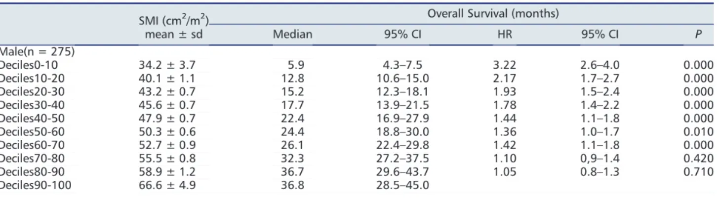
Introduction: Low skeletal muscle area and density, as deter-mined by computed tomography (CT), have yet to be exadeter-mined and compared in terms of prognostic capability in patients re-quiring open cardiovascular surgery. This study was performed
to examine whether psoas muscle area and density are
associated with postoperative mortality and physical
performance in patients undergoing cardiovascular surgery.
Methods: We reviewed the findings in 773 consecutive
patients undergoing preoperative CT imaging including the level of the third lumbar vertebra for clinical purposes. Skeletal muscle area was calculated from psoas muscle cross-sectional area (CSA) on preoperative CT images at the level of the third lumbar vertebra divided by the square of the patient’s height to give the skeletal muscle index (SMI:
cm2/m2). Skeletal muscle density determined by muscle
attenuation (MA) was calculated by measuring the average
Hounsfield units of the psoas muscle CSA. Quadriceps
strength and 6-minute walking distance were examined as indices of physical performance.
Results: The mean age of the study population was 68.6 ± 14.0 years, and 64.7% of the patients were male. Mul-tivariate Cox regression analysis showed that low MA (hazard ratio [HR], 2.18; 95% confidence interval [CI], 1.14–4.16, P = 0.018), but not low SMI (HR, 1.35; 95% CI, 0.71–2.56, P = 0.361), was significantly associated with all-cause mortal-ity. Kaplan–Meier analysis showed that low MA, but not low SMI, predicted poor prognosis (P = 0.014). Correlation analy-sis indicated that MA was more strongly associated with quadriceps strength and 6-minute walking distance than SMI. Conclusions: Low skeletal muscle density, but not skeletal muscle area, predicted survival in patients undergoing car-diac surgery.
2-04
The usefulness of body weight for predicting
skeletal muscle mass in congested state of heart
failure outpatients
Shunichi Doi1,Norio Suzuki1,Keisuke Kida2,Chikayuki Ito3,
Kohei Ashikaga3,Kengo Suzuki2,Hisao Matsuda1,Tomoo Harada2&
Yoshihiro J. Akashi2
1
Division of Cardiology, Department of Internal Medicine, St. Marianna University School of Medicine Yokohama City Seibu Hospital, Yokohama, Japan,2Division of Cardiology, Department of Internal Medicine, St. Marianna University School of Medicine, Kawasaki, Japan, 3Division of Cardiology, Department of Internal Medicine, Kawasaki Municipal Tama Hospital, Kawasaki, Japan
Introduction: Body mass index is cited as an index to recognize significant correlation with skeletal muscle mass. However, body weight changes due to edema are often observed in chronic heart failure (CHF). We investigated the usefulness of body weight for evaluating skeletal muscle mass wasting in congested CHF outpatients.
Methods: Totally 45 CHF outpatients with brain natriuretic peptide (BNP)≥200 pg/ml were enrolled. Total skeletal muscle mass was measured at the level of the third lumbar vertebra using available preoperative computed tomography images (Cutoff value: male 36.2cm2/m2, female 29.6cm2/m2). It was investigated on the relationship between skeletal muscle mass and each nutritional indicator.
Results: The mean age was 75.6 ± 6.4 years old, body mass index (BMI) was 22.4 ± 2.9 kg/m2and left ventricular ejection fraction was 44.3 ± 18.9%. Median BNP was 417.5 pg/ml (in-terquartile range, 271.1–590.8). Of the study patients, 53.3% patients were male, 26.7% patients had ischemic heart fail-ure, 57.8% patients had New York Heart Association (NYHA) classification ≥2, and 68.9% patients had Mini Nutritional
As-sessment Short Form (MNA-SF) score ≤ 11. Correlation
be-tween skeletal muscle mass and each index was BMI (r = 0.51, p < 0.01), Geriatric Nutritional Risk Index (GNRI; r = 0.42, p = 0.04), MNA-SF(r = 0.28, p = 0.15) and serum al-bumin value (Alb; r = 0.06, p = 0.77). The logistic regression analysis indicated that the odds ratio, in BMI, was 0.66 (95% confidence interval; 0.48–0.85, p < 0.01) and area under the receiver operating characteristic curve (AUC) was 0.76, sug-gesting that BMI might be independent predictors for muscle mass wasting.
Conclusions: Though the congested state of CHF outpatients, it was suggested the usefulness of body weight for predicting skeletal muscle mass.
2-08
Comparison of height-, weight-, body surface
area-, and body mass index-adjusted muscle mass
indices for prediction of physical performance in
Korean hemodialysis patients
Jun Chul Kim1,Jun Young Do2,Kyu Hyang Cho2&Seok Hui Kang2
1
Division of Nephrology, Department of Internal Medicine, CHA Gumi Medical Center, CHA University, Gumi, Gyeongsangbuk-do, Republic of Korea,2Division of Nephrology, Department of Internal Medicine, Yeungnam University Hospital, Daegu, Republic of Korea
Introduction: Our study aims to evaluate the association between height-, weight-, body surface area- (BSA), or body mass index- (BMI) adjusted muscle mass indices and physical performance in Korean hemodialysis patients.
Methods: Patients were included if they were on HD for ≥6 months (n = 84). Each patient’s appendicular skeletal mus-cle mass (ASM, the sum of both upper extremities and lower extremities) was measured by dual X-ray absorptiometry. ASM was adjusted to body weight (BW, kg), height2 (Ht2, m2), BSA (m2), or BMI (kg/m2). Low muscle mass was defined
as muscle mass of 2SD below sex-specific means of healthy
young adults (20–29 years). Each participant performed a gait speed test (GS), a hand grip strength (HGS) test, a sit-to-stand test performed 5 times (5STS), a sit-to-stand for 30 second test (STS30), a 6-minute walk test (6MWT), a timed up and go test (TUG), and an average steps count (AS).
Results: In men, Pearson’s correlation coefficients for GS, 5STS, STS30, 6-MWT, TUG, and AS were highest in ASM/Ht2. Results from partial correlation or linear regression analyses
displayed similar trends to those derived from Pearson’s
correlation analyses. ASM/Ht2 had the greatest negative
had significantly lower physical performances (HGS, 5STS, 6MWT, TUG) than those with normal muscle mass by
ASM/Ht2. In women, the association between muscle mass
indices and physical performance was lack.
Conclusions: Height adjusted muscle mass may be the best for predicting physical performance in men on hemodialysis.
2-12
Prevalence and progression of limb contractures
amongst long-term care residents: data from a
5-year observational study
Kuen Lam1,Joseph Kwan2,Chi Wai Kwan3&Iris Chi4
1
Cheshire Home, Shati, Hong Kong,2Department of Medicine, The University of Hong Kong, Hong Kong SAR,3Department of Statistics and Actuarial Science, The University of Hong Kong,4Suzanne Dwork-Peck School of Social Work, University of Southern California, Los Angeles, CA, USA
Background: Limb contractures are associated with poor outcomes and quality of life in long-term care facility (LTCF) residents. We aimed to study the prevalence and progression of limb contractures over a 5-year follow-up period amongst LTCF residents in Hong Kong.
Methods: From the Hong Kong Longitudinal Study on LTCF Residents between 2005 and 2015, we analyzed the data for residents who had assessment from the 1st up to 5th year since admission. Trained nurses, social workers and therapists
utilized the Minimum Data Set Resident Assessment
Instrument (MDS-RAI 2.0) in 10 residential LTCFs. Limb
contractures were defined as functional limitation in the
range of motion involving the upper or lower limbs. Primary outcomes were annual prevalence and time trend of limb contractures over 5 years.
Results: We analyzed the data for 1,736 older residents
(611 men, mean age 83.2 years). During the first 5 years
since admission, the annual prevalence of upper limb tractures increased from 30% to 36%, and lower limb con-tractures increased from 41% to 56%. Time trends were as follows: the proportion of residents who had no contrac-tures on admission remained contracture-free after 5 years was 59.7% for upper limbs and 39.8% for lower limbs, while the proportion of residents who had developed new contractures after 5 years was 15.1% for upper limbs and 26.5% for lower limbs. The proportion of residents who had unilateral contractures on admission which had im-proved after 5 years was 4.1% for both upper limbs and lower limbs, and the proportion of residents who had ei-ther unilateral or bilateral contractures on admission which did not change after 5 years was 21.2% in upper limbs and 29.6% in lower limbs.
Conclusions: Joint contractures are highly prevalent amongst residents admitted to the LTCF, and many residents develop new or worse contractures during the first 5 years of their admission. Further studies are needed to identify the poten-tial strategies to prevent functional decline in this vulnerable group.
2-13
Validating the Care Assessment Need (CAN) tool
for frailty screening
Shivani Priyadarshni1,Zubair Rahaman1,Kimberly Cabrera1,
Stuti Dang1,2,Willy Valencia1,Ramankumar Anam1,
Michael J. Mintzer1&Jorge G. Ruiz1,2
1Miami VAHS Geriatric Research Education and Clinical Center,2Clinical Center and
University of Miami Miller School of Medicine
Introduction: Frailty is a state of vulnerability to stressors resulting in higher morbidity, mortality and healthcare utilization in older adults. Multiple instruments are used to measure frailty; most are time-consuming. The Care Assess-ment Need (CAN) score is automatically generated from elec-tronic health record data using a statistical model; it is expressed as a percentile, ranging from 0 (lowest risk) to 99 (highest risk). The CAN score is a known predictor of high risk for hospitalization and mortality at 90 days and one year. The purpose of the study was to validate the CAN score as a screen-ing tool for frailty among older adults in clinical practice. Methods: This cross-sectional study compared the CAN score with a reference standard. The reference standard, a 40-item Frailty Index, was generated using retrospective data collected during a Comprehensive Geriatric Assessment (CGA) performed by geriatric medicine physicians. To assess the ability of the CAN score to correctly identify frailty, we calculated the sensitivity, specificity, positive predictive value (PPV), negative predictive value (NPV) and diagnostic accuracy (assessed by the area under the receiver operating characteristic (ROC) curve).
Results: 184 patients over age 65 were included in the study: 98% male, 61% White, 80% non-Hispanic. Our CGA-based
Frailty Index defined 13% as robust, 55% as prefrail and
32% as frail. For the frail, statistical analysis demonstrated that a threshold CAN score of 52.5 provides sensitivity, specificity, PPV and NPV of 91%, 40%, 27%, and 95%, respec-tively. Area under the ROC curve was 0.749 (SD = 0.038, p = 0.0005, 95% CI = 0.674–0.823).
Conclusions: CAN score is a potential screening tool for frailty among older adults as it is generated automatically and provides acceptable diagnostic accuracy. Hence, CAN score may offer useful information to Primary Care Providers for early clinical interventions.
2-14
Age-related variations of skeletal muscle mass and
strength among Italian and Taiwanese
community-dwellers: results from the Milan-EXPO
survey and the I-Lan Longitudinal Aging Study
Francesco Landi1,Matteo Tosato1,An-Chun Hwang2,3,
Liang-Kung Chen2,3,Li-Ning Peng2,3,Riccardo Calvani1,Anna Picca1,
Roberto Bernabei1&Emanuele Marzetti1
1
Department of Geriatrics, Neurosciences and Orthopedics, Catholic University of the Sacred Heart, Rome, Italy,2Aging and Health Research Center, National Yang Ming University, Taipei, Taiwan,3Center for Geriatrics and Gerontology, Taipei Vet-erans General Hospital, Taipei, Taiwan
Background: Age- and gender-specific curves of muscle mass and strength, using data from large samples of community-dweller people, need to be better established and so are pos-sible differences among ethnic groups. The aims of the pres-ent study were to analyze age- and gender-specific changes in measures of muscle and strength among community-living persons and to identify differences between Caucasian and Asiatic individuals.
Methods: The Italian survey (“Longevity Check-up”),
con-ducted during EXPO 2015 in Milan, consisted of a population
assessment aimed at evaluating the prevalence of specific
health metrics in persons outside of a conventional research setting (n = 1924), with a special focus on muscle mass and strength. The Taiwanese survey used thefirst-wave sampling from the I-Lan Longitudinal Aging Study (ILAS) collected from August 2011 to August 2013 (n = 1839). Muscle mass was es-timated by using calf circumference of the dominant side. Muscle strength was determined through handgrip strength testing.
Results: The mean age of the 1924 Italian participants was 62.5±8.3 years, of whom 1031 (53.6%) were women.
Simi-larly, the mean age of the Taiwanese sample was
63.9±9.3 years with 966 (52.5%) women. Overall, cross-sectional observations suggest that calf circumference de-cline with age in both genders. The calf circumference was
significantly greater among Italian participants compared
with Taiwanese people in all age groups. A similar effect of age was observed for muscle strength. As for calf circumfer-ence, muscle strength was significantly greater among Italian persons relative to Taiwanese participants.
Conclusions: Muscle mass and strength curves for Caucasian and Asiatic people may be used to derive reference values for subsequent use in research and clinical settings. In particular, the analyses of trajectories of muscle parameters may help identify cutoffs for estimating risk of adverse events as well as the optimal timing for intervening.
2-15
Associations between lean mass, strength and
mortality in the elderly: the EXERNET study
Lucía Sagarra-Romero1,2,Alejandro González-Agüero3,4,5,6,
David Navarrete-Villanueva3,5,Alejandro Gómez-Bruton3,4,7,
Angel Matute-Llorente3,4,José A. Casajús3,4,5,6,Ignacio Ara2,8,
Germán Vicente-Rodriguez3,4,5,6&Alba Gomez-Cabello3,4,5,8,9
1VALORA Reseach Group, Universidad San Jorge, Spain,2GENUD Toledo Research
Group, Universidad de Castilla-La Mancha, Toledo, Spain, 3GENUD (Growth,
Exercise, NUtrition and Development) Research Group, Universidad de Zaragoza, Spain, 4Centro de Investigación Biomédica en Red de Fisiopatología de la
Obesidad y Nutrición (CIBERObn), Spain, 5Instituto Agroalimentario de Aragón
(IA2),6Faculty of Health and Sport Sciences (FCSD), Department of Physiatry and
Nursing, University of Zaragoza, Zaragoza, Spain,7Universidad Isabel I, Burgos,
Spain,8Centro de Investigación Biomédica en Red de Fragilidad y Envejecimiento
Saludable (CIBERFES), Spain,9Centro Universitario de la Defensa, Zaragoza, Spain
Introduction: Negative changes in lean mass (LM) and muscle strength (MS) have been shown to occur across the aging process1. These changes are linked with morbidity and have a negative effect on physical ability and independence in older adults and also increase risk of falls. However, the link between LM and mortality in the elderly is not clear. Purpose: The aim of this study was to investigate the rela-tionship between LM, MS and mortality in the elderly. Methods: In this prospective longitudinal study a total of 223 seniors (64 men and 159 women) (age 73 ± 5.8 years) were evaluated in Zaragoza-Aragon (Spain) during 2008, as part of the elderly EXERNET multi-centre study2. Whole body mus-cle mass was measured with dual energy X-ray absorptiome-try (kg). MS of upper and lower extremities was assessed using two tests from the“Senior Fitness Test”: “Chair Stand Test” and “Arm Curl Test”. Access to mortality data was ob-tained from the Spanish Statistical Office (INE) register in 2017. The Mann–Whitney U test was used to compare differ-ences between groups (deceased vs alive).
Results: There were 19 deaths among the original partici-pants. The mean total LM was 55.5 kg in men and 39.9 kg in women. No association was found between LM and mor-tality. However, MS in lower and upper body were statistically
lower (p < 0.05) in the deceased group (12.7 ± 4.4 and
13.3 ± 4.9, respectively) compared with those still alive (14.3 ± 3.1 and 16.2 ± 3.8, respectively). These differences were maintained after adjustment for daily sitting time. Conclusions: Lean mass is not associated with mortality in el-derly people. However, there is an inverse association be-tween muscular strength and mortality suggesting that functionally rather that the size of the muscle seems related to mortality.
Acknowledgements: The elderly EXERNET multi-centre study has been supported by IMSERSO (104/07 and 147/2011), Uni-versity of Zaragoza (UZ 2008-BIO-01), Centro Universitario de la Defensa de Zaragoza (UZCUD2016-BIO-01), Ministerio de
Economía, Industria y Competitividad (DEP2016–78309-R),
Biomedical Research Networking Center on Frailty and Healthy Aging (CIBERFES) and FEDER funds from the Euro-pean Union (CB16/10/00477). The authors are also grateful
to all the volunteers and to the Community Center for Seniors Pedro Laín Entralgo (Zaragoza), whose cooperation and dedi-cation made this study possible.
References:
1. Keller K, Engelhardt M. Strength and muscle mass loss with aging pro-cess. Age and strength loss. Muscles, ligaments and tendons journal. 2013 Oct;3(4):346–50. PubMed PMID: . Pubmed Central PMCID: 3940510.
2. Gomez-Cabello A, Pedrero-Chamizo R, Olivares PR, Luzardo L, Juez-Bengoechea A, Mata E, et al. Prevalence of overweight and obesity in non-institutionalized people aged 65 or over from Spain: the elderly EXERNET multi-centre study. Obesity reviews : an official journal of the International Association for the Study of Obesity. 2011 Aug;12(8):583–92.
2-16
Prevalence of sarcopenia in elderly residents in the
urban and rural area of the south region of Brazil
Letícia Mazocco1,Maria Cristina Gonzalez2,Thiago G. Barbosa-Silva3
&Patrícia Chagas1,4
1Postgraduate Program in Gerontology at the Universidade Federal de Santa Maria
(UFSM), Santa Maria, RS, Brazil,2Postgraduate Program in Health and Behavior at
the Universidade Católica de Pelotas (UCPEL), Pelotas, RS, Brazil,3Postgraduate
Program in Epidemiology of the Universidade Federal de Pelotas (UFPel), Pelotas, RS, Brazil, 4Department of Food and Nutrition at the Universidade Federal de
Santa Maria (UFSM), Santa Maria, RS, Brazil
Introduction: The sarcopenia is a syndrome characterized by the progressive and generalized loss of skeletal muscle mass and strength. The aim of this study was to evaluate the prevalence of sarcopenia in a convenience sample of elderly women submitted to bone densitometry and living in the urban and rural area in the South of Brazil.
Methods: This is a cross-sectional study with elderly (over 60 years old) who performed bone densitometry. Sarcopenia
was defined according to the criteria recommended by the
European Working Group on Sarcopenia in Older People
(EWGSOP): muscle mass was evaluated through the
Dual-energy X-Ray absorptiometry, muscle strength was measured by using the handgrip strength, and muscular performance was assessed through the 4 m gait speed test.
Sociodemographic data was evaluated through a specific
questionnaire.
Results: A total of 205 elderly women with a mean age of 67.3 ± 5.9 years were included in the study, the majority living in rural areas (65.9%), aged 60–69 years (66.3%), Caucasian (71.2%), with 4 to 8 years of schooling (47.3%), with partner (61.5%) and retired (92.2%). The prevalence of sarcopenia was 2.4% of the total sample (5 subjects), with a significant higher prevalence in the urban area (5.1%) when compared to the rural area (0.7%), (p = 0.047). There was a significant association with the living area (urban × rural) and schooling (p < 0.001), occupation (p = 0.010), socioeconomic status (p = 0.001) and smoking (p = 0.006). The environment in which the elderly women lived was independently associated
with sarcopenia OR = 9.561 (95%CI: 1.021–89.523)
(p = 0.048). The prevalence of sarcopenia was significantly higher in the urban elderly than in rural women. After multivariate analysis, the residence was still independently associated with sarcopenia.
Conclusions: In a sample of elderly women of the south region of Brazil, 2.4% of the total sample presented sarcopenia. Residence was independently associated with sarcopenia, showing a greater chance of sarcopenia in the urban elderly.
2-17
Gender difference in anemia association with
physical function in community-dwelling Korean
elders: results from the Korean Longitudinal Study
on Health and Aging (KLoSHA)
Hoon Hoon Lee1,Seung Yeol Lee2,Nam-Jong Paik1,3&
Jae-Young Lim1,3
1
Department of Rehabilitation Medicine, Seoul National University Bundang Hospital, Seongnam, South Korea, 2Department of Rehabilitation Medicine, Soonchunhyang University Bucheon Hospital, Soonchunhyang University College of Medicine, Bucheon, South Korea, 3Department of Rehabilitation Medicine, Department of Rehabilitation Medicine, Seoul National University College of Medicine, Seoul, South Korea
Introduction: Anemia is common in old age, and the preva-lence increases with aging. It is known that the physical performance declines with aging, and anemia is associated with the frailty, but whether gender factor affects functional decline is still controversial. The aim of this study is tofind out the different associations of anemia with muscle strength and physical performance between old-men and old-women. Methods: We recruited baseline data from a
population-based cohort study on old-aged Koreans, known as ‘the
Korean Longitudinal Study on Health and Aging’ (KLoSHA). A total of 542 people aged 65 years and above were included. Data regarding age, gender, hemoglobin level, body weight, height, body mass index (BMI), physical activity score, muscle strength, muscle mass, pain related dysfunction (WOMAC-K), depressive symptoms (GDS-K), global cognition (MMSE), comorbid conditions and physical performance measure with short physical performance battery (SPPB) were included and analyzed.
Results: Skeletal muscle mass, strength and SPPB were highly associated with anemia. SPPB of old-women with ane-mia was lower than those without aneane-mia (7.76 ± 2.47 vs 8.93 ± 2.53), but there’s no definite difference in strength whether anemia presents or not (43.1 ± 19.5 Nm vs 48.4 ± 16.9 Nm). In contrast, old men with anemia and with-out anemia showed similar SPPB (9.44 ± 2.19 vs 9.95 ± 2.26), but the strength of men with anemia was significantly lower
than that without anemia (62.4 ± 22.8 Nm vs
79.8 ± 27.1 Nm). In the multivariate linear regression analysis, SPPB of both men and women with anemia related with only WOMAC-K, while one without anemia has multiple related variables with such as MMSE, Age and physical activity other than WOMAC-K.
Conclusions: We found out that anemia associated differ-ently with muscle strength and physical performance be-tween old men and old women. Additionally, managing pain along with treatment of anemia in both old men and women may be key source in developing model for promoting better health.
2-18
Sarcopenia, obesity and metabolic syndrome
Gloria Gabriela Peña Ordóñez1,Lilia Patricia Bustamante Montes2,
Ninfa Ramirez Duran3&Alfonso José Cruz Jentoft4
1Universidad Autonoma del Estado de Mèxico, Toluca50180, Mexico,2Universidad
Autonoma de Guadalajara, 3Universidad Autonoma del Estado de Mexico, 4Hospital Universitario Ramon y Cajal
Introduction: Sarcopenia is a geriatric syndrome that in-creases the risk of falls and severe fractures, physical depen-dence and death. The relationship between obesity, muscle and its function, as well as the effect on its metabolism are important to understand sarcopenia because the body fat af-fects the metabolism and muscle. In Mexico over 70% of the adult population is overweight. The objective of this study is to evaluate the association between sarcopenia, obesity and metabolic syndrome.
Methods: Observational, analytical, prospective study of cases and controls incident in 2016 and 2017. Sampling is done for convenience in subjects older than 60 years of the Geriatric Care Clinic (ISEM). Clinical records were reviewed to obtain biochemical and sociodemographic data; diagnostic tests for Sarcopenia (percentage of bioelectrical impedance muscle mass, hand grip and short battery of physical tests, SPPB) were performed.
Data were analyzed using non-conditional multiple logistic re-gression models to obtain the odds ratios (OR), (confidence intervals calculated at 95%).
Results: 97 patients were recruited, of which 62 were cases and 35 controls. The higher weight was found in controls
(72.9 kg DE +/ 11.4 vs. to 68.8 kg DE +/ 12.2). However,
mean body mass index (BMI) and fat percentage were higher in cases than in controls. In cases with severe sarcopenia, it was observed more fat than muscle (mean of 8.6 kg).
There was a cut of the total patients (35 cases/35 controls) to obtain preliminary results, founding that for each unit that increase the fat percentage, the risk to sarcopenia increase 30%. The rest of the 27 controls will be added to the study in August.
Conclusions: Excess body fat reduces the quality of muscle function, especially strength, probably by fat infiltration in the muscle. However sarcorpenia in elderly is related with overweight and desnutrition.
2-19
Elderly quilombolas: prevalence of sarcopenia
using algorithm proposed by the European
working group on sarcopenia in older people
Luiz Sinésio Silva Neto,Margô Gomes de Oliveira Karnikowski,
Neila Barbosa Osório,Leonardo Costa Pereira,Liana Barbaresco Gomide&João Paulo Chieregato Matheus
University of Federal Tocantins, Palmas, Brazil
Introduction: Sarcopenia is considered a geriatric syndrome. Currently, there is no agreed definition of sarcopenia, so it is still a challenge to establish the actual prevalence of sarcopenia in the elderly in different races/ethnicities, espe-cially in elderly quilombolas.
Objective: Identify sarcopenia in the elderly living in maroon settlement using the algorithm proposed by the European Working Group on Sarcopenia in Older People.
Patients and Methods: This is a cross-sectional study with 70 participants (SD 65.58 ± 6.67 years) men and women living in the Quilombo communities called Malhadinha and Córrego Fundo, located in the city of Brejinho Nazaré-Tocantins-Brazil. For the diagnosis of sarcopenia we used the recommendations proposed by the European Working Group on Sarcopenia in Older People. Muscle mass was analyzed by the Dual-energy X-ray absorptiome-try and the handgrip strength by hand dynamometer. The physical performance was analyzed using the walking speed test. We used the SF-36 questionnaire to analyze the quality of life.
Results: We identified a prevalence of sarcopenia of 10% in
the sample. The sarcopenic individuals were classified as
low handgrip strength. All individuals in the sample had adequate physical performance.
Conclusions: We conclude that the identification of the
prevalence of sarcopenia in the elderly maroons was high. The algorithm proposed by the European Working Group on Sarcopenia in Older People had clinical applicability in the study population.
2-20
Association between sarcopenia and quality of life
in quilombola elderly in Brazil
Luiz Sinésio Silva Neto,Margô Gomes de Oliveira Karnikowski,
Neila Barbosa Osório,Leonardo Costa Pereira,
Liana Barbaresco Gomide&João Paulo Chieregato Matheus
University of Federal Tocantins, Palmas, Brazil
Sarcopenia has a direct and indirect impact on the quality of life of elderly populations of different races and ethnicities. No study has yet analyzed these variables in populations of elderly people of the“quilombola” ethnic group.
Objective: We aimed to verify the association between sarcopenia and quality of life in quilombola elderly using the Baumgartner and the EWGSOP criteria.
Methods: This was a cross-sectional study of 70 male and female participants. Quality of life was evaluated using the multidimensional SF-36 of the Medical Outcomes Study. Sarcopenia was diagnosed according to the Baumgartner cutoff for appendicular skeletal muscle mass and the criteria recommended by the EWGSOP. Muscle mass and fat mass percentages were analyzed by DXA, while handgrip strength (HGS) was evaluated using a hand-held dynamometer. Physi-cal performance was assessed through a gait speed test. Results: The prevalence of sarcopenia was 15% according to the Baumgartner cutoff and 10% according to EWGSOP criteria. Quilombola elderly classified as physically active or very active were at least six times less likely to develop sarcopenia than those classified as irregularly active or seden-tary. HGS was negatively associated with a diagnosis of sarcopenia according to both sets of criteria. Subjects with sarcopenia reported lower scores than those without the condition on the physical role functioning and bodily pain domains of the SF-36.
Conclusion: In this sample of quilombola elderly, quality of life was negatively associated with sarcopenia, regardless of the classification criteria used. Additionally, the results showed that diagnostic criteria for sarcopenia should include reductions in lean mass in addition to measures of function-ing and physical performance because some subjects showed the former symptom without any alteration of the latter two variables.
2-21
Quantitative assessment of muscle in cats using
ultrasound
Lisa M. Freeman,James Sutherland-Smith,Charles O. Cummings&
John E. Rush
Cummings School of Veterinary Medicine, Tufts University, North Grafton, MA, USA
Background and Aims: Cachexia occurs in cats with naturally occurring heart failure, kidney disease, cancer, and other chronic diseases. Muscle loss can be assessed subjectively using a muscle condition score, but is difficult to quantify in cats as CT and DEXA require general anesthesia. Therefore, non-invasive methods for quantifying muscle loss are
needed. We previously quantified an ultrasound technique
of muscle normalized with vertebral size in dogs. The goal of this study was to assess this technique in cats.
Methods: Thirty healthy pet cats between 1 and 6 years of age were enrolled in the study. All cats were neutered and in ideal body condition. Transverse ultrasound images of epaxial muscle height (mean, 3 measurements each side) were obtained at the 13th thoracic vertebra using a 12–5 Mhz linear transducer. Fourth thoracic vertebral (T4) length was measured from a thoracic radiograph and the ratio of the epaxial muscle height to T4 length was calculated
and compared to body weight using the Pearson correlation coefficient.
Results: One cat was identified to have a cardiac murmur, so 29 cats completed the study. Mean body weight was 5.05 ± 1.40 kg (range, 2.23–8.05 kg). Mean epaxial muscle height = 1.27 ± 0.13 cm which was significantly correlated with body weight (r = 0.65; P< 0.001). The ratio of epaxial muscle height to T4 length was not correlated with body weight (r = 0.18; P = 0.34). The range of values for the ratio of epaxial muscle height to T4 length in healthy pet cats was 0.93–01.55.
Conclusions: Since the ratio of epaxial muscle height to T4 was independent of body weight, it appears to be a good way of normalizing muscle size across cats of different body weights. Studies assessing this method in cats of different levels of adiposity and in those with cachexia and sarcopenia are warranted.
2-22
Decreased muscle CT attenuation in old dogs
James Sutherland-Smith,Dana Hutchinson&Lisa M. Freeman
Cummings School of Veterinary Medicine, Tufts University, North Grafton, MA01536, USA
Background and Aims: Sarcopenia has been documented in aging dogs by CT, DEXA, and ultrasound. In addition to reduc-tions in muscle mass, qualitative changes in muscle also occur during aging in humans, but changes in muscle density have not been documented in dogs. Therefore, the objective of this study was to measure muscle CT attenuation of healthy young and old dogs.
Methods: Healthy young (1–5 years old) and old (>8 years old) pet Labrador Retrievers of optimal body weight were el-igible for the study. A CT was performed under mild sedation, and the mean muscle attenuation of the left epaxial (paravertebral) muscles over the 13th thoracic vertebral body was measured. To determine the mean values, multiple free-hand regions of interest were drawn on the axial images and histogram analysis was performed.
Results: Nine young dogs (5 female, 4 male; mean age = 2 ± 1 years) and 11 old dogs (9 female, 2 male; mean age = 9 ± 1 years) were enrolled. Body weight was not differ-ent between groups [mean weight for all dogs = 30.7 ± 5.1 kg). Mean CT attenuation of the epaxial muscles was significantly lower in old compared to young dogs (P = 0.005). There was a significant negative correlation between CT attenuation and age (r = 0.74, P< 0.001).
Conclusions: Qualitative and quantitative muscle changes oc-cur in aging in both dogs and humans. Additional studies evaluating functional muscle changes in this canine model are warranted, as are studies to evaluate potential benefits of exercise and nutritional modifications.
2-23
Comparative inpatient outcome of fragility
fracture integrated rehabilitation management
between sarcopenic and non-sarcopenic patients
after hip fracture
Seung-Kyu Lim1,Sang Yoon Lee2,Jae Won Beom3&Jae-Young Lim1
1
Department of Physical Medicine & Rehabilitation, Seoul National University Bundang Hospital, Seoul National University College of Medicine, Bundang, South Korea, 2Department of Physical Medicine & Rehabilitation, Seoul Metropolitan Government Seoul National University Boramae Medical Center, Seoul, South Korea,3Department of Physical Medicine & Rehabilitation, Chung-Ang University College of Medicine, Seoul, South Korea
Introduction: Hip fracture and sarcopenia are critical causes of mortality and loss of function in elderly patients, and com-prehensive rehabilitation is important for preventing disabil-ity and maintaining health. This study evaluates the effects of Fragility Fracture Integrated Rehabilitation Management (FIRM) on functional outcome in sarcopenic and non-sarcopenic patients in inpatient care following hip fracture surgery.
Methods: Patients over 65 years who underwent hip surgery for fragility hip fracture at two university hospitals in Korea from July 2016 to May 2017 were entered the prospective study. After surgery, all patients received FIRM of advanced physical, occupational therapy and education for 2 weeks. The patients were divided into two groups according to the presence of sarcopenia based on Asian Working Group for Sarcopenia (AWGS) criteria. The demographic and functional characteristics were analyzed before and after FIRM to evalu-ate the short-term outcome during inpatient care.
Results: Overall, 56 patients were eligible for the study and patients satisfied the criteria of sarcopenia were 18 (32.1%). There were no significant differences in premorbid ambula-tory status between the groups. Berg Balance Scale (BBS),
Modified Rivermead Mobility Index (MRMI) and Korean
Instrumental Activities of Daily Living were significantly differ-ent between the groups at baseline. KOVAL, MRMI, BBS, Mini
Mental State Examination and Modified Barthel Index were
significantly improved, and there were no differences in the change between in the both groups after FIRM, but func-tional ambulatory category was significantly more improved in non-sarcopenic group.
Conclusions: The functional level of hip fracture patients with sarcopenia was lower than that of the patients those without sarcopenia. FIRM may be effective for short-term functional recovery in elderly patients who suffered fragility hip fracture with or without sarcopenia.
2-24
Appendicular skeletal muscle mass index adjusted
by height correlated more practically with burn
size in muscle wasting of post burn
Yoon Soo Cho,So Young Joo&Chong Hoon Seo
Department of Rehabilitation Medicine, Hallym University, College of Medicine, Seoul, South Korea
Introduction: Severe burns are inevitably accompanied by a hypermetabolic and hypercatabolic response. These result in a loss of skeletal muscle mass, bone mineral mass and fat mass. Appropriate appendicular skeletal muscle mass indi-ces (ASMIs) is needed in burn patients for proper nutritional intervention. This paper aims to compare diverse ASMIs and to suggest more suitable index for burn survivors.
Methods: We conducted retrospective study with 162 burn patients. Appendicular skeletal muscle mass (ASM) mea-sured by dual-energy X-ray absorptiometry was divided by height squared (kg/m2), weight (Wt)(%) and body mass
in-dex (BMI) to obtain ASMIs. Sarcopenia was defined as
the ASMI of 1 SD below the sex-specific mean for young
Korean adults.
Results: The mean age of patients was 31.17 ± 5.08 years old. ASM loss was dependent on the burn percentage of total body surface (β = 0.23, P = 0.001) with adjusting for age, sex,
height (Ht), weight(Wt) and BMI. ASM/Ht2 in the group of
≥20% burn surface area (BSA) was significantly lower than in <20% BSA group (P = 0.005), while ASM/Wt(%) and ASM/BMI were not different between two groups (P = 0.14, P = 0.36). Independent association between BSA and sarcopenia was showed in multiple logistic regression analysis after adjustment for sex, age, Ht, Wt and BMI in≥10% BSA
(ad-justed ORs (AORs) = 1.23 [95% CI 0.42–3.57]), ≥20% BSA
(AORs = 1.41 [0.53–3.76]) and ≥30% BSA group (AORs = 3.47 [1.26–9.06]).
Conclusions: This study demonstrates ASM/Ht2is related more closely to burn injury. The burn groups were at higher risk for sarcopenia depending on the size of BSA, especially≥30% BSA.
2-25
Handgrip strength and falls among
community-dwelling older adults
Beatriz Fernandes1,3,Alejandro Mercant-Galán2,3&Teresa Tomás1,3
1Escola Superior de Tecnologia da Saúde de Lisboa
– Instituto Politécnico de Lisboa, Portugal, 2Universidad de Jáen, 32GHRG - Gerontology and Geriatric Health
Research Group
Introduction: Falls prevention in older adults includes early screening for fall risk; risk factor assessment and specialized intervention. Several variables such as balance, gait speed and mobility have been used to assess the risk of falling.
More recently, handgrip strength has also been identified
for this purpose. The aim of this study was to investigate the relationship between handgrip strength and balance, gait speed and mobility in community-dwelling older adults.
Methods: A sample of 45 community-dwelling older adults (16 M; 30F), aged 76.9 ± 8.6 was enrolled in the study. Inclusion criteria were age 65 and over; ability to walk auton-omously (with or without assistive devices) and to understand and perform the tests. Participants were excluded if they had limitations interfering with the performance of tests and med-ical contraindications for exercise. Balance was assessed with the Berg Balance Scale (BBS), mobility with the 8-foot-up-and-go-test, gait speed with the 4 meter walk test and handgrip strength with the hydraulic dynamometer Jamar®.
A Spearman’s correlation was run to investigate whether
there were associations between variables.
Results: A strong positive correlation was found between handgrip strength and balance (rs = 0.645, p = 0.000) and handgrip strength and gait speed (rs = 0.593, p = 0.000). Results from the 8-foot-up-and-go-test (7.8 ± 3.4 s) did not revealed increased risk of falling; however, there was a strong
negative correlation between mobility and balance
(rs = 0.758, p = 0.000), gait speed (rs = 0.681, p = 0.000) and handgrip strength (rs = 0.632, p = 0.000).
Conclusions: For our participants as handgrip strength increases gait speed and balance also increase. Decreases in balance, gait speed and handgrip strength are related to mobility decline which is related to an increased risk of falling. Our results point-out handgrip strength as a valuable measure to identify the risk of falling. Further studies with larger samples are needed to confirm these results.
IPL/2016/SFQ2017_ESTeSL [email protected]
2-26
High risk of fall, fracture and rapid bone
microstructure deterioration in men with poor
physical performance
– the prospective STRAMBO
study
Pawel Szulc,Philippe Wagner&Roland Chapurlat
INSERM UMR 1033, University of Lyon, Hôpital Edouard Herriot, Lyon, France
Introduction: Poor physical leg capacity is associated with poor bone microarchitecture (Blaizot, Osteopor Int, 2012) and may increase the risk of fall and fracture. We assessed the association of physical performance of lower limbs with subsequent changes in distal tibia bone microarchitecture and the risk of fall and fracture in older men.
Methods: A cohort of 817 men aged 60–87 was followed up
prospectively for 8 yrs. At baseline, physical performance was assessed using a score based on the clinical tests (chair tests, standing with feet in side by side position with eyes open or closed, 10-step tandem walk forward and back-ward). The score accounted for the ability and time needed to perform each test (Szulc, JBMR, 2009). Every year, men replied to questionnaires covering incident falls and frac-tures. Distal tibia bone microarchitecture was assessed by
high-resolution pQCT (XtremeCT Scanco) at baseline and af-ter 4 and 8 yrs. Statistical analyses were adjusted for rele-vant confounders.
Results: Compared with men having good physical perfor-mance (best quartile), men with poor perforperfor-mance had greater decrease in cortical thickness ( 2.05 ± 0.19 vs.
0.82 ± 0.11%/yr, p < 0.001) and cortical density
( 0.79 ± 0.06 vs.–0.42 ± 0.03%/year, p < 0.001). Poor phys-ical performance was associated with greater decrease in
central trabecular density and trabecular thickness
(p< 0.001). Compared with men having good physical per-formance, men with poor physical performance had higher risk of recurrent falls (≥1 fall/year, n = 65, HR = 2.3, 95%CI: 1.1–5.1, p < 0.05) and of falls requiring hospitalization (n = 71, HR = 3.9, 95%CI: 1.7–9.0, p < 0.005). Men having poor physical performance had higher risk of non-spine frac-ture (n = 61, HR = 2.9, 95%CI: 1.2–7.1, p < 0.05).
Conclusions: In older home-dwelling men, poor physical per-formance of the lower limbs was associated with greater de-cline in distal tibia bone microarchitecture, higher risk of fall and higher risk of non-spine fracture.
2-27
Utility of phase angle as marker of low muscle
mass, impaired muscle function and muscle quality
in colorectal cancer patients
Nilian Carla Silva Souza1,2,Maria Cristina Gonzalez3,
Renata Brum Martucci1,2,Viviane Dias Rodigues2,
Nivaldo Barroso de Pinho2&Carla Maria Avesani1
1Rio de Janeiro State University– Rio de Janeiro, Brazil,2Brazilian National Cancer
Institute– Rio de Janeiro, Brazil,3Catholic University of Pelotas– Pelotas, Brazil
Introduction: Cancer is a catabolic disease that leads to mus-cle loss, impaired musmus-cle function and quality and, ultimately, to sarcopenia. Phase angle (PA) is a well-known marker of muscle mass, but its applicability as a marker of impaired muscle function and muscle quality in oncologic patients has not yet been investigated. Therefore, we aimed to inves-tigate the association between PA with measurements of skeletal muscle mass, muscle function and muscle quality in colorectal cancer patients.
Methods: This study included 194 colorectal cancer patients (age: 60 ± 11 years; 58% men). The skeletal muscle mass (SMM) and skeletal muscle attenuation were assessed by computed tomography (CT) at the third lumbar vertebra. Muscle quality was assessed by skeletal muscle gauge (SMG) obtained as SMM × skeletal muscle attenuation, where high SMG indicates good muscle quality. Muscle func-tion was assessed by handgrip strength (HGS) and gait speed (GS). The association between PA and the variables studied were assessed by Spearman/Pearson’s test. In order to inves-tigate the association between SMM, SMG, HGS and GS with PA, after adjustments for confounding variables (sex, age and BMI), linear regression models were constructed.
Results: Most patients (62%) were overweight and obese (BMI ≥ 25 kg/m2). The PA was significantly associated with BMI (r = 0.36), with SMM (r = 0.66), SMG (r = 0.68), HGS (r = 0.53) and with GS (r = 0.32). In the regression models, the PA remained as a significant and independent predictor
(p < 0.05) of SMM (r2 = 0.75), SMG (r2 = 0.70), HGS
(r2= 0.62) and GS (r2= 0.16).
Conclusions: PA was independently associated with skeletal muscle mass, muscle function and muscle quality in colorec-tal cancer patients. These results suggest that PA is a marker muscle mass, function and quality in this subset of patients.
2-28
Low muscle radiodensity is associated with
pre-existing comorbidities in early stage colorectal
cancer patients
Jingjie Xiao1,Bette J. Caan2,Erin Weltzien2,
Elizabeth M. Cespedes Feliciano2,Candyce H. Kroenke2,
Jeffrey A. Meyerhardt3,Vickie E. Baracos4,Marilyn L. Kwan2,
Adrienne L. Castillo2&Carla M. Prado1
1
Human Nutrition Research Unit, Department of Agricultural, Food and Nutritional Sciences, University of Alberta, Edmonton, AB, Canada,2Division of Research, Kaiser Permanente Northern California, Oakland, CA,3Department of Medical Oncology, Dana Farber Cancer Institute, Harvard Medical School, Boston, MA,4Department of Oncology, University of Alberta Cross Cancer Institute, Edmonton, AB, Canada
Introduction: Comorbidities and computerized tomography (CT)-measured muscle abnormalities are both common in cancer patients and evident in diseases such as diabetes and obesity. This is thefirst study to examine the association between comorbidities and muscle abnormalities in patients with colorectal cancer (CRC).
Methods: This cross-sectional study included 3051 patients with stage I-III CRC. Muscle abnormalities were defined as low skeletal muscle mass index (SMI) or low skeletal muscle
radiodensity (SMD) quantified using diagnostic CT images.
Charlson comorbidities were ascertained. Chi-square
tests were used to compare the prevalence of comorbidities by the presence/absence of each muscle abnormality. Logistic regression analyses were performed to evaluate which comor-bidities predicted muscle abnormalities adjusting for age, sex, body mass index, weight change, stage, race/ethnicity, and smoking.
Results: Mean age was 63 years; 50% of patients were male. The prevalence of low SMI and low SMD were 43.1% and 30.2%, respectively. Comorbidities examined were more prevalent in patients with low SMD than in those with normal SMD (58.6% vs. 34.4%, p < 0.001), and most remained significant predictors of low SMD, including myocardial infarction (odds ratio [OR] = 1.82, p = 0.017), congestive heart failure (OR = 3.34, p < 0.001), peripheral vascular disease (OR = 2.20, p < 0.001), diabetes with or without complications (OR = 1.61, p = 0.008; OR = 1.47, p = 0.003, respectively) and renal disease (OR = 2.23,
p < 0.001). No comorbidities were associated with low
SMI except for diabetes with complication which was asso-ciated with a lower likelihood of low SMI (OR = 0.64, p = 0.007).
Conclusions: Prevalence of muscle abnormalities was high in early stage CRC. Pre-existing comorbidities were most commonly associated with low SMD, suggestive of a potential
shared mechanism between fat infiltration into muscle and
each of these comorbidities.
2-29
Relation between cachexia, patient-generated
Subjective Global Assessment and hand grip
strength in patients with advanced cancer in
palliative care unit in Brazil
Nathália Masiero Cavalcanti de Albuquerque1,2,Juliana Rodrigues1 ,
Emanuelly Varea Maria Wiegert1,Larissa Calixto-Lima1,
Livia Costa de Oliveira1,Mayane Marinho Esteves Pereira1&
Wilza Arantes Ferreira Peres2
1Palliative Care Unit, National Cancer Institute José Alencar Gomes da Silva (INCA),
Rio de Janeiro, RJ, Brazil,2Federal University of Rio de Janeiro (UFRJ), Rio de Janeiro,
RJ, Brazil
Cachexia is frequently observed in advanced cancer patients, and it is associated with muscle mass loss and maybe strength. Therefore, we aimed to correlate cancer cachexia with Subjective Global Assessment Short Form (PG-SGA SF) and hand grip strength (HGS) in patients with palliative care. This is an observational study comprising 525 patients attended for thefirst time, presenting different locations of tumor. Cachexia was defined by Fearon et al. (2011) criteria: 1) weight loss>2% in 6 months (A) + body mass index (BMI) <20 kg/m2
; or 2) A + Mid-upper arm muscle area (low mus-cle mass) <32 cm2for male and <18 cm2for female. Low muscle strength was characterized by HGS<20 kg for female and<30 kg for male. Associations between clinical and nutri-tional parameters with cachexia were tested by univariate and multivariate regressions.
Patients presented a median age of 63 (54;72, IQR) years, 57.5% were female. Half of then presented Karnofsky
Per-formance Status (KPS) < 50% and 60% had albumin
<3.5 g/dl and CRP ≥10 mg/dl. Low BMI and cachexia was observed in 37% and 61% of the patients, respectively. Reduced HGS in 79% of women and 74% of men. Patients with cachexia had worst nutritional status by PG-SGA SF and lower HGS compared with those without (Figure 1). In the univariate regression gastrintestinal tract tumor, CRP and KPS <50%, albumin (<3.5 g/dL), HGS and PG-SGA SF
(≥9) presented an elevated Odds Ratio (OR) for cachexia
(p < 0.02). In the multivariate analysis PG-SGA (≥9) pre-sented the highest OR (2.43 [CI 95%: 1.52; 3.90]) against gender, GI tract tumor, and HGS. In conclusion, nutritional risk by PG-SGA SF, low muscle strength and GI tract tumor, but not KPS were associated with an increased odds to present cachexia.