Plasmodium
-Hepatocyte interactions:
implications for protection against Malaria
Patrícia Rodrigues Saavedra Leirião
U
NIVERSIDADE
N
OVA DE
L
ISBOA
Instituto de Higiene e Medicina Tropical
Plasmodium
-Hepatocyte interactions:
implications for protection against Malaria
Patrícia Rodrigues Saavedra Leirião
A dissertation submitted to obtain a Doctor of
Philosophy degree in Biomedical Sciences, Parasitology
speciality, by the Universidade Nova de Lisboa,
Instituto de Higiene e Medicina Tropical.
Superviser: Doutora Maria Manuel Mota
Instituto de Medicina Molecular - FML Instituto Gulbenkian de Ciência
Co-Superviser: Doutora Ana Rodriguez
Medical and Molecular Parasitology Departament - NYU
Preface
This thesis assembles data obtained during my PhD research project developed at the New York University School of Medicine, Department of Medical and Molecular Parasitology; and at the Instituto Gulbenkian de Ciência, from August 2001 to July 2005. The work was supervised by Doctor Maria Manuel Mota and co-supervised by Doctor Ana Rodriguez. The financial support was provided by Fundação para a Ciência e Tecnologia with a PhD fellowship grant (SFRH/BD/3230/2000).
This thesis is structured in 7 chapters, which are preceded by a summary, both in Portuguese and English, outlining the aims, results and outcomes, of this Malaria research project. The first chapter places our work within the Malaria scientific field giving a background and significance of its input, and also specifies the objectives that we proposed to accomplish. A general introduction to Malaria ant its current world situation is presented in chapter two, together with a literature review of the late insights to liver stage biology and immunity, in addition to the present knowledge concerning apoptosis at host-pathogen interface. A description of the methods and materials employed to carry out the present work is done in chapter three. The results obtained throughout this research project are presented in the next three chapters from four to six. Each one is organized as follow: a short specific introduction to the particular subject, data observed and analyzed, and a discussion of those. Finally, chapter seven encloses an overall discussion and conclusion of the studies performed, together with an additional perspective of the recent highlights to what lies ahead within this scientific area of Malaria research. In Appendix are included the publications that derived from this project.
The data presented in this dissertation is the result of my own work and it is stated in the text whenever data or reagents produced by others as part of collaborations were used. This work has not been previously submitted for any degree at this or any university.
Cover Legend| A cell undergoing apoptosis with generation of apoptotic bodies that are engulfed by a phagocyte. (adapted from Kerr,1995)
Acknowledgements
Once I heard someone state that “Better than doing Science itself, is who you do it with!” This complies what I believe has been one of the greatest accomplishes of these last years, all of you that I had the privilege to live, work and learn from. Looking back I now realize that it really made all the difference and therefore I take this opportunity to acknowledge every one.First I would like to thank Maria Manuel Mota for accepting me as student and giving me the opportunity to perform this research project. Your supervision, ideas, discussions, patience, friendship and your special endless optimism and enthusiasm towards Science (everything is always fantastic!), has contributed to the very rewarding experience that turn out to be these last years. I also would like to thank you for your efforts in providing all of us a top scientific environment to work. I learn a lot and I will be always grateful to you.
Several thanks to Ana Rodriguez for receiving me in your lab and accepting to co-supervise this project. Thank you for introducing me to the fascinating dendritic cell world and for showing me the beauty of cell biology. Your guidance, opinions, and discussions, contributed to make this life time experience.
To Professor Virgílio E. do Rosário and to the Centro de Malária e outras Doenças Tropicais, for all your support, patience with the administrative dealings, and for offering and persuading me into starting this PhD. Thank you also for accepting to be my university advisor and being part of my scientific committee. It was from you that I first hear about the importance of working within the malaria research field.
To Professor Dr Maria Amélia Grácio, for accepting me as a PhD student at the Instituto de Higiene e Medicina Tropical, giving me the opportunity to present and submit this research work for a doctoral degree. I also would like to acknowledge Dr Ruth Nussenzweig for welcoming me at the NYU Medical and Molecular Parasitology Department. Her sympathy and example of perseverance were extraordinary. To Dr Michael Parkhouse and Dr Sukalyan Chatterjee, for accepting to be part of my IGC committee and for the superb discussion of my work and an overall look at Science as a big picture.
To Sónia, my colleague and friend, your interest, work and dedication, contributed directly to the outcome of the HGF work presented in this thesis. Thank you so much for your precious help and endless hours at the microscope…without you I would never had finished this. Love your unique and exquisite sense of humour and your attitude (please do not ever change girl! Big kiss!).
To the rest of my past and present colleagues of the Malaria Cell Biology group, Dani, Laurita, Cris, Mar, Sa, Ana, Bruno, Susana, Marta, Miguel, Nuno, Silvia, Iana, Ricardo, Cristina, Carina and Ana Roberto; I am in debt to each one of you for all your help, support, enthusiasm, incentive, ideas, suggestions, discussions, patience and friendship. I sincerely think that we have one of kind group of people that is rare to find. I do not take you for granted, so therefore I will always cherish these years and hope to continue to do so with you near by. Mil obrigadinhos ppl!!!
For a liver stage malaria lab, the most precious and indispensable gift is our invertebrate friends. Thus, enormous big thanks is entitle to all of the fantastic people that produce, maintain and infect our superb mosquitoes. Without them this entire project would have not been possible. So here is my acknowledge to Ivette Caro-Aguilar, Jean Noonan (amazing Jamaica smile!), Claudia Canasto, Catarina Alves, Catarina Casimiro, Geert-Jan van Gemert and Robert W. Sauerwein. Many thanks also to Simona Corso and Silvia Giordano for establishing a great collaboration with our group, and especially for providing the HGF/MET cell lines and useful advices. To Susana Constantino for a precious technical help.
To Carlos (Latin music rules of course!), Jullius, Urvashi, Purnima, Kathy, Alexandre, Gen, Photini, Gerard, Jessica, Karine, and Kristine; a big thanks to every one for your help, technical assistance, friendship and for making the department a fun set to work. To my big apple Portuguese acquaintances: Margarida Carrolo (the unbeatable morning energy), it was great having you around, thanks for everything we shared; and Catarina Figueiredo, huge thanks for all your help, caring and for just being like you are!
To the Instituto Gulbenkian de Ciência and to its crowd, especially to the members of the Malaria groups and our neighbour groups in ala Gil Eanes, thank you for providing an exceptional environment to work and having fun doing it, and also for your permanent availability to help. As they say the penthouse is always the best (just teasing, lols)! But, of course in any rule there are exceptions, so a big thanks to the Stress and Transcription group, Célia, Filipa, Susaninha, Nuno, Marisa, and everyone else of course.
I would like to thank Carina, Miguel and Tiago for having the patience of reviewing this thesis and some of you that also gave your opinion at certain point of this work. To Fundação para a Ciência e Tecnologia, and to the PRAXIS XXI program for funding me with a PhD fellowship.
To Cristy, Michelle and Kathy, the crazy Latin girls that gave a whole new meaning to the Big Apple my life there without you…many thanks for your friendship. To Heloísa, for sharing her strength, sincerity and incredible alto
astral. Your presence and friendship was crucial at historic moments. Also to Amanda, Mike, Lulu, Aisha, David, Elena (simply a brit blast!), Damara (you really gave a new meaning to R&B groove girl), Carole, and Yvonne; thank
you for the loads of fun and terrific moments.
To a fantastic Mary Jane, my scientific partner, for your constant presence and continuous motivation. To Dinora (the number one responsible for the beginning of this story) for all your help and support. To the rest of the gang, João, Cati, and Célia; many thanks for being my friends. To Beta, Carina, Filipa, Sandra, Carlos, Rafael and Zé, for your presence, support, incentive and sharing quality time. To Paula, our eternal conversations, emails, discussions, and your understanding were amazing. The music that we make together renovates my soul. I will always be grateful to you all!
To each and every one of my friends and colleagues that were not mention here, I deeply appreciate all your precious help that in someway allowed this thesis to be carry out, and for all the sympathy with which you have done it. Finally, to my parents and sister, for their unconditional support, love, caring, comfort, values and sense of responsibility towards the others! I can never thank you enough! Muito Obrigada!
Sumário
A malária é uma das doenças infecciosas mais importantes a nível mundial, sendo anualmente responsável por mais de 1 milhão de mortes. O agente causador da doença é o parasita intracelular, denominado Plasmodium, que possui um ciclo de vida bastante complexo. A infecção tem início com a inoculação de esporozoítos através da picada de um mosquito fêmea Anopheles, o vector de transmissão da doença. Uma vez na corrente sanguínea, o esporozoíto migra até ao fígado onde infecta a célula hospedeira, o hepatócito. No fígado, o parasita replica-se e desenvolve-se até atingir o seu próximo estado de maturação – o merozoíto.
Dada esta complexidade, é natural que o Plasmodium provoque no hospedeiro uma variedade de mecanismos distintos, especialmente ao nível imunitário. Por isso, a resposta imune desenvolvida pelo hospedeiro, contra o parasita, é caracterizada como sendo complexa e específica em relação à espécie e ao estádio do mesmo.
De forma a adquirir uma resposta imune protectora contra a malária é necessário que o indivíduo seja infectado consecutivamente durante a vida. Mesmo assim o resultado obtido é apenas uma imunidade parcial contra o parasita.
Melhorias significativas têm sido registadas, no que diz respeito à compreensão dos mecanismos de protecção envolvidos na doença, assim como na identificação de novas moléculas que possam ser utilizadas no desenvolvimento de novas vacinas. No entanto, ainda não está disponível uma vacina que seja eficaz conferindo uma protecção total.
O uso de esporozoítos irradiados em imunizações induz uma protecção total contra a doença, que é mediada pela activação de linfócitos T CD8+ específicos para antigénios do parasita. O início desta
resposta é mediado por células dendríticas, embora a origem dos antigénios intervenientes seja ainda desconhecida.
Os esporozoítos irradiados conseguem infectar os hepatócitos. Contudo, não são capazes de progredir para a fase sanguínea da doença. Este desenvolvimento incompleto da fase hepática é uma característica fundamental para que ocorra imunidade. Embora alguns dos mecanismos protectores induzidos pela infecção com esporozoítos irradiados já tenham sido identificados é, ainda, necessário proceder a uma caracterização detalhada dos mesmos.
Sendo o fígado um local de extrema importância durante o ciclo de vida do parasita da malária, qualquer descoberta ao nível das interacções que se estabelecem entre o Plasmodium e o hepatócito, terá uma repercussão no melhoramento do processo de indução de uma resposta imune contra a doença.
Utilizando um modelo murino, demonstrou-se que os hepatócitos infectados com esporozoítos irradiados entram em apoptose logo após o início da infecção. Durante esta fase as células
dendríticas são recrutadas para o fígado, local onde fagocitam os corpos apoptóticos provenientes dos hepatóctios que entraram em morte celular. Uma vez que estas células são capazes de apresentar antigénios exógenos e ainda induzir o priming e activação das células T, os resultados por nós obtidos sugerem que os hepatócitos infectados apoptóticos são a fonte de antigénios do parasita utilizada pelas células dendríticas durante a iniciação de uma resposta imune contra a malária.
Durante o curso de uma infecção, a morte celular possui um papel fundamental no estabelecimento de uma resposta imune contra um agente patogénico. Os parasitas possuem a capacidade de modular esta resposta através da indução ou inibição da morte da célula hospedeira, de forma a possibilitar o seu desenvolvimento e sobrevivência.
Previamente, foi demonstrado que durante a migração dos esporozoítos através dos hepatócitos, as células atravessadas secretam um factor de crescimento específico, o HGF -“hepatocyte growth factor”, que aumenta a susceptibilidade celular à infecção. Esta via de sinalização iniciada pelo HGF através do seu receptor, o MET, provoca uma série de efeitos em diversos tipos de células. Entre eles destaca-se a protecção contra a morte celular programada. Considerando tal facto, estudou-se qual o efeito desta protecção durante a infecção por Plasmodium, tendo como hipótese que a activação da via de sinalização do HGF/MET induziria uma protecção da apoptose nas células infectadas. Os resultados por nós obtidos confirmaram esta teoria. A inibição desta via de sinalização induziu um aumento na quantidade de morte celular observada.
Tendo em conta que, usualmente, a activação da sinalização do HGF ocorre segundo o sinal de transdução do PI3K/Akt, testou-se se o bloqueio desta via produzia algum efeito na infecção. De facto, os resultados observados indicam que esta via de sinalização é utilizada durante a infecção quando o HGF/MET é activado. Estas observações demonstram que a inibição da apoptose da célula hospedeira durante a infecção por Plasmodium é necessária para ocorrer doença.
Na parte final deste trabalho, procurou-se ainda identificar um gene do parasita responsável pela inibição da morte do hepatócito. Algumas observações preliminares levaram-nos a sugerir que a proteína HSP70 do parasita possa exercer uma função neste processo, daí que sugerimos que no futuro este envolvimento seja mais aprofundado.
Assim, os resultados apresentados nesta tese contribuem para um maior esclarecimento e compreensão das interacções que se estabelecem no fígado aquando da infecção por Plasmodium, e
para um conhecimento mais alargado da relação entre o parasita e o hepatócito.
Palavras-chave: malária; apoptose; células dendríticas; infecção hepática; Plasmodium esporozoíto; resposta imune.
Abstract
Malaria is one of the most predominant infectious diseases worldwide, accounting for more than 1 million deaths annually. The intracellular parasite Plasmodium is the causative agent of malaria which undergoes a complicated life cycle. Infection is initiated by inoculation of sporozoites through mosquito bite, which journey to the liver where they must migrate and invade hepatocytes in order to replicate and mature.
Immunity to malaria is complex and is essentially both species and stage specific, thus a wide variety of distinct immune mechanisms are provoked by the parasite in the host.
The generation and maintenance of protective immune responses requires repeated infections over the lifetime of an individual and even though only partial immunity is achieved against the disease. Despite the significant advances in understanding mechanisms of protection and identifying new targets for vaccine design, an effective protection against malaria is still not available.
However, immunization with irradiated Plasmodium sporozoites induces antigen-specific CD8+ T
cells immune response that confers complete protection against malaria. The initiation of this response is mediated by dendritic cells, but the source of parasite antigens intervening in this response remains unknown. Irradiated sporozoites are capable of infecting hepatocytes but do not progress into blood stages forms. Both this incomplete liver development and the hepatic stage itself are indispensable steps for the outcome of a successful malaria protection. Although some protective mechanisms conferred by irradiated sporozoites have been identified, a thorough characterization is still needed.
The liver plays a key role in the life cycle of the malaria parasite and therefore insights into
Plasmodium-hepatocyte interactions will have a promising effect in improving the process of
triggering an immune response against the disease.
Using a rodent malaria model, we show that hepatocytes infected with irradiated Plasmodium sporozoites undergo apoptosis shortly after infection. In addition, after infection dendritic cells are recruited to the liver where they phagocytose apoptotic bodies derived from infected hepatocytes. Given that dendritic cells are capable of cross-presenting exogenous antigens and elicit the priming and activation of T cells, our results suggest that the apoptotic Plasmodium infected hepatocytes provide a source of parasite antigens for the initiation of the protective immune response against the disease.
Cell death plays a central role in the course of an infection helping establish an immune response against a pathogen. Furthermore, some parasites have the capacity to modulate this response by apoptosis induction or inhibition of the infected host cell, in order to survive and develop within the host.
Previously it was shown that wounding of hepatocytes by sporozoite migration induces the secretion of hepatocyte growth factor (HGF) by traversed cells, which renders neighbor hepatocytes susceptible to infection. The signaling initiated by HGF through its receptor MET has multifunctional effects on various cell types. Survival signals and protection of host cells is one of these features of HGF/MET signaling. The role of this protection on Plasmodium infected hepatocytes was also a subject of study in this thesis.
Therefore, we hypothesize that HGF/MET would induce in infected host cells protection from apoptosis, which in turn would lead to an increased infection. Our data confirms that HGF/MET signaling protects infected cells from apoptosis, since an increase in apoptosis of infected cells was observed when the signaling pathway was inhibited.
Given that HGF inhibits cell death primarily through the PI3-kinase/Akt signal transduction pathway, we tested if the infection susceptibility increase was impaired by inhibition of this pathway. In fact, inhibition of PI3-kinase completely abrogates the HGF effect on malaria infection. Taken together, these results implicate that the permissive effect of HGF for susceptibility to malaria infection is, at least in part, mediated by its anti-apoptotic signal. To our knowledge, these results demonstrate for the first time that active host’s cell apoptosis inhibition during infection by
Plasmodium is required for a successful infection.
Finally, an attempt at identifying a Plasmodium candidate gene responsible for the apoptosis inhibition of the host cell was carried out. Preliminary results evidence a promising role for Plasmodium heat shock protein 70 which broad function should be studied in the future.
In summary, data presented in this thesis contributes to a wider understanding of the events that occur in the liver during a malaria infection and expand our knowledge within the interactions established between the malaria parasite and its host.
Keywords: malaria; apoptosis; dendritic cells; hepatocyte infection; Plasmodium sporozoite; immune response.
Abbreviations
Ab antibody
APC antigen presenting cell CS circumsporozoite protein CTL cytotoxic T Lymphocyte DAPI 4'6-diamidino-2-phenylindole DC dendritic cell
DMEM Dulbeco’s modified eagles medium EEF exo-erythrocytic form
FCS Foetal calf serum
FADD Fas-associated death domain GFP green fluorescence protein Hepa 1-6 mouse hepatoma cell line HepG2 human hepatoma cell line HGF hepatocyte growth factor HSP heat shock protein
IFN interferon
IL interleukin i.p. intraperitoneal
i.v. intravenous
MAPK mitogen-activated protein kinase MET tyrosine kinase receptor
MHC major histocompatibility complex NFkB nuclear factor kappa B
NK natural killer cell PBS phosphate buffered saline PCR polymerase chain reaction PFA paraformaldehyde
PI3K phosphoinositide 3-Kinase
RT room temperature
RT-PCR reverse transcriptase polymerase chain reaction
Spz Sporozoite
TNF tumour necrosis factor
TRAP thrombospodin-related adhesive protein
Table of Contents
Preface
iii
Aknowledgements
v
Sumário
vii
Abstract
ix
Abbreviations
xi
Table
of
Contents
xiii
Index of Figures
xv
Index of Tables
xvi
Chapter one
Background, Significance & Objectives
1
1.1 Background and Significance 3
1.2 Objectives 6
Chapter Two
General Introduction
7
2.1 Malaria: historical aspects and current global picture 9
2.2 Plasmodium life cycle 11
2.3 Liver stage biology 12
2.3.1 From skin to liver 13
2.3.2 Reaching the liver 14
2.3.3 Hepatocyte invasion 16
2.3.4 Intrahepatic development 17
2.4 Immunity to malaria liver stage 17
2.4.1 Natural infection versus immunization 18
2.4.2 Exo-erythrocytic antigens 19
2.4.3 Initiation of host immune response 20
2.4.4 Host immune responses after immunization 20
2.5 Apoptosis at the host-pathogen interface 23
2.5.1 Cell death 23
2.5.2 Pathways of apoptosis 25
2.5.3 Intracellular living 27
2.5.4 Modulation of apoptosis and parasite survival/death 27
2.5.5 Modulation of apoptosis and immunity 30
Chapter Three
Materials and Methods
31
3.1 Cells 33
3.2 Parasites, mosquitoes and mice 33
3.3 Sporozoite isolation and purification 33
3.4 In vitroinfections 33
3.5 Immunofluorescence assays 34
3.6 Cell treatment during sporozoite infection 35
3.7 Apoptosis induction assays 35
3.8 Detection of apoptosis 35
3.9 Dendritic cell recruitment to the liver 36
3.10 In vivo infection, isolation and staining of liver mononuclear cells 36 3.11 Detection and quantification of in vivo Plasmodium sporozoite infection using Reverse
Transcription and Real-Time PCR 37
3.12 Inhibition of PI3-kinase pathway during Plasmodium infection 37 3.13 Analysis of AKT expression during Plasmodium infection 37
3.14 Immunopreciptitation and Western blot analysis 38
3.15 MET down-modulation by siRNA in hepatoma cell line 38
3.16 Assembly of DN MET-GFP hepatoma cell line 39
3.17 Isolation of plasmid DNA 39
3.18 DNA restriction enzyme digestion 39
3.19 DNA Electrophoresis 40
3.20 DNA Extraction from agarose gels 40
3.21 Cloning PCR products 40
3.22 HSP70 construct 40
3.23 In vitro transient transfection system 40
Chapter Four
Results I - Role of dendritic cells during liver stage malaria and the initiation of
an immune response
41
4.1 Introduction 43
4.2 Results 45
4.2.1 Irradiated Plasmodium sporozoites induce infected hepatocytes apoptosis in vitro 45 4.2.2 The role of dendritic cells during P. yoelii infection 48 4.2.2.1 Recruitment of dendritic cells to the liver after malaria infection 49
4.2.2.2 Plasmodium antigens are phagocytosed by dendritic cells and macrophages 50
4.2.2.3 Establishment of a cross-presentation assay 53
4.3 Discussion 54
Chapter Five
Results II - Anti-Apoptotic HGF/MET signalling in malaria infection
59
5.1 Introduction 61
5.2 Results 63
5.2.1 Sporozoite infection protects cells from apoptosis 63 5.2.2 HGF/MET signalling enhances infection and confers protection 64 5.2.3 MET inactivation leads to apoptosis during infection 66
5.2.4 MET down-regulation leads to apoptosis during infection 68 5.2.5 Anti-apoptotic effect of HGF/MET signalling is mediated via
activation of PI3K pathway 69
5.2.6 Inhibition of PI3K pathway decreases Plasmodium infection in vivo 70
5.3 Discussion 70
Chapter Six
Results III - Does PyHSP70 inhibit infected hepatocyte apoptosis?
73
6.1 Introduction 75
6.2 Results 78
6.2.1 PyHSP70 expression protects hepatocytes from death 78
6.2.2 PyHSP70 knock out parasite 79
6.3 Discussion 79
Chapter Seven
Final Discussion & Conclusion
81
7.1 Final Discussion and Conclusion 83
7.2 Perspectives 91
Bibliography
93
Appendix
127
Index of Figures
Fig. 2.1|Global distribution of malaria. 11
Fig. 2.2|Plasmodium life cycle. 12
Fig. 2.3|Pathways of apoptosis. 26
Fig. 3.1|Schematic representation of the in vitro sporozoite infection system. 34 Fig. 4.1|Time-course of an in vitro infection with non-irradiated or
irradiated P. yoelii sporozoites. 46
Fig. 4.2|Irradiated P. yoelii sporozoites lead to the apoptosis of the infected hepatocyte. 47 Fig. 4.3|Quantification of apoptosis at early time points after infection. 47 Fig. 4.4|Apoptotic cell infected with irradiated P. yoelii sporozoites. 48 Fig. 4.5|Dendritic cells are present in the liver after infection with Plasmodium sporozoites. 49
Fig. 4.6|Dendritic cells are recruited to the liver after infection with P. yoelii sporozoites. 50 Fig. 4.7|Phagocytosis of apoptotic hepatocytes containing Plasmodium antigens by
dendritic cells and macrophages. 51
Fig. 4.8|Hepatocyte proteins are found in P. yoelii phagocytosed vesicles. 52 Fig. 4.9|Caspase-3 is active in P. yoelii phagocytosed apoptotic bodies. 52 Fig. 5.1|Schematic representation of the different pathways that are activated
by HGF/MET signalling. 62
Fig. 5.2|Plasmodium infection protects from death. 63
Fig. 5.3|Infected cells are more resistant to apoptosis. 64
Fig. 5.4|HGF-induced MET activation enhances infection and protects
infected cells from apoptosis. 65
Fig. 5.5|HGF agonists induce infection and host cell protection. 66 Fig. 5.6|Inhibition of caspase activityincreases infection. 66 Fig. 5.7|MET inactivation leads to early apoptosis of infected cells. 67 Fig. 5.8|Dominant negative MET-GFP infected cell undergoing apoptosis. 68 Fig. 5.9|MET expression on short interfering RNA cell line. 68 Fig. 5.10|Down-modulation of HGF/MET signaling leads to early apoptosis of
infected cells. 68
Fig. 5.11|Inhibition of PI3K/AKT pathway in vitro leads to a decrease in infection. 69 Fig. 5.12|In vivo inhibition of PI3K/AKT pathway during infection. 70 Fig. 6.1|Events regulated by HSPs in the mitochondrial and death
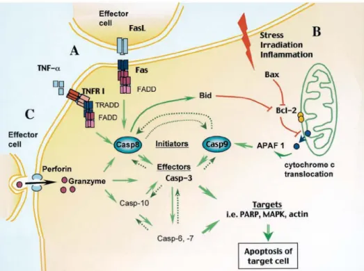
receptor-mediated apoptotic pathways. 76
Fig. 6.2|P. yoelii HSP70 protects hepatocytes from apoptosis. 79
Index of Tables
Table 3.1|Antibodies used in immunofluorescence stainings. 35
C
hapter One
Background,
Significance & Objectives
1.1 Background and Significance
Malaria is one of the most prevalent and severe human infectious diseases in the world. Recently its incidence has increased and it is estimated more than 300 million episodes of acute illness occur in endemic countries and at least 1 million people die per year from this disease (WHO, 2003). With 41% of the world’s population exposed to this threat, malaria also imposes an extreme burden onto affected populations as economy and development are deeply impaired resulting from the symptoms experienced by infected individuals. Applicable measures to control the disease vary according to each country’s endemicity but several factors contribute to malaria resurgence such as drug-resistant parasite strains and insecticide-resistant mosquitoes (Greenwood et al., 2005; WHO, 2003).
After decades of a relative lack of attention, new efforts are presently being made to address these challenges, such as new strategies that are being applied to the development of an effective vaccine (Moorthy et al., 2004; Tongren et al., 2004). Although in terms of public health vaccination has always been a priority, it still remains an elusive and complex research field. A comprehensive knowledge of the host-parasite interface would constitute a reinsured guarantee for attaining this goal successfully.
Malaria is caused by the intracellular Apicomplexan parasite Plasmodium spp., which holds a complex life cycle involving different hosts and stages of infection. The first step in malaria infection is the invasion of the liver by Plasmodium sporozoites. These are the infective form of the parasite, transmitted by female anopheline mosquitoes during a blood meal. Inside hepatocytes, sporozoites replicate and develop into a merozoite state, a process that constitutes the hepatic stage of the parasite life cycle. Although, being an obligatory step towards the establishment of a successful malaria infection, this stage of Plasmodium life cycle is poorly understood. This parasite specificity towards the liver cells indicates that parasite-encoded surface proteins and host surface receptors are implicated in the process of invasion and development, playing a key role in the establishment of infection. However, the mechanisms, as well as the host and parasite molecules, at play during the course of infection in the mammalian host by malaria sporozoites are not entirely known (Gruner et al., 2003; Miller et al., 2002; Plebanski and Hill, 2000).
The full requirements for Plasmodium development inside hepatocytes are still unknown. The lack of an adequate in vitro system capable of delivering sufficient material has deeply impeded research within the study of host/parasite interactions. Currently, in vitro cultivation methods have been developed, and hepatoma cell lines are being used as in vitro models for Plasmodium infection to study the molecular and cellular basis of invasion mechanisms and intracellular development of the parasite. Nevertheless, infection yields remain very low and the limited number of infective
mosquitoes available impairs a swift progress in the understanding of malaria liver stage biology (Mota and Rodriguez, 2000; Calvo-Calle et al., 1994; Hollingdale et al., 1983b).
The availability of the genome sequence of both several Plasmodium species and their host provides a genetic tool that greatly increases the possibilities of research in this area (Cooke and Coppel, 2004).
The liver, besides being the place where amplification and molecular changes of Plasmodium parasites take place, is also a unique organ in what regards the host’s immune responses (Knolle and Gerken, 2000). The antigenic pool generated within this organ is required for the induction and maintenance of a protective anti-malaria immune response. Thus, the hepatic stage may well hold the secret for understanding the parasite’s preference for this organ within the mammalian host and, simultaneously, providing effective immune targets against the disease (Baldacci and Ménard, 2004; Frevert, 2004; Krzych et al., 2000).
Unlike many other diseases in which a lifelong resistance to re-infection is induced, malaria causes only partial immunity after several years of recurring infections and illness. Nonetheless, a complete resistance to malaria can be achieved by vaccination with radiation-attenuated sporozoites in both mice and humans (Doolan and Hoffman, 2000; Weiss, 1990; Clyde et al., 1975; Nussenzweig et al., 1972; Nussenzweig et al., 1967).
Irradiated sporozoites infect hepatocytes as normal sporozoites, but they do not reach a merozoite stage (Scheller et al., 1995; Sigler et al., 1984). Additionally, it is known that it is essential that a hepatic stage occurs during infection for the effectiveness of irradiated-sporozoite protection (Scheller and Azad, 1995). Although, until now this is the only vaccine that confers complete protection, the mechanism behind the process of the establishment of the immune response associated is still unclear (Doolan and Hoffman, 2000).
Intracellular pathogens have the capacity to modulate the host cell response and exploit its resources in order to develop and replicate. Parasites can manipulate host cell behavior, including immune modulation and regulation of apoptosis (James and Green, 2004). While infected cells are capable of initiating their own death, a process called apoptosis, which can be used by the organism as a defense mechanism against pathogens, inhibition of host cell apoptosis is frequently used by parasites as a strategy for survival (Heussler et al., 2001; Luder et al., 2001).
During the course of infection, apoptosis of infected host cells may either be induced by the host cell response or be a direct result of pathogen invasion. In both cases, apoptotic death results in the formation of apoptotic bodies. These apoptotic bodies are taken up by phagocytes that rapidly recognize and phagocytose them, eliminating the parasite together with the remains of the infected cell. The pathogen-derived antigens included in apoptotic bodies can be presented by dendritic cells in the context of both class I and class II molecules, which are recognized by CD8+ and CD4+ T
cells respectively, and activate naïve lymphocytes for the initiation of an immune response (Schaible
et al., 2003; Savill et al., 2002; Rodriguez et al., 1999; Albert et al., 1998a).
Dendritic cells are key players in initiating immune responses because they are the only cell type that are able to prime naïve T cells efficiently as well as cross-present exogenous antigens (Banchereau et
al., 2000; Mellman et al., 1998).
It has been shown that dendritic cells are able to induce P. yoelii-specific CD8+ and CD4+ T cells
(Bruna-Romero and Rodriguez, 2001). Additionally, dendritic cells-depleted mice failed to induce cytotoxic T cell responses with a result in the loss of priming potential of dendritic cells during P.
yoelii infection (Jung et al., 2002).
Altogether, these observations suggest an important role of dendritic cells during the immune response initiated against liver stages. An extended comprehensive knowledge of these molecular and cellular events will provide promising immediate applications in the field of vaccine development.
1.2 Objectives
Hepatocyte infection by gamma-irradiated Plasmodium sporozoites is required to achieve complete protection against malaria. The basic trigger of this conferred immunity is still unknown. Conversely, apoptotic bodies derived from infected cells enclose a source of antigens that are processed by dendritic cells, which efficiently present pathogen antigens for the initiation of immune responses. The major goal of this project is to elucidate the role of hepatocyte apoptosis in the course of a malaria liver infection.
For this purpose, we focused on two main aspects. First, it was determined whether apoptotic death of hepatocytes infected with irradiated sporozoites would play a role in the initiation of an anti-malarial immune response. The immunological mechanisms that mediate this protection were studied, namely the origin of Plasmodium antigens, and the way in which its processing and presentation occur. Furthermore, the involvement of dendritic cells as key mediators of the protective immunity conferred by irradiated-sporozoites immunization was also examined.
Secondly, molecular aspects of parasite survival and development during the establishment of a successful liver infection were also addressed. The hypothesis of hepatocyte apoptosis modulation by the parasite was studied as well as the involvement of a host cell pathway.
Additionally, an attempt was made at identifying a Plasmodium gene product as a candidate responsible for the inhibition of apoptosis of hepatocytes. In this context, the role played by the parasite’s heat shock protein 70 during liver infection was studied.
C
hapter Two
GENERERAL
INTRODUCTION
2.1 Malaria: historical aspects and current global picture
The malaria situation deteriorated during the 90’s and recent estimates have shown an increase in numbers of morbidity and mortality as a result of overlooked prevention measures together with the widespread of resistance to drugs and insecticides. To overcome this situation new efforts are being undertaken with a focus on both cure and prevention of this disease. Attempts to address these challenges include the use of new technologies in research and the combination of different therapies in the field (Breman et al., 2004; Nchinda, 1998).
The fight against this menace is currently being pursuit by an international partnership launched by the World Health Organization (WHO), the Roll Back Malaria, whose goal is to attain a 50% reduction of the burden of malaria by 2010. The impact of this achievement will reach far beyond the disease burden itself since it will implicate several factors and success will result in a global development for the countries at risk and the world’s populations in general (WHO, 2003; Fig. 2.1). Malaria is a parasitic disease caused by the intracellular pathogen Plasmodium and transmitted by a mosquito vector. In ancient times it was believed that the disease had its origin in the injurious air inhaled from swamps and marshes, thus the name ‘mal aria’ (bad air). In 1880, Lavern’s discovery of the unicellular parasite P. falciparum in the blood of a French soldier put an end to this belief (Laveran, 1880). In 1897, the mode of transmission to humans was uncovered and attributed to the female Anopheles mosquito bite by Ronald Ross (Ross, 1897). The missing part of the malaria parasite’s cycle, the exo-erythrocytic liver schizont, was only elucidated in 1948 by Shortt and Garnham, when malaria parasites where found developing in livers of sporozoite-infected monkeys and, subsequently, in livers of human volunteers infected by mosquitoes carrying P. vivax (Shortt and Garnham, 1948).
Presently, around 40% of the world’s population is at risk of infection and more than a million people die each year, mostly children under five years of age and pregnant women. Symptoms appear within a week or two after transmission and consist of fever, nausea, vomiting, fatigue and headaches. They can progress into organ failure, coma and death, 90% of which occurs in tropical sub-Saharan African countries. Eradication of malaria from western European countries was achieved after the World War II when the anti-malarial drug chloroquine and the insecticide DDT became widely available (Hay et al., 2004; Greenwood and Mutabingwa, 2002).
The search for a safe and effective vaccine against malaria has become a case of endless failures. Years of vaccine research have produced few hopeful candidates and although scientists are doubling research efforts, an effective vaccine is, at best, years away. In terms of epidemiology, human malaria is extremely complex, depending on transmission levels and acquired immunity. The relationship between the prevalence of severe malaria phenotypes and higher transmission rates is
not linear. In fact, exposure during early childhood could account for a lower risk of severity as protection is stimulated against subsequent attacks (Snow et al., 1997).
There are four Plasmodium species that infect humans: P. vivax, P. falciparum, P.malariae, and P. ovale. The first two are the most common, with P. falciparum being the most deadly form of the parasite and responsible for the majority of the worst case scenarios of severe malaria infection. P. vivax accounts for the major cause of morbidity but is no longer a significant cause of mortality. A wide range of clinical symptoms, including fever, life threatening anemia, and coma in children and naïve adults characterize infection with P. falciparum (Greenwood et al., 2005; Trigg and Kondrachine, 1998).
The present crisis is mainly due to resistance developed against drugs by Plasmodium parasites and against insecticides by Anopheline mosquitoes. Additionally, national transmission control programs have weakened and increased migration and tourism contribute to the current situation (Greenwood and Mutabingwa, 2002). Malaria’s burden has an impact on the economical and political prospects of developing countries in which disease is endemic; therefore concerted funding for interventions and research priorities such as drug/vaccine development are being used to overcome this deteriorating situation (Sachs and Malaney, 2002).
In particular, vaccine development requires the achievement of several goals: induction of strong, durable and strain-transcending immune responses; identification of protective antigens for stage-specific immunity; and successful combination of candidate immunogens (Greenwood, 2005; Carvalho et al., 2002).
In humans, the first report of protection induced by a vaccine occurred in 1973 when volunteers where being submitted to infected-gamma-irradiated mosquito bites (Hoffman et al., 2002; Clyde et
al., 1973a). However, this approach was impracticable at a large-scale, and so a search for
molecules-based vaccines was initiated. Throughout the last two decades, success in protection of animal experimental models raised the enthusiasm about a few candidate molecules, but these met with failure during human trials. More recently, efforts were translated in a pack of potential candidates that are currently under clinical assessment (Moorthy et al., 2004; Moorthy and Hill, 2002).
A B
Fig. 2.1|Global distribution of malaria. (A) World’s malaria transmission risk in 2003 and (B) the estimated incidence of
clinical malaria episodes caused by any Plasmodium species, resulting from local transmission, country level averages in 2004. (adapted from Roll Back Malaria partnership report, 2005)
2.2 Plasmodium life cycle
Plasmodium presents an extremely complex life cycle involving two hosts, a vertebrate host, and the
invertebrate host, the female Anopheles mosquito vector. Interaction between them results in transmission and allows the infection to endure. Parasites enter the mammalian host through the bite of an infected female Anopheles mosquito during a blood meal. During such a meal, approximately five to twenty sporozoites will be injected into the host, which reach the liver within minutes (Vanderberg and Frevert, 2004; Ponnudurai et al., 1991; Rosenberg et al., 1990; Vanderberg, 1977). The sporozoites are deposited under the skin of the host, migrate into the bloodstream and aim directly to the liver, where they infect and develop inside hepatocytes (Matsuoka et al., 2002; Sidjanski and Vanderberg, 1997; Shin et al., 1982). This is known as the asexual exo-erythrocytic or hepatic stage of the parasite’s life cycle. For human malaria, this stage lasts 5 to 7 days on average while in rodent malaria it lasts only 2 days (Meis and Verhave, 1988). This is also the asymptomatic stage of the disease. At the end of the hepatic stage, 10.000 to 30.000 merozoites per invading sporozoite will be released into the blood-stream from where they disseminate systemically. Each merozoite invades an erythrocyte and divides mitotically to form an erythrocytic schizont, containing up to 20 daughter merozoites. These merozoites can re-infect fresh erythrocytes, giving rise to a cyclical blood stage infection with a periodicity of 48-72 hours, depending on the Plasmodium species. This constitutes the asexual blood stage of a malaria infection. In this stage, disease symptoms occur and infected individuals can get sick. In order to complete the cycle, there are some merozoites that develop into sexual parasite stages, the male and female gametocytes, which will be taken up by mosquitoes during blood meals. Gametocytes undergo fertilization and maturation in the mosquito midgut, forming an infective ookinete form that migrates into the mosquito hemocele
and develops into an oocyst form where sporozoites are developed (Miller et al., 2002; Gysin, 1998; Landau and Gautret, 1998). When fully matured, oocysts burst and release sporozoites which migrate into the mosquito’s salivary glands, completing Plasmodium’s life cycle (Fig. 2.2).
Fig. 2.2|Plasmodium life cycle.
When an infected female anopheline mosquito takes a blood meal, sporozoites enter the host bloodstream and travel through the circulation to the liver (A). Sporozoites invade hepatocytes and enter a phase of asexual reproduction in which they amplify their number thousands of times by the production of merozoites (B). The liver cell ruptures and the merozoites are released into the blood, attaching to and invading erythrocytes, beginning the erythrocytic cycle. Each merozoite invades an erythrocyte and replicates initiating a cycle that ends with the burst of the mature erythrocytic schizont and the release of new merozoites, which will infect new erythrocytes. Illness starts when the mature asexual erythrocytic schizont, ruptures (C). Other blood stages differentiate into male and female sexual stages parasites called gametocytes. These gametocytes enter a mosquito as it takes a blood meal. Sexual reproduction occurs in the mosquito midgut, and gametes fuse forming a motile zygote, the ookinet, that mature and migrates through the mosquito midgut developing into an oocyst, within which sporozoites develop. After being released, novel sporozoites travel to the salivary glands, making the mosquito infective (D). A B D C A B D C
2.3 Liver stage biology
The discovery of a mammalian malaria exo-erythrocytic stage was the last missing piece to be filled in the parasite’s life cycle, when liver schizogony was described in African monkeys (Shortt and Garnham, 1948). Since no symptoms are associated with this parasite stage, little attention was given to the events that occur during sporozoite invasion of hepatocyte and the intrahepatic development. However, this scenario changed when in 1967, Nussenzweig reported that immunizing mice with radiation-attenuated Plasmodium berghei sporozoites protected them against challenge with fully infectious sporozoites (Nussenzweig et al, 1967). These rodent studies provided the impetus for human studies, and, during the 1970s, Clyde, Rieckmann and colleagues established that immunizing human volunteers with the bites of irradiated mosquitoes carrying P. falciparum sporozoites in their salivary glands could protect volunteers against challenge with fully infectious P. falciparum
sporozoites (Edelman et al., 1993; Herrington et al., 1991; Rieckmann, 1990; Rieckman et al., 1979; Clyde et al., 1975; Rieckman et al., 1974; Clyde et al., 1973 a, b)
Still until the mid-1980s, knowledge of the biology of malaria liver stage was restricted to the fact that these forms of the parasite could arrest in the liver and later be responsible for relapses during infections with certain Plasmodium species, the so-called uninucleated “hypnozoite” form (Cogswell et
al., 1991; Krotoski et al., 1980).
Since then, experimental data from several laboratories began to elucidate the numerous steps taken by sporozoites from the starting point of invasion to their development inside the liver as well as the immune response that occurs during different immunization strategies with special emphasis for the immunization with attenuated parasites (Engwerda and Good, 2005; Baldacci and Menard, 2004). The study of the cell biology of the Plasmodium liver stage is particularly difficult due to lack of a high infectious in vitro culture system, isolation and number of sporozoites needed. Still, the recent advance in direct live observation of parasites within the liver represents a useful resource, promising an outbreak of insights into this field (Frevert et al., 2005; Hollingdale et al., 1998; Calvo-Calle et al., 1994; Hollingdale et al., 1983b).
The reasons why Plasmodium has elected the liver and the hepatocyte as a first cellular home inside mammalian hosts are not fully elucidated. However, it is possible that the reason is related to the hepatocyte’s highly complex metabolism, which is capable of fulfilling all parasite replication needs (Frevert et al., 2004; Saliba and Kirk, 2001). Others support the idea that the immunologic characteristics of this organ allow parasites to survive and pursue infection, either by minimal immune responses that are thought to occur or by the induction of immune tolerance. In addition, hepatocytes are capable of expressing major histocompatibility complex (MHC) class I and class II, while erythrocytes are not (Crispe, 2003; Krzych et al., 2000; Rajan, 1997). Another aspect to take into account is the morphology of the liver itself and the fact that hepatocytes are heterogeneous and allow easy access to venules and arteries separated by the space of Disse (Enomoto et al., 2004; Wisse et al., 1985).
2.3.1 From skin to liver
To reach the first and essential stop of its journey, the hepatocyte, Plasmodium sporozoites have to travel from the skin inoculation site to the liver. Being a highly vascularized organ, the skin is the perfect place for a mosquito blood meal, which usually last for 30s in Anopheles and occurs after a single probe of blood taken in small pools originated by capillary damage. Mosquito injections are accompanied by saliva, which has an anti-coagulant activity, facilitating blood digestion. In spite of the hundreds of sporozoites present in the mosquito’s salivary glands only a small number are
transmitted during the bite of an infected mosquito (Matsuoka et al., 2002; Ponnudurai et al., 1991; Rosenbergh et al., 1990; Vanderberg, 1977; Griffiths andGordon, 1952; Boyd and Kitchen, 1939). The migration of the sporozoite from the site of bite to the liver has been an issue under discussion. Previous studies with P. yoelii-infected mosquitoes allowed to feed in mice, provided evidence that mosquitoes deposit sporozoites in an avascular skin tissue area and, within 10 minutes post-inoculation, sporozoites could be found in the host’s bloodstream, long enough to find a blood vessel (Sidjanski and Vanderberg, 1997). A recent study applying intravital observations of P. berghei-infected mice at the site of mosquito bite revealed that parasites migrate widely across the skin covering distances of many micrometers for several minutes before reaching circulation (Vanderberg and Frevert, 2004). Anti-sporozoite acquired immunity reduces motility speed and could interfere with skin crossing (Frevert et al., 2005; Vanderberg and Frevert, 2004). Either because they find themselves in a new rough environment when they change from salivary glands to connective tissue or to prevent hostile encounters with the host immune system defenses, sporozoites have a short life span of approximately of 20 minutes to leave circulation in liver sinusoids and enter the parenchyma to infect hepatocytes.
The gliding motility presented by sporozoites is used during infections in vivo for their migration through the dermis, as was previously demonstrated in vitro (Vanderberg and Frevert, 2004; Vanderberg, 1974). Plasmodium gliding movement in host cells is characterized by trails of circumsporozoite (CS) protein that are left behind, similar to what occurs when they are placed in contact with artificial surfaces (Frevert et al., 1998; Stewart and Vanderberg, 1991). The released CS protein is distributed throughout the cytoplasm of the cell and it has been proposed thatis capable of inhibiting translation at the initiation step of protein synthesis, as it binds to RNA-associated binding sites on ribosomes (Frevert et al., 1998; Hugel et al., 1996). Nevertheless, mosquito-transmitted sporozoites are more infective than sporozoites injected intravenously, and are even capable of avoiding the antibody response mounted against them (Krettli and Dantas, 1999; Vaughan et al., 1999; Beier et al., 1991; Ponnudurai et al., 1991).
2.3.2 Reaching the liver
The liver is organized in lobules formed by connective tissue with branches for the portal venule and the hepatic arteriole. It possesses a diverse population of cells such as specialized endothelial cells, Kupffer cells (liver resident macrophages) and stellate cells (fat-storing cells) (Enomoto et al., 2004; Sinnis, 1996).
There are several interactions established between sporozoite proteins and the liver cells components. Malaria sporozoites possess an apical complex constituted by secretory organelles, unique to Apicomplexa parasites and essential for the invasion process. The major component of
the surface coat of Plasmodium sporozoites is the CS protein, which is responsible for the oocyst maturation and sporozoite morphogenesis (Kappe et al., 2004; Nussenzweig and Nussenzweig, 1989). It is mainly stored in the micronemes (sporozoites secretory organelles) and is continuously exported to the cell surface and discarded at the parasite posterior pole (Thathy et al., 2002; Stewart and Vanderberg, 1991; Posthuma et al., 1989). It also possesses a glycosyl-phosphatidylinositol (GPI) sequence as anchor at the C-terminus of the CS protein, which also contains a species-specific central repeat region and two conserved motifs. Specific regions within these motifs are responsible for the linkage to the glycosaminoglycan of liver sulfated proteoglycans and sporozoite motility and invasion (Tewari et al., 2002; Ying et al., 1997).
Another protein present in the micronemes that is also expressed at the sporozoite membrane surface upon secretion induced by cell-contact is the thrombospodin-related adhesive protein (TRAP) (Bhanot et al., 2003; Gantt et al., 2000; Templeton and Kaslow, 1997). Besides being involved in sporozoite invasion of salivary glands and the hepatocyte, its vital role was assessed when P. berghei mutant TRAP parasites were shown to lose their gliding motility (Matuschewski et al., 2002; Wengelnik et al., 1999; Sultan et al., 1997).
On the host side, the molecules that seem to play an important role during infection by allowing sporozoites to recognize and reach the liver are the proteoglycans. These molecules, which are highly sulfated and abundant, are involved in the molecular mechanism of sporozoite adhesion, where CS protein binds to hepatocyte membranes (Frevert et al., 1993; Cerami et al., 1992). Both CS and TRAP are capable of recognizing in particular the glycosaminoglycan chains (GAG) in the sulfated proteoglycans expressed by liver cells (Ying et al., 1997; Robson et al., 1995).
When entering the liver sinusoids either via the hepatic arteriole or the portal venule, sporozoites glide along the endothelial cell layer through the endothelia, interacting with extracellular matrix proteoglycans that protrude from the space of Disse until they encounter a Kupffer cell and recognize the proteoglycans expressed on its surface (Pradel et al., 2002; Pradel and Frevert, 2001). Although previous work with P. berghei in rats suggested that these cells were responsible for parasite phagocytosis removing them from circulation, recently Kupffer cells were shown to be the liver portal entry to sporozoites from a set of experimental data based on in vitro models with P. berghei and P. yoelii sporozoite infections and Kupffer cells isolated from rat livers (Pradel and Frevert, 2001; Meis et al., 1983). Moreover, the development of most exo-erythrocytic forms close to the liver portal venules, where the Kupffer cells are located, also suggests that the these cells could well function as access gates to the liver (Verhave et al., 1985; Sleyster and Knook, 1982).
Thus, the current model proposes that sporozoites recognize and bind to proteoglycans expressed on the surface of Kupffer cells using their major surface proteins, CS protein and TRAP (Pinzon-Ortiz et al., 2001; Cerami et al., 1992). They actively invade, safely traverse and successfully exit
Kupffer cells protected by a non-fusogenic vacuole through the space of Disse (Frevert et al., 2005; Meis et al., 1983c).
2.3.3 Hepatocyte invasion
Hepatocyte infection occurs after sporozoites exit the space of Disse and gain access to the liver parenchyma. Sporozoites first migrate through several hepatocytes before invading a final one within a parasitophorous vacuole. The traversed cells have their plasma membrane wounded by the parasite and can either survive, undergoing a resealing process, or die (Mota et al., 2001). Experimental evidence demonstrates that when sporozoites invade host cells without formation of a vacuole the parasites do not develop, meaning that migration is a feature that precedes infection (Mota et al., 2001). This migration of parasites leaves behind wounded cells that could be needed to potentiate infection, which would be beneficial for the parasite’s development within infected hepatocytes. On the other hand, one could also suppose that wounding would increase the generalized inflammation detectable shortly after inoculation (Khan and Vanderberg, 1992). This particular sporozoite feature can be observed either in vitro or in vivo in mice livers, and it is unspecific as is shown by the fact that a range of cell types are suitable for hosting Plasmodium migration (Mota et al., 2001). Moreover, during parasites in vivo journey from the skin to the liver they encounter different tissues and probably migrate through them (Mota and Rodriguez, 2004). The molecular mechanism is still not fully elucidated, but recent reports revealed a newly discovered sporozoite protein that plays a role in migration. The SPECT protein (sporozoite microneme protein essential for cell traversal), which is localized in sporozoites micronemes, is required for the parasite’s migration in vitro. Yet, exo-erythrocytic development was maintained, and a lower infectivity was shown in vivo, suggesting that host cell migration is mandatory for sporozoite access into the liver parenchyma (Ishino et al., 2004). Recently, another SPECT protein was described and a similar function was shown for a protein present in mosquito ookinete invasion into the midgut epithelium (Ishino et al., 2005a; Kadota et al., 2004).
The final invasion is accompanied by the secretion of TRAP and the parasite finds itself surrounded by a parasitophorous vacuole, inside which it replicates and develops (Silvie et al., 2004; Mota et al., 2001; Meis et al., 1983b). Detailed information about how this event takes place is scarce, in part because of the low infectivity of sporozoites in vitro. By analogy with related parasites such as
Toxoplasma gondii, it is believed that sporozoite internalization with formation of a parasitophorous
vacuole occurs within a few seconds and is dependent on the plasma membrane-associated motor that also drives parasite gliding motility (Soldati and Meissner, 2004). In addition, there is an associated intense secretory activity in their apical end, which is either a constitutive exocytosis of
molecules from within the apical organelles, or a regulated exocytosis restricted to the formation of a cap (Mota and Rodriguez, 2004; Mota et al., 2002).
Moreover, migration induces secretion of “hepatocyte growth factor” (HGF) that render the surroundings more adequate to parasite growth and are imperative in order to obtain a successful infection (Carrolo et al., 2003). Experimental evidence has suggested that other host cell factors are involved in sporozoite infection in the liver, for example, the interaction of the CS protein with the low-density lipoprotein-related protein receptor or the requirement of CD81 tetraspanin for P.
falciparum and P. yoelii sporozoites to invade hepatocytes (Silvie et al., 2003; Shabkibaei and Frevert,
1996).
2.3.4 Intrahepatic development
Knowledge concerning parasite development inside hepatocytes is still very limited considering the large range of morphological characteristics that occurs during this intrahepatic development.
Hepatic schizogony, where parasites grow, maturate and replicate, lasts 2 days for rodent species and 5 to 7 days for primates, resulting in 10.000 to 30.000 of merozoites from each invading sporozoite (Meis et al., 1985 a, b; Meis et al., 1983 a, b). From seize a single generation, merozoites individualize in the cytoplasm, and then separate as islets within which membrane formation occurs before rupture into the bloodstream. An infective mosquito bite leads to the formation of around 12 exo-erythrocytic schizonts in a few days, reaching 100 µm in size, contrasting with blood stages that only enlarge up to 12 µm, replicating into 8 to 24 erythrocytic schizonts. Thus, the parasite’s needs for membrane and nucleic acid synthesis during the liver stage are enormous, almost three times more than for blood stages schizogony (Hollingdale, 1985). Nutrients are obtained from the hepatocyte which as it harbors glycogen and serum protein factories, allowing the maturation process to progress ending in the release of merozoites that will invade erythrocytes (Frevert, 2004; Meis et al., 1985 a, b).
However, sporozoites are able to undergo partial development to early exo-erythrocytic forms in the absence of host cells or other cell types, and preserve some morphological and molecular features presented by regular ones (Wang et al., 2004; Kaiser et al., 2003).
2.4 Immunity to malaria liver stage
When naturally exposed to malaria, humans gradually acquire immunity to the parasite, although repeated infections are required in order to maintain it. Naturally acquired immunity is characterized by being short-lived and strain-specific (Hviid, 2005; Day and Marsh, 1991). The reasons for such General Introduction|17
scarce protection are not known. It is mediated by blood-stage specific antibodies and is partially T cell-based (Stevenson and Riley, 2004; Good and Doolan, 1999). But the contribution of each of the different stages to the general immunity-related protection remains poorly understood. During the blood stages it is clear that the host develops mechanisms either to neutralize the effects of parasite-released toxins or to kill the parasite or even to inhibit its replication. The reduced acquisition of malaria immunity in naturally exposed populations has also been explained by the fact that the parasite actively modulates the immune system of the host during blood stages, preventing the development of specific immune responses (Ocana-Morgner et al., 2003; Urban et al., 1999; Plebanski et al., 1997). Concerning the liver, the fact that the gut is located close to the liver is one of the reasons why this organ has such a peculiar immunologic profile, predominantly tolerogenic when responding to foreign antigens. Liver dendritic cells, Kupffer cells and sinusoidal endothelia are mature antigen-presenting cells that undertake the task of maintaining this immune environment (Doherty and O’Farrelly, 2001; Knolle and Gerken, 2000; Lohse et al., 1996). Together with anti-inflammatory cytokine secretion, the liver controls the inflammation induced by influx of bacteria and endotoxins from intestines, and reaches a state of portal vein tolerance. Antigen-specific activated CD8+ T cells are eliminated by Fas-induced apoptosis considering the model of
activation-induced cell death, thus accounting for liver tolerance (Crispe, 2003).
Nevertheless, naturally acquired protection observed in infected individuals and several experimental data records, raised hopes for the development and feasibility of an effective vaccine against this disease (Carvalho et al., 2002).
In what regards the liver stage of infection, major hopes were raised based on the fact that sterile protective immunity was obtained both in mice, monkeys and humans; against P. berghei, P. yoelli, P.
knowlesi, P. falciparum or P. vivax sporozoite challenge induced by immunization with
radiation-attenuated sporozoites (Hoffman et al., 2002; Gwadz et al., 1979; Clyde, 1975; Clyde et al., 1973 a, b; Nussenzweig et al., 1969; 1967).
Protective mechanisms against the liver stage of malaria infection have been the aim of many studies, since knowledge concerning the type of immune responses induced towards the infected hepatocyte are still very limited. The advantages of inducing exo-erythrocytic stage immunity would not only be a decrease in mortality and disease transmission, but also a prevention of symptoms, since the parasite would be arrested before reaching the erythrocytic stage (Tdryk and Walther, 2005; Tsuji and Zavala, 2003).
2.4.1 Natural infection versus immunization
Mimicking features occurring in naturally infected individuals is the general aim of vaccines which, in turn, means that they must be capable of inducing antibodies and T cell responses, if possible to
more than one antigen. In particular, a greater magnitude in responses than the one achieved in individuals that have been infected, together with transcendence both in time and strains, would be desirable for the development of a vaccine against malaria. Administration of a cocktail of several antigens has been the prevailing hypothesis for many years now, but an expensive and complex product would be the likely result. Ideally, a vaccine against the exo-erythrocytic stage of the parasite should fulfill two roles: the induction of high titers of functional antibodies against sporozoites inoculated by the infectious mosquito, in order to stop them from entering the liver; the establishment of potent cytotoxic T lymphocyte immunogenicity against the liver stage to kill all infected hepatocytes while not harming the host (Hill, 2006; Doolan and Hoffman, 2000; Doolan and Hoffman, 1997).
In that respect, the discovery that immunization with radiation-attenuated sporozoites could lead to full protection paved the way to determine the full range of factors involved in this protection, namely Plasmodium antigens, initiation of host immune response and immune response types (Nardin
et al., 1999). Gamma-irradiated sporozoites infect the liver but are arrested there without pursing to
blood stages. Later, it was also found that the targets of this protection were not only the sporozoite but mainly the infected hepatocyte (Scheller and Azad, 1995; Suhrbier et al., 1990; Weiss, 1990; Hoffman et al., 1989). In fact, sporozoites irradiated with a dose that did not allow them to reach the liver were not protective any more (Silvie et al., 2002; Nussler et al., 1989). Still, is not clear if this relies on the presentation of different and specific antigens or on the way that antigens are presented to the host immune system (Langhorne et al., 2004; Krzych et al., 2000).
2.4.2 Exo-erythrocytic antigens
Some sporozoite antigens are also expressed within the newly formed liver stage forms. In fact, a limited number have been already identified and characterized as targets for CD8+ T cell responses.
Among these are the CS protein and the TRAP protein (Khusmith et al., 1994; Rogers et al., 1992; Weiss et al., 1992). Another protein that has also been shown to be a target for T cells is the P. yoelii hepatocyte exported protein 17 (PyHEP17), an homolog of the P. falciparum exported protein 1 (PfEXP) (Doolan et al., 1996).
The first strictly specific malaria liver stage antigen discovered was the P. falciparum liver stage antigen 1 (PfLSA-1), which is expressed in the vacuole lumen and is solely expressed during the hepatic stage (Guerin-Marchand et al., 1987). The LSA-3 antigen was characterized through antibody recognition in immunized and non-immunized individuals (Daubersies et al., 2000; Connelly et al., 1997). A few other parasite proteins appear at this stage of the parasite’s life cycle, such as glutamine-rich protein and parasite heat shock protein 70 (Kumar et al., 1993). More parasite