%'-,.)*(%*-#/"-#)'*%*0%-",.,1#)*
!!"#$%&'%(#)'*!"#!+,*-*.,%
! ! ! !23456789:;43*9<6*05:48=5>73;3
?**
3487>7@;=*4A;64<=4*7B*;<B4=C;7<*;<*D;>6*:798*E7E5>9C;7<3*;<*
375CF493C*278C5@9>*9<6*9337=;9C46*8;3G3*B78*#:48;9<*>H<I*
=7<348A9C;7<*
! !$9<4339*)>4I9<689*A;4;89*'74;87*
*
!*/0123456/7!$/681996/!$4:;6!<=;06!$1/10/4!(4/3029!%;>19
!<6?6/0123456/7!"6:36/!<@/093042!.6/3AB4/!CD@E053!
!
! ! J43C8967*4K*L;7>7@;9*69*-7<348A9MN7* !"0991/34FG6!! ! ! H>6/4I!JKLM! !%'-,.)*(%*-#/"-#)'*%*0%-",.,1#)*
!!"#$%&'%(#)'*!"#!+,*-*.,%
! ! ! !23456789:;43*9<6*05:48=5>73;3
?**
3487>7@;=*4A;64<=4*7B*;<B4=C;7<*;<*D;>6*:798*E7E5>9C;7<3*;<*
375CF493C*278C5@9>*9<6*9337=;9C46*8;3G3*B78*#:48;9<*>H<I*
=7<348A9C;7<*
! !$9<4339*)>4I9<689*A;4;89*'74;87*
*
!*/0123456/7!$/681996/!$4:;6!<=;06!$1/10/4!(4/3029!%;>19
!<6?*/0123456/7!"6:36/!<@/093042!.6/3AB4/!CD@E053!
!
! ! J43C8967*4K*L;7>7@;9*69*-7<348A9MN7* !"0991/34FG6!! ! ! H>6/4I!JKLM! ! !Agradecimentos
Aos meus orientadores Prof. Dr. Paulo Célio Alves e Prof. Dr. Christian Gortázar Schmidt pela sua disponibilidade.
Ao Parque Biológico de Gaia na pessoa do Dr. Nuno Gomes Oliveira pelo tempo dispensado para chegar ao fim de mais esta etapa.
Aos gestores de zonas de caça, organizadores de montarias e caçadores que permitiram e algumas vezes colaboraram na recolha de amostras dos javalis abatidos.
Aos meus colegas de trabalho de campo, Nuno Santos, João Fragoso, Filipa Cabecinhas, aos veterinários inspectores sanitários de caça maior, assim como aos colaboradores do IREC que tão bem me acolheram, ajudando sempre que possível.
Ao Prof. Dr. José Alberto Gonçalves e Prof. Dr. António Múrias pela ajuda e disponibilidade.
A todos aqueles que me incentivaram, acompanharam, ajudaram, e porque não dizê-lo, me aturaram, nomeadamente a minha família, Joana Reis, José Paulo Pires, José Potes Sara Lóio, Hugo Oliveira, Henrique Alves, Joaquim Peixoto, Engª Joana Almodôvar, António Coelho e Pedro Pinto, o meu enorme agradecimento.
Pseudoraiva e Tuberculose: evidência serológica de infecção
em Javali no Sudeste de Portugal e riscos associados para a
conservação do Lince-ibérico
Resumo
Em Portugal foi recentemente iniciado um programa que visa o restabelecimento da população de Lince-ibérico. O rastreio do estado sanitário das populações selvagens é essencial para qualquer programa de reintrodução. O presente estudo teve como principal objetivo estimar em javalis caçados a taxa de contacto com os agentes etiológicos da pseudoraiva (também conhecida por doença de Aujesky) e da tuberculose bovina, na área com maior potencial para a reintrodução de Lince-ibérico no sudeste de Portugal. Para o efeito foram realizados dois testes diferentes de ELISA para deteção de anticorpos contra o vírus da pseudoraiva (PrV) e o complexo Mycobacterium tuberculosis. Os resultados evidenciaram uma taxa de seroprevalência para PrV de 45,9% que está entre as mais altas documentadas até à data. Os anticorpos contra
Mycobacterium bovis foram detetados em 9,9% das amostras, o que se encontra
dentro do intervalo de valores para taxas de infeção por M. bovis previamente referidos. A prevalência destas doenças numa espécie reservatório, abundante, simpátrica e, ocasionalmente, presa do Lince-ibérico, pode constituir um importante problema para o programa de reintrodução devendo, por esse facto, ser implementado um sistema de vigilância sanitária e exploradas opções de prevenção.
Palavras-chave: Doença de Aujeszky; Complexo Mycobacterium tuberculosis;
Pseudorabies and Tuberculosis: serologic evidence of infection
in wild boar populations in Southeast Portugal and associated
risks for Iberian lynx conservation.
Abstract
An important effort has been recently endeavoured to promote the reestablishment of Portuguese Iberian lynx population. The awareness of the sanitary status of wild populations is essential in any reintroduction program, and the present study aimed to estimate the rates of contact with causative agents of pseudorabies (also known as Aujeszky’s disease) and bovine tuberculosis in hunted wild boar from the foremost Iberian lynx potential releasing site in Southeast Portugal. To achieve this purpose two different ELISA tests were performed in order to detect serum antibodies against pseudorabies virus (PrV) and Mycobacterium tuberculosis complex. Results evidenced a seroprevalence rate for PrV of 45,9%, which is amongst the highest so far documented.
Mycobacterium bovis antibodies were detected in 9,9% of the samples, which is
within the range of previously reported M. bovis infection rates. The prevalence of contact with both disease agents in a sympatric, abundant, reservoir and occasionally prey species can represent a serious problem for the Iberian lynx reintroduction program, and thus a monitoring scheme should be implemented and prevention options explored.
Key words: Aujeszky’s disease; ELISA; Iberian lynx; Mycobacterium
Contents
List of figures v
List of tables vi
Abbreviations vii
1.
Introduction 12.
Materials and methods 152.1 Study area 15
2.2 Sampling 17
2.3 Serology testing 19
2.3.1 Detection of serum antibodies against
Pseudorabies/ Aujeszky’s disease virus 19 2.3.2 Detection of serum antibodies against
Mycobacterium tuberculosis complex 20
2.4 Statistical analysis 21
3.
Results 234.
Discussion 285.
Conclusions 33List of figures
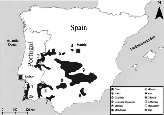
Figure 1: Historical distribution of the Iberian lynx, adapted from
Sarmento et al. (2009a): 1 - Algarve-Odemira-Sado valley; 2 - Gata-Malcata-San Pedro-S. Mamede; 3 - Western Sierra Morena-Guadiana; 4 - Alberche; 5 - Gredos; 6 - Subbéticas; 7 - Doñana; 8 -
Central Sierra Morena; 9 - Central population 3
Figure 2: Current Iberian lynx distribution in Spain adapted from
Sarmento et al. (2009a): 1 - Doñana; 2 – Cardeña-Andujár 4
Figure 3: Geographic localization of the study area and sampled
municipalities 15
Figure 4: Characteristic “Montado” landscape within the study area 16
Figure 5: Permanent water source in the study area 16
Figure 6: Hunted wild boars and procedures to collect blood
samples 18
Figure 7: Tissue sample collection 18
Figure 8: Submandibular lymph node collection 19
Figure 9: Two 96 well plates ready after colorimetric reaction 21
Figure 10: Seroprevalence distribution, in each sampled
municipality. Seropositivity for antibodies against Pseudorabies virus (Pr+) and bovine tuberculosis agent (bTB+); Absence of
seropositivity (NEG). 24
Figure 11: Wild boar serology results for pseudorabies virus
antibodies depending on age and gender classes 25
Figure 12: Wild boar serology results in which antibodies against
bovine tuberculosis agent (Mycobacterium bovis) were detected
List of tables
Table 1: Number of samples, number of seropositive samples to
pseudorabies virus (PrV) and bovine purified protein derivative (bPPD) and respective prevalence (%) with 95% confidence
intervals (CI 95%), in each sampled municipality 23
Table 2: Log-Linear Analysis results for pseudorabies ELISA test in
sampled wild boar 26
Table 3: Log-Linear Analysis results for bovine tuberculosis ELISA
Abbreviations
AD Aujeszky’s disease ADV Aujeszky’s disease virus
bPPD Bovine tuberculin purified protein derivative bTB Bovine tuberculosis
CI Confidence interval df Degrees of freedom
E% ELISA percentage
e. g. Exempli gratia
ELISA Enzyme-Linked Immunosorbent Assay FelV Feline leukaemia virus
G2 Chi-square value calculated by the log-linear method
IUCN International Union for Conservation of Nature
neg Negative
OD Optical density
P Probability
PBS Phosphate buffered saline
pos Positive
PrV Pseudorabies virus
SSC Species Survival Commission TMB Tetramethylbenzidine
Unrec. Unrecorded
%IN Percentage of inhibition
µl Microliter
1. Introduction
The role of captive breeding as a tool for the recovery of endangered species is a controversial subject. Rather than a long-term or prophylactic solution, Snyder et al. states that such approach should be viewed as the last resort in a species recovery strategy. Nevertheless in several conservation programs captive breeding for reintroduction1 is considered an option (despite its
known limitations) and therefore justifiable when other alternatives are not available (Snyder et al., 1996).
Difficulties in keeping self-sustainable captive populations, strict management conditions settings not to condition behaviour (loss of ability to survive in the wild – wild fitness), disease monitoring and other more political and financial features should be evaluated before deciding whether a recovery plan should or should not include any animal movement. Moreover the reduced survival probability amongst captive-born individuals, when in contact with endemic diseases in the releasing site is a significant risk (due to decreased immunological capacity to survive diseases that prevail in the wild). Jule and co-authors found evidence that the survival rates were higher for wild caught individuals than for captive-born ones, particularly in carnivore reintroduction programs. Captive-born carnivores are more susceptible to starvation with a rate of over 50% of fatal incidents being directly associated with human activities (Jule
et al., 2008). This data is in agreement with authors who describe high mortality
rates in captive-born Canada lynx (Lynx canadensis) caused by starvation early after release into the wild, apparently due to the lack of hunting competencies (Shenk et al., 2009). !!!!!!!!!!!!!!!!!!!!!!!!!!!!!!!!!!!!!!!!!!!!!!!!!!!!!!!!!!! # !$%%&'()*+!,&!,-.!/0123441!5#6678!'.)*,'&(9%,)&*!):!;<*!<,,.=>,!,&!.:,<?@):-!<!:>.%).:!)*!<*!<'.<!A-)%-! A<:!&*%.!<!><',!&B!),:!>'.C)&9:!-):,&')%<@!'<*+.DD!! !
It is, therefore, of great concern when the conservation efforts towards an endangered species rely almost exclusively on captive breeding for reintroduction, because along with it, efforts should be made in order to save the wild population and restore or preserve its habitat (Millán et al., 2009). Consequently both in situ and ex situ actions must be engaged, to promote adequate measures to improve species viability within the historical distribution area. Furthermore, reintroduction is considered a high risk activity for wildlife, thus, surveillance and monitoring programs should be designed in order to gather reliable information. Disease surveys and monitoring must be comprised in reintroduction programs.
Until recently diseases were considered a secondary factor in the dynamics and decline of wild animal populations. Small, fragmented and isolated wildlife populations are defiantly threatened by any stochastic event such as a disease outbreak, therefore more vulnerable to extinction (Delahay et al., 2009). To better understand the effects and dynamics of infectious agents in wild populations, it is fundamental to integrate the study of demographic, reproductive and mortality aspects associated with disease surveillance and the host-pathogen interaction.
Disease surveillance can be more objectively applied when focused on more abundant species or prey ones, being easier to establish management actions for disease control and risk reduction (Artois et al., 2009).
The Iberian lynx (Lynx pardinus) is an interesting case study since a captive-born reintroduction program is being considered for this endemic species in the Iberian Peninsula. It is also the world’s most endangered felid species. This wild felid was distributed throughout the Iberian Peninsula (Palomares, 2009), but by 1950 its distribution area was reduced and it almost disappeared from the northern middle of and eastern regions of Spain (Rodríguez, 2004). By this time, in Portugal it was already extinct from the northern and central regions of the country and a drastic reduction in the remaining population also occurred in the
same decade. Figure 1 reports to the historical distribution of the Iberian lynx after the mentioned period.
Figure 1: Historical distribution of the Iberian lynx, adapted from Sarmento et al. (2009a): 1 -
Algarve–Odemira–Sado valley; 2 - Gata–Malcata–San Pedro–S. Mamede; 3 - Western Sierra Morena–Guadiana; 4 - Alberche; 5 – Gredos; 6 - Subbéticas; 7 – Doñana; 8 - Central Sierra Morena; 9 - Central population.
During this century’s first decade a new Iberian census was conducted and no evidence of the species presence was found in Portugal, placing the Iberian lynx population in this country in the verge of extinction (Sarmento et al., 2009b).Thus, currently this species only occurs in Spain.
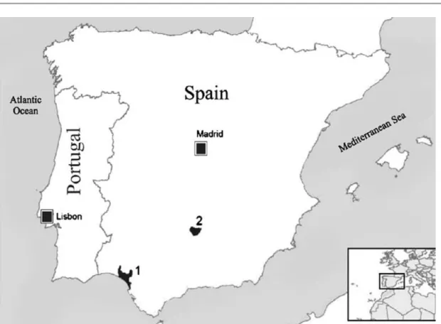
More recently this species population in the Iberian Peninsula was estimated in less than two hundred individuals, mostly grouped in two subpopulations in the region of Andaluzia (Palomares et al., 2010; Gil-Sánchez & McCain, 2011), both in Doñana and Eastern Sierra Morena (Palomares, 2009) (Figure 2).
Figure 2: Current Iberian lynx distribution in Spain adapted from Sarmento et al. (2009a): 1 -
Doñana; 2 - Cardeña–Andujár.
The Iberian lynx’s basic prey is the wild rabbit (Oryctolagus cuniculus) and both species evolved together and inhabit the same landscape (Real et al., 2009; Rodríguez et al., 2011). Habitat changes (due to altered agricultural practices and subsequent habitat loss) and viral diseases, namely myxomatosis in the 1950s and rabbit hemorrhagic disease (RHD) at the end of the 1980’s, caused consecutive abrupt declines in wild rabbit numbers (Cabezas-Díaz et al., 2009; Calvete, 2009). Wild rabbit populations have declined 55% in three decades in Spain (1973-2002) (Cabezas-Díaz et al., 2009) and in Portugal this reduction is estimated in the last decade in 30% (Ferreira & Alves, 2009). According to Ferreras and co-authors, the reduction of Iberian lynx’s staple prey, has had a direct impact in this predator abundance and led to high rates of non-natural mortality (Ferreras et al., 1992).
The confluence of events such as rabbit number decline, lynx population fragmentation, habitat loss and non-natural mortality, confined the Iberian lynx species to the present situation (Meli et al., 2009a; Palomares, 2009). Moreover the observed change in forestry management affected negatively both predator and prey species, which in association with other economic activities (artificial feeding for hunting purposes) enhanced ungulate species such as the wild boar (Sus scrofa) and the red deer (Cervus elaphus) occupying/ destroying the rabbit’s territorial patches (Cabezas-Díaz et al., 2009).
A widespread decrease in environmental favorability for the rabbit within lynx’s areas, as described by Real et al. (2009), led to the scarcity of its basic prey and probably forced the Iberian lynx to look for alternative resources (Gortázar et al., 2007). Since ungulates populations are currently increasing, they can easily become alternative or complementary prey (Briones et al., 2000).
The Iberian lynx is also known for its habitat specificity. Apart from the shrubland cover structure, dominant in Mediterranean scrubland landscape where the Iberian lynx still occurs and breeds (Fernández et al., 2007), rocky areas with some scrubland are also described as suitable for this species to establish its territory (Fernández et al., 2006). Furthermore, important elements (other than food availability) must be present, namely breeding dens (such as old hollow trees or rock cavities) (Fernández & Palomares, 2000), and permanent water sources during the dry season (Palomares et al., 2001).
Habitat loss and fragmentation has also been determinant in this top predator constraining range. Changes on soil uses, related to agricultural and forestry altered practices as well as the construction of large infrastructures disrupted the landscape structure and connectivity, consequently narrowing potential lynx territories (Ferreras, 2001; Ferreras et al., 2004). Moreover the inadequate management practices of livestock and hunting estates are over pressuring this species distribution (Calzada et al., 2007). Regarding the dispersal of young lynxes outside the National Park in Doñana, the 85% mortality rate was mostly due to predator control practices (Ferreras et al., 1992), and the
stable population within this National Park may be attributable to the protection status and cessation of predator persecution (Rodríguez, 2004).
Another study comprised radio-tracking of more than 50 individuals and revealed that 21% of the deaths were due to direct shooting, another 21%,to illegal trapping with leg-hold traps and snares and hunting with dogs, 17% were road accident casualties and 4% drowned in water wells (Palomares, 2009). This reflects the relevance of non-natural mortality over the Iberian lynx population, even after the legal protection of the species (Rodríguez & Delibes, 2004). Furthermore, the recent evidence that local extinctions have occurred before disease outbreaks within the wild rabbit populations, suggests that the primary cause of Iberian lynx decline was directly or indirectly related to human persecution (Gil-Sánchez & McCain, 2011).
Despite this catastrophic scenario, recent data (2001 – 2006) are more optimistic, indicating some improvements, particularly regarding the increase of population in Sierra Morena (Simón et al., 2009; Simón et al., 2012).
Concerning to the Portuguese claimed extinction of the Iberian Lynx (an unfortunate but expected outcome), the importance of a National Conservation Action Plan was highlighted, and it was legally approved in 2008 (Despacho n" 12697/2008, de 6 de Maio). The contemplated guidelines in this document aim to implement in situ and ex situ actions in order to preserve both habitat and prey populations, to develop pre-release activities (IUCN /SSC, 1998) and strengthen reintroduction viability in Portugal. For this purpose these actions should be carefully planned, and the multidisciplinary team, committed to establish the necessary connection between in situ and ex situ actions (Vargas et al., 2009).
As in any long-term conservation plan, the evaluation of disease prevalence is an important aspect to consider, especially in small and isolated populations, in which the vulnerability to stochastic events such as infectious diseases is increased (Gortázar et al., 2007; Simón et al., 2009) and genetic variability is reduced, potentially compromising individuals immunocompetence (Godoy et al., 2009).
Within the scope of disease epidemiology, it is less likely, due to Iberian lynx’s solitary social system (Palomares, 2009) that disease transmission occurs during intra-species, rather than inter-species contacts, particularly when reservoir species are maintaining the disease in the wild (Power & Mitchell, 2004; Millán et al., 2009). In order to better understand the role of sympatric wild and domestic species, potentially acting as disease reservoir, several studies regarding disease prevalence in those species were endeavoured within the Iberian Peninsula (Aranaz et al., 2004; Martin-Atance et al., 2005; Millán et al., 2007; Sobrino et al., 2007; Millán et al., 2009; Santos et al., 2009a). That is the case of wild ungulates which have been extensively studied for their role in tuberculosis epidemiology (Vicente et al., 2007; Gortázar et al., 2008; Naranjo et
al., 2008; Santos et al., 2009b). On other hand, for pseudorabies (also named
Aujeszky’s disease), the major concern is the economic loss as an outcome from disease transmission to livestock, but the risks to carnivore conservation are also in evidence (Vicente et al., 2005).
Therefore, it is mandatory to establish an epidemiological surveillance program for the Iberian lynx and associated species in its current range and in potential reintroduction sites (Roelke et al., 2008; Martínez et al., 2009).
Different pathogens have been identified in Iberian lynxes, namely: parasitic (Luaces et al., 2005; Millán et al., 2007; Sobrino et al., 2007; Roelke et
al., 2008; Millán et al., 2009; Acosta et al., 2011), bacterial (Briones et al., 2000;
Aranaz et al., 2004; Martin-Atance et al., 2005; Willi et al., 2007; Meli et al., 2009a; Millán et al., 2009) and viral agents (Roelke et al., 2008; López et al., 2009; Meli et al., 2009b; Meli et al., 2010; López et al., 2011; Palomares et al., 2011). Amongst these, few are known to be lethal to infected individuals, but the scrutiny of all possible diseases affecting wild and domestic felids, as well as other sympatric species, comprise an important tool to the Iberian lynx conservation plan.
Within the Iberian Peninsula the infectious diseases described with utmost relevance in lynx’s mortality rate are from bacterial and viral origin, respectively:
tuberculosis (Briones et al., 2000; Pérez et al., 2001) and feline leukaemia (López et al., 2009; Meli et al., 2009a). The death of five Iberian lynxes between 1998 and 2007, in Spain, was reported to be due to bovine tuberculosis (Meli et
al., 2009a). This disease was also described in free ranging lions, cheetahs and
leopards in South Africa (Keet et al., 1996) and other free-ranging and captive carnivore species (Carbin, 1982; Bruning-Fann et al., 1998; Helman et al., 1998; Lantos et al., 2003) and in these cases feeding on infected carcasses or meat was probably the primary infection route. Emaciation and multiple granulomas within the lungs and regional lymph nodes are common findings.
Even though considered a rare disease in nondomestic felids, feline leukaemia virus (FelV) infection has been reported in free-ranging and captive felids (Jessup et al., 1993; Leutenegger et al., 1999; Sleeman et al., 2001; Ostrowski et al., 2003; Cunningham, 2005) and recently a severe epidemic outbreak struck the Doñana’s Iberian lynx population; starting in December 2006, and during a 6 month period, 7 individuals died (López et al., 2009; Meli et al., 2010; López et al., 2011). This was evidence to the susceptibility of small sized populations associated with low genetic variability regarding the introduction of new diseases by other sympatric animals.
Pseudorabies or Aujeszky’s disease (AD) is also a viral infection already confirmed and reported in a dead Florida panther (Felis concolor coryi) (Glass et
al., 1994). Despite the scarce information about this particular disease in wild
felids, it was fairly studied in domestic cats (Hagemoser et al., 1980), hunting dogs (Müller et al., 2010; Müller et al., 2011), racoons (Thawley & Wright, 1982), reported in captive carnivores (Raymond et al., 1997; Zanin et al., 1997), and surveyed in other wild free–ranging carnivore species (Garcelon et al., 1992; Roelke et al., 1993; Mainka et al., 1994; Dunbar et al., 1998; Bakker et al., 2006). The members of suidae family are the natural hosts for pseudorabies virus thus acting as main potential reservoirs.
Within the suidae from the Old World, the wild boar is the species with the widest distribution. It is the second most abundant ungulate in Europe (Müller et
2004).This is particularly true in areas where there is food availability all year round and quality vegetation cover is present (Fonseca et al., 2004; Santos et al., 2006).
In Portugal the wild boar populations experienced a severe bottleneck in the middle of 19th century, mainly due to African swine fever outbreak and over
hunting (Ferreira et al., 2009). The increase in wild boar abundance started after the abandonment of agricultural lands (1960), and the banning of wild boar hunting in 1967. A notorious recovery in this species population was observed in 1980’s, being currently found throughout all of Portugal (Lopes & Borges, 2004; Fonseca, 2006). In 1986 a new hunting law was published (Lei n" 30/86, de 27 de Agosto) and along with the increase of game areas, the interest of hunters in this species also grew (Lopes & Borges, 2004), acquiring an economically important value as game species (Lopes & Borges, 2004; Ferreira, 2005; Fonseca, 2006). This economic relevance is present in all Iberian Peninsula, but particularly more noticeable in South and Central Spain where an important commercial hunting industry is developing (Vicente et al., 2006). In these areas, artificial management of this ungulate populations either by fencing or by providing feeding and watering in artificial points, as well as translocation of individuals2, have been suggested as possible causes of re-emergence of some
diseases (Vicente et al., 2007).
In contrast, wild boar breeding farms have little importance in Portugal. Nevertheless, wild ungulate translocation for hunting purposes is increasing and circumstantial evidence was found of the introduction of the bovine tuberculosis infectious agent in two areas by ungulate translocation, out of its previous distribution (Santos et al., 2009b).
Diseases like tuberculosis and pseudorabies are fairly studied and it was evidenced that overabundance (“overpopulation”) boosts infectious disease transmission risk, because of its effect in host aggregation and consequent increase in contact rate, either intra-species or inter-species (Gortázar et al.,
!!!!!!!!!!!!!!!!!!!!!!!!!!!!!!!!!!!!!!!!!!!!!!!!!!!!!!!!!!!
E$%%&'()*+!,&!,-.!/0123441!5#6678F!,'<*:@&%<,)&*!):!,-.!;(.@)?.'<,.!<*(!=.()<,.(!=&C.=.*,!&B!A)@(!
2006). Therefore, high disease prevalence and incidence can be expected in areas where a given wildlife species is overabundant. Artificial management with fencing, creation of feeding and watering points promotes aggregation, consequently enhancing infection transmission (Vicente et al., 2004; Gortázar et
al., 2006). These and other human interventions (such as predator control and
preventative treatments), interfere with natural mechanisms of wildlife population control, thus, in predator absence, hunting is an important regulation mechanism for these ungulate populations (Gortázar et al., 2006).
Diseases can affect not only the fitness of a given species, but also public health, livestock health, and the conservation of endangered species (Gortázar et
al., 2006; Roelke et al., 2008). The aforementioned, underlines the importance of
disease surveillance, wildlife abundance monitoring and ecology. The wild boar is an excellent example of direct correlation between overabundance and disease prevalence, and is also an interesting species for wildlife diseases epidemiological research (Ruiz-Fons et al., 2008a).
The increasing range and density of this species contributed to the spread of diseases like classical swine fever (Ruiz-Fons et al., 2008a), pseudorabies (Ruiz-Fons et al., 2008b), post weaning multisystemic wasting syndrome caused
by the porcine circovirus type 2 (Vicente et al., 2004), and bovine tuberculosis (Santos et al., 2012). This wild host’s role in these diseases epidemiology still endeavours scientific investigations, suggesting or confirming the species as wildlife reservoir (Acevedo et al., 2007; Ruiz-Fons et al., 2008a). Whenever confirmed, serious complications arise for diseases eradication programs in livestock (Cunha et al., 2011; Vieira-Pinto et al., 2011; Boadella et al., 2012). As some previous studies have suggested, disease prevalence is most probably related to wildlife’s farm management schemes promoting overabundance and aggregation (Vicente et al., 2004), also interfering with natural regulation mechanisms of wildlife populations (Gortázar et al., 2000; Gortázar et al., 2006).
Pseudorabies is a viral disease caused by an alphaherpesvirus that infects not only natural hosts as domestic pig, feral pig and wild boar, but also other domestic and wild mammals (Romero et al., 2001; Vengust et al., 2005;
Ruiz-Fons et al., 2007; Müller et al., 2011). This virus is distinguished by its rapid lytic growth in cell culture, its neurotropism, and its latency in neurons (Mettenleiter, 2000). After oronasal infection, the first viral replication occurs in the upper respiratory epithelium, with a later infection of the tonsils and lungs, from where the virus disseminates to the entire organism, either in a free form or associated with leucocytes. The virus easily progresses through the nervous terminations of the trigeminus and olfactive nerves to the Central Nervous System (Mettenleiter, 2000; Ruiz-Fons et al., 2007; Allepuz et al., 2008). Pigs and the wild boar are the only animals capable of surviving a productive infection, becoming infection reservoirs and carriers (Vengust et al., 2005; Allepuz et al., 2008). Major sites of pseudorabies virus latency are the trigeminal ganglion, the olfactory bulb, and the tonsil (Mettenleiter, 2000).
The reactivation of the latent viral replication can occur when animals are subjected to any stimulus, including stressful situations (Allepuz et al., 2008). The infection can result in neurological signs (like encephalitis) and respiratory signs, being more frequent in young and adult animals respectively (Vengust et al., 2005; Vicente et al., 2005; Ruiz-Fons et al., 2007). Even though very young wild swine seem to be more resistant to pseudorabies virus infection than domestic ones (probably due to earlier maturation of the CD8 T cells), high mortality rates have been reported in young wild individuals (Müller et al., 2011).
The clinical presentation of an Aujeszky’s disease outbreak in a free-ranging wild boar population was described as typical nervous affliction (inability to stand on hind legs, tremor and incoordination) (Gortázar et al., 2002).
Disease transmission occurs by several possible routes, namely:
• respiratory route (favoured by social gregariousness) among females and offspring:
• the venereal route that may be particularly important for males during the breeding season;
• the digestive infection route can occur by consumption of infected carcasses (Romero et al., 2001; Müller et al., 2011).
The gastrointestinal route is of most concern for wild boar predators like Iberian wolf, Iberian lynx, and other opportunistic scavengers (Vicente et al., 2005).
Pseudorabies is nowadays as distributed as the wild boar is, and once present it raises economic concerns due to pig industry and international pig product trade. For this reason several serosurvey studies were conducted all over Europe in countries with ongoing eradication plans (Müller et al., 2011).
Tuberculosis is a bacterial disease generally chronic, progressive, caused by mycobacteria from Mycobacterium tuberculosis complex, affecting a broad range of species including humans. Some of the members of this complex like
Mycobacterium tuberculosis, M. africanum and M. canetti are predominantly
human pathogens, M. microti affects rodents, whereas M. bovis and M. caprae have a broader host selection (Good & Duignan, 2011). Within this complex the
Mycobacterium bovis species is the one that presents a wider range of hosts
(Biet et al., 2005; Mostowy et al., 2005). In the infected host the disease is characterized by the development of pyogranulomatous lesions in lungs and lymph nodes, but has potential to disseminate to other organs (Bengis, 1999).
For its chronical and progressive character, clinical signs are only evident in advanced stages of infection and include anorexia, weakness, depression and progressive emaciation. In advanced pulmonary disease, cough, reduced exercise tolerance and progressive dyspnea may be present (Bengis, 1999).
Even though the direct contact amongst animals is the most efficient route of infection, this obligate intracellular organism can survive for long periods out of the animal host in the environment, directly or indirectly contaminated by infected animal discharges. This suggests other possible transmission routes (Biet et al., 2005). According to Bengis, the bovine tuberculosis agent can be transmitted by:
• respiratory route by aerosolized infectious particles;
• digestive route by ingestion of contaminated food (e.g. infected carcasses) or water;
• vertical or intra-uterine transmission and pseudo-vertical transmission by infected milk in lactants.
Respiratory route infection requires a lower number of infectious agents when compared with digestive route (Bengis, 1999).
Several authors refer that the tuberculosis re-emergence and the failure in its eradication in livestock in many countries is related with the maintenance of reservoirs of disease in wild populations (Alexander et al., 2002; Pavlik et al., 2002; Naranjo et al., 2008; Cross et al., 2009).
According to Biet et al., (2005), a host species can become a maintenance species through the confluence of aspects like physiopathogenesis (capacity to excrete the infectious agent), ethology (e.g. social behaviour) and ecology (feeding, population density and interactions between species). In reservoir hosts the infectious agent can be horizontally transmitted in the absence of other sources.
The wild boar and red deer (Cervus elaphus) are referred by some authors as being nowadays reservoir hosts, while formerly they were only affected by bovine tuberculosis in a sporadic way (Aranaz et al., 2004; Santos, 2006; Trcka
et al., 2006; Vicente et al., 2006; Gortázar et al., 2007; Naranjo et al., 2008).
Wild felids are very sensitive to Mycobacterium bovis infection (Gortázar et
al., 2007), and the severe course of pseudorabies infection in domestic cats is
well known (Hagemoser et al., 1980). It is thus logical to speculate that wild felines are also susceptible to pseudorabies virus.
This can be a problem for the Iberian lynx reintroduction program since both of these diseases etiological agents are present in other sympatric species, but furthermore important because they are prevalent in an abundant, reservoir and occasionally prey species – the wild boar. These diseases’ transmission to top predators such as the Iberian lynx will probably occur when they feed over infected carcasses.
The main goals of the present study are:
• to conduct a survey on antibody seroprevalence against pseudorabies virus and to monitor bovine tuberculosis agent within a wild boar population in a potential area for Iberian lynx reintroduction of the Portuguese portion of the Guadiana valley.
• the evaluation of the adequacy of the chosen sampling and test methodology (the use of hunting bags for serum collection and ensuing ELISA testing) for large scale surveys on the sanitary status of a given wild boar population.
• to use the wild boar as an indicator species of the risk of infection with both disease agents for Iberian lynx, as well as other wild carnivore species.
• to contribute to other long-term ongoing disease monitoring studies by collecting tissue samples.
To our knowledge this was the first study on pseudorabies virus seroprevalence in this geographical area.
2. Materials and methods
2.1 Study area
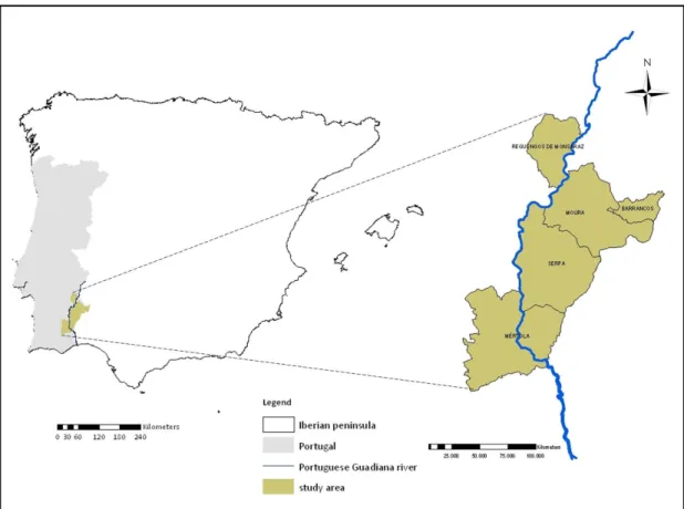
This study was conducted in four municipalities within Beja’s district and in one of the Évora’s district, all included in Alentejo’s region in southeast Portugal (Figure 3).
Figure 3: Geographic localization of the study area and sampled municipalities.
This region is characterized by its habitats diversity, with shrublands, scrublands and well preserved forest, with either cork-oaks (Quercus suber) “montados” or holm-oaks (Quercus ilex subsp. rotundifolia) predominance with or without shrubby undercover. The sub-shrubby vegetation cover is mostly composed by rockroses (Cistus spp.), as well as brooms (Genista spp.), heathers (Erica spp.), among others. These areas are intercalated by open areas with pastures and cereal cultures creating a mosaic (Figure 4 and 5).
Figure 4: Characteristic “Montado” landscape within the study area.
The study area has Mediterranean characteristics with hot summers and cold, rainy winters. Economic activities in the area are mainly agriculture, livestock, and hunting.
2.2 Sampling
The chosen sampling area was defined based on the targeted areas for future reintroductions.
Serum samples were collected from hunted wild boars in five municipalities in Guadiana river valley between November 2010 and February 2011. The eleven sampling sites were grouped according to their belonging municipalities and all except one were located in the left margin of the Guadiana River. In every hunting estate sampled the wild boar was free-living except in a fenced one where animals were artificially managed.
Sampled wild boars were classified in two different age classes. Animals less than 24 months were classified as juveniles and those over 24 months as adults based on external characteristics (Ruiz-Fons et al., 2007).
Pertaining to field work constraints, for some of the animals sampled it was not possible to determine either sex or age classes or both. Therefore only 50 samples had complete data and the remaining 61 were incomplete.
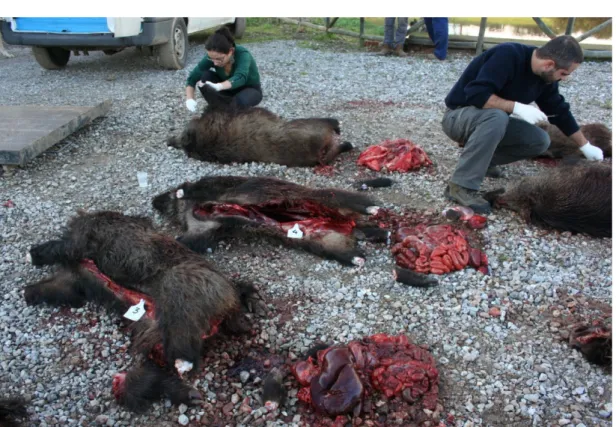
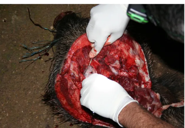
Blood samples were collected directly from the thoracic cavity into 15 ml sterile tubes, after the animal evisceration (Figure 6). They were kept refrigerated until arrival to laboratory. Serum was obtained by centrifugation and stored at -20"C in 0,5 ml aliquots until use.
Alongside with blood collection other tissues were collected, namely submandibular and mesenteric lymph nodes in order to contribute to other long-term ongoing studies. (Figure 7 and 8)
Figure 6: Hunted wild boars and procedures to collect blood samples
Figure 8: Submandibular lymph node collection.
2.3. Serology testing
Two different Enzyme-Linked Immunosorbent Assay (ELISA) tests were used to detect specific antibodies against pseudorabies virus and Mycobacterium
tuberculosis complex infectious agents in the collected serum samples. Positive
and negative controls were tested in duplicate in each plate and XREAD PLUS program version 2.10 was used to read the plates. This test is based in the interaction between antibody and antigen.
2.3.1 Detection of serum antibodies against Pseudorabies/ Aujeszky’s disease virus
The ELISA test chosen to perform pseudorabies (gpI antigen) antibodies detection is commercially available as a kit (IDEXX HerdCheck Anti-ADV gpl, IDEXX Inc., USA).
The protocol was followed according to the manufacturer’s instructions. Briefly, the sample was diluted (1:2) in sample solvent. This diluted sample (100 µl) was deposited in each well (except the control ones) in a PrV-antigen coated plate and incubated for 1 hour at room temperature. After a washing step, an anti-PrV gpI monoclonal antibody conjugate (100 !l) was added and left to incubate for 20 minutes at room temperature. The wells were washed again and a Tetramethylbenzidine (TMB) chromogen solution (100 !l) was added and incubated for 15 minutes at room temperature, generating a blue colour when interacting with the enzyme. This reaction was then interrupted by the deposition of 50 !l of Stop solution in each well. Optical density (OD) was measured in a spectrophotometer at 650 nm. The percentage of inhibition (%IN) was calculated to express the results (%IN) value using the following formula: [%IN = (mean negative control OD - mean sample OD/mean negative control OD) × 100]. The quantity of antibodies against PrV gpI was inversely proportional to the OD and directly proportional to the %IN. According to manufacturer’s instructions samples were considered positive when respective %IN values were equal to or greater than 40%. For presented %IN values between 30 and 40%, samples were considered doubtful and samples with %IN values under 30% were negative. According with the manufacturer this test has a sensitivity of 95-98% and a specificity of 97–99% for domestic pigs. It also has been broadly used in wild boars.
2.3.2 Detection of serum antibodies against Mycobacterium tuberculosis complex.
The selected method for bovine tuberculosis antibody detection was based on previously validated Indirect ELISA (Aurtenetxe et al., 2008).
Bovine tuberculin purified protein derivative (bPPD)3 was used as antigen to coat the wells of 96 well plates by incubating for 18 hours at room temperature. The wells were then washed with PBS and later blocked unspecific binding sites
!!!!!!!!!!!!!!!!!!!!!!!!!!!!!!!!!!!!!!!!!!!!!!!!!!!!!!!!!!!
3CZ Veterinaria SL, Porriño, Lugo, Spain. !
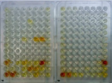
by incubating at 37"C for 1 hour with 5% skim milk in PBS with 0.05% Tween 20. The plate was washed again and then the serum samples were directly added to the wells (100 µl/ well), at a concentration of 1:200 in PBS. Again, the wells were washed with PBS. It was then added the conjugate solution of immunoglobulin-binding protein G and horseradish peroxidase4. After washing 100 µl per well of o-phenylenediamine dihydrochloride (SIGMAFAST™ OPD) was added as revelation solution. The reaction is interrupted by adding 50 µl per well of sulphuric acid (H2SO4; 3N). The colour intensity (Figure 9) was then measured at 450 nm. The following formula expresses sample results as an ELISA percentage (E%): [sample E% = (mean sample OD/2 x mean negative control OD) x100]. Samples with E% values greater than 100 were considered positive. This test sensitivity was 79.2% and the specificity 100% (Boadella et al., 2011a).
Figure 9: Two 96 well plates ready after colorimetric reaction.
2.4 Statistical analysis
Data was organized in an excel database and statistical analysis performed using VassarStats: Website for Statistical Computation, a statistical computation tool, available freely on line in http://vassarstats.net/. The 95% confidence intervals (CI 95%) were calculated using a method without a
!!!!!!!!!!!!!!!!!!!!!!!!!!!!!!!!!!!!!!!!!!!!!!!!!!!!!!!!!!!
ISigma-Aldrich Quimica SA, Madrid, Spain. !
correction for continuity. In order to test differences in seroprevalence, between different sexes and age groups and their interactions, Log-Linear Analysis was used. Three-Way Contingency Tables were built using two rows for factor A (Gender: Female/Male), two columns for factor B (Age: Adult/Juvenile) and two layers for factor C (Antibodies: Positive/Negative). All variables investigated were included in the initial model (saturated model) and then removed in a stepwise way. ABC represents the 3-way interaction between factors A, B and C. AB(C), AC(B) and BC(A) represent interactions between pairs of variables when the effects of the third variable are removed. For example, AB(C) represents the interaction between gender and age when the effects between each and the presence/absence of the antibodies are removed.
3. Results
From the 111 sampled wild boars, 32 were females, 23 were males and 56 with undetermined sex. Thirty eight animals were adults; twenty three were juveniles and the remaining 50 of undetermined age. The eleven sampling sites were grouped according to their belonging municipalities and the respective seroprevalence of antibodies against pseudorabies virus and bovine tuberculosis are shown in Table 1.
Table 1: Number of samples, number of seropositive samples to pseudorabies virus (PrV) and
bovine purified protein derivative (bPPD) and respective prevalence (%) with 95% confidence intervals (95% CI), in each sampled municipality.
Municipality Samples (no.) Pseudorabies + (PrV) Bovine tuberculosis + (bPPD) N %(95% CI) N %(95% CI) Barrancos 35 20 57,1 (40-70) 2 5,7 (0-20) Mértola 5 0 ! 0 ! Moura 45 22 48,9 (40-60) 8 17,8 (10-30) Serpa 22 8 36,4 (20-60) 1 4,5 (0-20) Reguengos 4 1 25,0 (0-70) 0 ! Total 111 51 45,9 (40-60) 11 9,9 (10-20)
Regarding to the distribution of seroprevalence for pseudorabies antibodies in a municipal level, Barrancos presented the highest prevalence, followed by Moura, Serpa and Reguengos. In Mértola, no tested sample presented antibodies for this disease agent.
Concerning the bovine tuberculosis agent distribution, the municipality that presented higher antibodies seroprevalence was Moura, followed by Barrancos and Serpa. Mértola and Reguengos showed no positive samples (Figure 10).
Figure 10: Seroprevalence distribution, in each sampled municipality.
Seropositivity for antibodies against Pseudorabies virus (Pr+) and bovine tuberculosis agent (bTB+); Absence of seropositivity (NEG).
Seventeen out of 32 females were seropositive to pseudorabies antibodies, as well as 11 out of 23 males and 23 out of 56 of unrecorded sex. Regarding the 11 seropositive samples for bovine tuberculosis (bTB) antibodies, 4 belonged to females; 2 were from males and 5 from undetermined sex. Seropositivity rates for gender classes were in females for PrV and for bTB agent respectively 53,1% (CI 95%= 40-70) and 12,5% (CI 95%= 5-28), in males 47,8% (CI 95%= 29-67) and 95%= 40-70) and 12,5% (CI 95%= 5-28), in males 47,8% (CI 95%= 29-67) and
8,7% (CI 95%= 2-26) and for unrecorded gender 41,1% (CI 95%= 30-54) and 8,9% (CI 95%= 4-2) (Figures 11 and 12).
Figure 11: Wild boar serology results for pseudorabies virus antibodies depending on
age and gender classes.
Figure 12: Wild boar serology results in which antibodies against bovine tuberculosis agent
Of 38 adult animals, 22 tested positive to PrV and 4 to bTB antibodies. Of the 23 juveniles, 9 were seropositive to PrV and 2 to bTB antibodies. Within the animals with unrecorded age (n=50), 20 were seropositive to pseudorabies and 5 to bovine tuberculosis antibodies. Seropositivity rates for age classes were for PrV and bTB respectively 57,9% (CI 95%= 42-72) and 10,5% (CI 95%= 4-24) in adults; 39,1% (CI 95%= 22-59) and 8,7% (CI 95%= 2,4-2,6) in juveniles and finally 40% (CI 95%= 27-53) and 10% (CI 95%= 4-21) in undetermined age samples.
In the overall 111 serum samples, 51 tested positive for pseudorabies virus antibodies, 11 for bovine tuberculosis antibodies, with seropositivity prevalences of 45,9% (CI95%= 40-60) and 9,9% (CI95%= 10-20), respectively. Five blood samples tested positive for contact with both diseases agents (prevalence was 4,5% and CI95%= 0-10).
To perform the Log-Linear Analysis only the samples with complete data for all variables were used. Log-Linear Analysis results for evidence of contact with pseudorabies and bovine tuberculosis agents are shown in Tables 2 and 3, respectively.
Table 2: Log-Linear Analysis results for pseudorabies ELISA test in sampled
wild boar Source G2 df P ABC 4.68 4 0.3217 AB(C) 3.34 2 0.1882 AC(B) 0.76 2 0.6839 BC(A) 0.74 2 0.6907
Table 3: Log-Linear Analysis results for bovine tuberculosis ELISA test in
sampled wild boar
Source G2 df P
ABC 3.82 4 0.4309
AB(C) 3.38 2 0.1845
AC(B) 0.46 2 0.7945
BC(A) 0.22 2 0.8958
The results obtained showed that there are no significant interactions among any of the variables (gender, age, and antibodies), since each one is independent from the others, for the established level of significance of "=0.05.
4. Discussion
This work is the first reported survey for pseudorabies virus seroprevalence in the wild boar in the Guadiana valley, the principal area for the reintroduction of Iberian lynx in Portugal.
The decision to study this particular geographic area was related to its high support capacity (Sarmento et al., 2009b), not only for the Iberian lynx, but also for other carnivores like the wild cat (Felis silvestris).
Therefore, considering that there is already an Action Plan in place in the Natura 2000 Network in Moura-Barrancos (Special Conservation Zone); the Adiça area, contiguous to Contenda-Barrancos has a high habitat suitability and high lynx’s staple prey density; and the Iberian lynx corridor definition and its presence in western Sierra Morena, it is reasonable to recognise that these border areas in the Guadiana valley are probably the ones with more adequate characteristics to attend to the reintroduced Iberian lynx needs. As a result, it is of the utmost importance to be aware of the sanitary status of wildlife within this area. This is an important aspect, considered in the Iberian Lynx Conservation Strategy II (Grupo de trabajo del Lince Ibérico, 2008). In this work, both pseudorabies and bovine tuberculosis are referred as important infections to be surveyed in sympatric animals, namely wild and domestic ungulates as well as wild and domestic carnivores.
Pseudorabies and bovine tuberculosis have been reported in a wild potential reservoir (wild boar) in several European countries (Meng et al., 2009). In areas like the Mediterranean region of Spain, where the hunting industry promoted a more intensive management of game ungulates for economic reasons, and in some patches of the Portuguese territory, special epidemiological circumstances arose and are associated with higher prevalences of these diseases (Aranaz et al., 2004; Cunha et al., 2011; Müller et al., 2011).
A wide range of pseudorabies seroprevalence (0,3% to 66%) is reported in different regions of Europe (Müller et al., 2011). The seroprevalence obtained in the present study area (45,9%) is amongst the highest rates so far documented. According to Müller et al. (2011) the countries that reported higher seropositivity rates using ELISA method were Croatia (54,55%), Romania (55,18%), France (up to 54% in some continental areas and 53% in Corsica) and Spain. In this latter country the seroprevalence in South-central region is at the same level (45,95%) as the one observed in Portuguese Guadiana valley. Nevertheless, pseudorabies virus seroprevalence has a higher value (66,06%) when the surveyed animals come from intensively managed populations for hunting purposes (Ruiz-Fons et al., 2007). Probably, due to residual or absent management actions concerning the increase of game populations, Canado et al. (2012) reported lower pseudorabies seroprevalence (8%) in a Portuguese northern municipality (Vinhais).
Considering the sample from this work representative of the hunting bags’, a higher proportion of contact rate with the causative agent occurs in adults (57,9%) than in juveniles (39,1%). The present results are consistent with other researches (Vicente et al., 2005; Lari et al., 2006; Pannwitz et al., 2012) and are most likely to be justified by the fact that older animals have had a longer time frame for possible exposure.
The difference in pseudorabies seroprevalence between sexes is not noticeable in this work but still the value in females (53,1%) is higher than in males (47,8%). Higher seroprevalence in females have also been reported in Spain (Vicente et al., 2005), even though this difference has not been described for other European countries (Lari et al., 2006; Pannwitz et al., 2012).
Within the considered study area, wild boar and Iberian pig share the same landscape, thus, the high contact rate with pseudorabies virus evidenced in the present work may raise economical concerns.
Bovine tuberculosis monitoring in wildlife has become a major concern since wildlife reservoirs can maintain the disease among livestock despite the eradication plans implemented in several countries (Pavlik et al., 2002; Cross et
al., 2009).
In Portugal until recently, bovine tuberculosis in cattle (subject of a national eradication plan) was decreasing, but since 2008 the number of infected animals and herds has grown, increasing concern about the success of the control program (Cunha et al., 2011). This is probably the reason why potential wildlife reservoirs are being subject of attention by sanitary authorities.
The information concerning bovine tuberculosis prevalence in the wild boar in Portugal is scarce, but an overall prevalence of 11,1% and high rate (63%) of Mycobacterium bovis isolation in Eastern-central region have been reported (Santos et al., 2009b; Cunha et al., 2011).
In the present study the seroprevalence found for Mycobacterium bovis antibodies (9,9%) is within the range of previously reported M. bovis infection rates in tuberculosis epidemiological risk area in Eastern South-central Portugal (Santos et al., 2009b; Vieira-Pinto et al., 2011). Also, an higher evidence of exposure to bovine tuberculosis agent was found for females (12,5%) than for males (8,7%), which is in agreement with Santos’ results. This is coherent with the more gregarious nature of females, increasing the probability of infection. Age differences in seroprevalence rates (although slight) were also present, and adults had higher antibody prevalence (10,5%) than juveniles (8,7%). This fact can be explained by the chronical character of the disease (Bengis, 1999). This data is in compliance with results from other surveys (Vicente et al., 2006) but contrasting with others (Santos et al., 2009b).
The present results should be interpreted with caution. Despite the small sample size, sample collection from hunted wild boars can be considered as random and representative of the study area, even though for larger sample
sizes differences between variables could be statistically significant. Misinterpretation of the seroprevalence results can be induced by biased sampling, serological assay or investigation period, being desirable a longer study period and a larger sample size.
Tests based on antibody detection may fail to detect some of the recently infected animals (Ruiz-Fons et al., 2007), as well as those with delayed seroconversion (Müller et al., 2011) or in a latency period. So, under-detection of infected individuals is a concern (Ruiz-Fons et al., 2007). In contrast crossed reactions with related members of the Mycobacterium tuberculosis complex may occur increasing the proportion of positive detected samples (Boadella et al., 2011b). Regarding the ELISA test used for detection of bovine tuberculosis antibodies in this work, cross reactions between different mycobacteria (Mycobacterium bovis, and Mycobacterium avium) have already been studied and a high specificity was confirmed (Boadella et al., 2011a).
Serologic tests are easy to perform, sensitive, specific, reproducible and not expensive to perform, thus highly recommendable for large-scale research (Sedlak et al., 2008; Boadella et al., 2011b; Boadella et al., 2012). The implementation of large-scale serosurveys with potential wild reservoir species may bring enlightenment about the diseases occurrence in Portuguese territory, with benefits both economical and conservationist.
In this work the wild boar was chosen as the targeted species for several reasons:
• its abundance in the destination reintroduction site; • the possibility to sample carcasses from hunting bags;
• the possibility to become an alternative prey for the Iberian lynx;
• for its already recognizable role as a maintenance or reservoir host for Pseudorabies virus and Mycobacterium bovis, and possibility to spread these disease agents to other wildlife species.
The evidence of exposure to pseudorabies and bovine tuberculosis agents within a potential reservoir species, suggests the presence of both diseases in
the environment. This can pose considerable risks of infection to some endangered species mainly concerning top predators like the wild cat and lynxes or other carnivores like bears and wolves (Lari et al., 2006; Sedlak et al., 2008; Boadella et al., 2012).
The control of these diseases in wildlife is most certainly related with game management control. The adopted measures should focus on balanced abundance thresholds, the discouragement of practices that promote aggregation and strict supervision of any animal translocation. Notwithstanding, to reduce the risk that the presence of diseases within the lynx reintroduction site represents, it is fundamental to discuss a significant reduction in wild boar population. Nevertheless, to assure these measures effectiveness it is important to gather information about the species diversity, its populations, as well as its sanitary status.
Although vaccination is not an option at present, it will probably acquire greater importance in future years.
5. Conclusions
Despite the relative small number of animals sampled in some municipalities to get any reliable conclusion about these diseases’ agents seroprevalence, as a whole it is possible to have a spatial perspective of its distribution.
It is reasonable to assume that pseudorabies is highly prevalent in wild boar population within the study area (the main potential place for the Iberian lynx reintroduction) and the observed seroprevalence data is amongst the highest reported in Europe. This study also reports seroprevalence rates for bovine tuberculosis similar to those previously described for the same geographical area.
Notwithstanding the use of other more sensitive methods in those previous studies, the serologic test performed in this work confirms the appropriateness for continuous regular surveys at a lower cost and may be a valuable tool for disease surveillance and consequently for the establishment of control measures and evaluation of their effectiveness.
Considering the study area as a possible Iberian lynx reintroduction site, there is an increased risk of infection for future reintroduced animals. Given the similarity of ecological characteristics of the bordering Spanish territory, inference about both studied diseases epidemiological cycles can be made to implement a disease control program.
Hunting bags can represent important and convenient resources of information although field constraints were particularly disappointing, whereas in some of the hunting bags it was very difficult to catch up and get all necessary data for each animal. It is therefore advisable for future studies that imply sample collection on hunted animals to have a larger working team.
In this work the study of these two economically important diseases focus on its conservation implications. Nevertheless the role of the wild boar in these diseases’ epidemiology should be assessed both in livestock and public health. Even if not in the scope of this work, it is noticeable that the low standards of health and safety procedures among hunters and carcass handlers may place them in major infection risk.
Future prospects
Portugal should develop an effective nationwide disease monitoring program, enabling the integration of information in an international epidemiological surveillance network. To accomplish this task, a multidisciplinary working team should be assigned to establish specific priorities, to design surveillance programs, to establish adequate protocols for sample collection and testing and to keep the highest standards in scientific production.
This study results suggest the need to intensify sample collection by enlarging the sampling area and sample size. The present work also reinforces the usefulness of serological tests in fieldwork. These practices when performed in a regular basis may provide long-term surveillance information about both diseases’ dynamics. It is also urgent to determine wild boars role in these diseases’ epidemiology. The surveillance programmes developed in the future should also encompass other ungulate and non-ungulate species, when pseudorabies and tuberculosis are considered.
6. References
Acevedo P, Vicente J, Höfle U, Cassinello J, Ruiz-Fons F & Gortázar C. (2007). Estimation of European wild boar relative abundance and aggregation: a novel method in epidemiological risk assessment. Epidemiology &
Infection 135, 519-527.
Acosta L, Leon-Quinto T, Bornay-Llinares F, Simon M & Esteban J. (2011). Helminth parasites in faecal samples from the endangered Iberian lynx (Lynx pardinus). Veterinary Parasitology 179, 175-179.
Alexander K, Pleydell E & Williams M. (2002). Mycobacterium tuberculosis: An emerging disease of free-ranging wildlife. Emerging Infectious Diseases 8, 598-601.
Allepuz A, Saez M, Alba A, Napp S & Casal J. (2008). Exploratory spatial analysis of Aujeszky's disease during four phases of the eradication programme in Catalonia, Spain (2003-2007). Preventive Veterinary
Medicine 86, 164-175.
Aranaz A, de Juan L, Montero N, Sánchez C, Galka M, Delso C, Álvarez J, Romero B, Bezos J, Vela A, Briones V, Mateos A & Domínguez L. (2004). Bovine tuberculosis (Mycobacterium bovis) in wildlife in Spain. Journal of
Clinical Microbioly 42, 2602-2608.
Artois M, Bengis R, Delahay R, Duchêne M, Duff J, Ferroglio E, Gortázar C, Hutchings M, Kock R, Leighton F, Mörner T & Smith G. (2009). Wildlife disease surveillance and monitoring. In Management of Disease in Wild
Mammals, ed. Delahay RJ, Smith GC & Hutchings MR, pp. 187-213.
Aurtenetxe O, Barral M, Vicente J, de la Fuente J, Gortázar C & Juste R. (2008). Development and validation of an enzyme-linked immunosorbent assay for antibodies against Mycobacterium bovis in european wild boar. BMC
Veterinary Research 4, 43.
Bakker V, Van Vuren D, Crooks K, Scott C, Wilcox J & Garcelon D. (2006). Serologic survey of the island spotted skunk on Santa Cruz Island.
Western North American Naturalist 66, 456-461.
Bengis R. (1999). Tuberculosis in free-ranging mammals. In Zoo and Wild Animal
Medicine Current Therapy 4, 5 edn, ed. Fowler M & Miller R, pp. 101-114.
W. B. Saunders Company, Philadelphia.
Biet F, Boschiroli M, Thorel M & Guilloteau L. (2005). Zoonotic aspects of
Mycobacterium bovis and Mycobacterium avium-intracellulare complex
(MAC). Veterinary Research 36, 411-436.
Boadella M, Acevedo P, Vicente J, Mentaberre G, Balseiro A, Arnal M, Martínez D, García-Bocanegra I, Casal C, Álvarez J, Oleaga Á, Lavín S, Muñoz M, Sáez-Llorente J, de la Fuente J & Gortázar C. (2011b). Spatio-temporal trends of Iberian wild boar contact with Mycobacterium tuberculosis Complex detected by ELISA. EcoHealth 8, 478-484.
Boadella M, Gortázar C, Vicente J & Ruiz-Fons F. (2012). Wild boar: an increasing concern for Aujeszky's disease control in pigs? BMC Veterinary
Research 8, 7.
Boadella M, Lyashchenko K, Greenwald R, Esfandiari J, Jaroso R, Carta T, Garrido JM, Vicente J, de la Fuente J & Gortázar C. (2011a). Serologic tests for detecting antibodies against Mycobacterium bovis and
(Sus Scrofa Scrofa). Journal of Veterinary Diagnostic Investigation 23, 77-83.
Briones V, Juan L, Sánchez C, Vela A, Galka M, Montero N, Goyache J, Aranaz A, Mateos A & Domínguez L. (2000). Bovine tuberculosis and the endangered Iberian lynx. Emerging Infectious Diseases 6, 189-191.
Bruning-Fann C, Schmitt S, Fitzgerald S, Payeur J, Whipple D, Cooley T, Carlson T & Friedrich P. (1998). Mycobacterium bovis in coyotes from Michigan.
Journal of Wildlife Diseases 34, 632-636.
Cabezas-Díaz S, Lozano J & Virgós E. (2009). The declines of the wild rabbit (Oryctolagus cuniculus) and the Iberian lynx (Lynx pardinus) in Spain: redirecting conservation efforts. In Handbook of Nature Conservation, ed. Aronoff JB, pp. 283-310. Nova Science Publishers, Inc.
Calvete C. (2009). Status and trends of rabbit populations in the Iberian Peninsula. In Iberian lynx ex situ conservation: an interdisciplinary
approach, ed. Vargas A, Breitenmoser C & Breitenmoser U, pp. 13-21.
Fundación Biodiversidad / IUCN Cat Specialist Group, Madrid.
Calzada J, Guzmán N & Rodríguez A. (2007). Ficha libro rojo. Lynx pardinus (Temminck, 1827),. In Atlas y libro Rojo de los Mamíferos Terrestres de
España, ed. Palomo L, Gisbert J & Blanco J. Atlas y libro Rojo de los
Mamíferos Terrestres de España., Madrid.
Canado M, Lourenço M & Vieira-Pinto M. (2012). Doença de Aujeszky em javalis (Sus scrofa) caçados no concelho de Vinhais. In IV Congresso da Fauna
Selvagem WAVES Portugal, ed. Teixeira A, Geraldes A, Cortez P &