“Enhanced harmonic endoscopic
ultrasonography (CH-EUS) in the evaluation
of pancreatic solid lesions: a systematic
review”
MESTRADO INTEGRADO EM MEDICINAINSTITUTO DE CIÊNCIAS BIOMÉDICAS ABEL SALAZAR, UNIVERSIDADE DO PORTO
ALUNO:
Hélder Xavier Martins Balouta
6º Ano Profissionalizante do Mestrado Integrado em Medicina Nº de aluno: 201100762
Endereço eletrónico: [email protected]
ORIENTADOR
Professor Doutor F. Castro Poças
Assistente Hospitalar Graduado Sénior de Gastroenterologia no Centro Hospitalar do Porto; Professor Catedrático Convidado no Instituto de Ciências Biomédicas Abel Salazar, Rua de Jorge Viterbo Ferreira, nº 228, 4050-213, Porto
CO-ORIENTADOR Dra. Daniela Ferreira
Assistente Hospitalar de Gastrenterologista no Centro Hospitalar do Porto;
Docente no Instituto de Ciências Biomédicas Abel Salazar, Rua de Jorge Viterbo Ferreira, nº 228, 4050-213, Porto
AUTOR:
(Hélder Xavier Martins Balouta)
ORIENTADOR:
(Fernando Manuel Castro Poças)
CO-ORIENTADORA:
(Daniela Armanda Gonçalves Ferreira)
DEDICATÓRIA:
Dedico a realização desta dissertação para obtenção do Grau de Mestre em Medicina aos meus pais, que em muito me ajudaram a chegar ao final desta etapa e, acima de tudo, à minha tia Odete, que sempre me apoiou e ajudou em tudo e que infelizmente perdeu a luta contra um adenocarcinoma pancreático, patologia muitas vezes mencionada neste trabalho.
i AGRADECIMENTOS:
Obrigado ao Professor Doutor F. Castro Poças e à Dra. Daniela Ferreira pela confiança depositada em mim para a realização desta que foi a minha primeira revisão sistemática. Sabemos que o caminho teve alguns imprevistos, mas deixo em agradecimento o apoio e a disponibilidade demonstrados.
ii
ABSTRACT
AIM: To do a systematic review about the applicability of CEH-EUS in the diagnosis of pancreatic solid lesions, clarifying its advantages and ability to differentiate this type of lesions.
METHODS: This study was based on the PRISMA Guidelines of 2009. The research was made
using PubMed and Cochrane Database of Collected Reviews, using all the possible combinations between the keywords described below. All types of articles were included and there was no restriction of the publication year or language. The exclusion criteria included articles which did not contain results about pancreatic solid masses, no references at the use of contrasts or harmonic endoscopic ultrasound, that concerned the Doppler mode, and that were only about the technical aspects of CEH-EUS.
References of selected articles were analyzed to obtain any other article that was not present on the previous search.
RESULTS: 108 articles were analyzed and 16 were included and presented according to the
different contrast agents used. 10 reported the use of SonoVue, 5 described results under Sonazoid and 1 was about Denifity. Pancreatic adenocarcinomas were described as having an hypoenhanced pattern on all of the studies with no differences between the various types of contrast. Neuroendocrine tumors were shown as having an hyperenhanced pattern and mass-forming pancreatitis had an isoenhanced pattern on all the studies. One article included de diagnostic of pancreatic metastasis, describing it as hyperenhanced in 61% and 28% as hypoenhanced.
CONCLUSIONS: SonoVue is more used worldwide and it seems that it has a better sensitivity and
specificity for the ability to distinguish the various types of pancreatic solid masses than Sonazoid. The appearance of the lesions had no differences between the use of SonoVue, Sonazoid or Definity. It appears that the index of the contrast uptake ratio and echo intensity reduction is a good marker as it can aid differentiation between malignant and benign masses but still cannot replace EUS-FNA.
Keywords: Contrast harmonic enhanced endoscopic ultrasound; ultrasonography; solid
iii
RESUMO
OBJETIVOS: Realizar uma revisão sistemática acerca da aplicabilidade da ecoendoscopia
harmónica com recurso a contrastes (CEH-EUS) para o diagnóstico de lesões sólidas do pâncreas, clarificando as suas vantagens na prática clínica e a sua habilidade em diferenciar os diversos tipos de lesões.
MÉTODOS: O estudo foi baseado nas Guidelines Prisma de 2009. A pesquisa foi baseada na
PubMed e na Cochrane Database of Collected Reviews utilizando todas as combinações possíveis entre as palavras-chave descritas. Incluíram-se todos os tipos de artigos, sem restrição do ano ou do idioma. Os critérios de exclusão incluíram artigos sem referência a lesões sólidas do pâncreas, sem menções ao uso de contrastes ou ecoendoscopia harmónica, alusivas ao modo Doppler, e que apenas descreviam os aspetos técnicos da CEH-EUS. Foram analisadas as fontes bibliográficas dos artigos selecionados de modo a obter artigos não incluídos na pesquisa inicial.
RESULTADOS: 108 artigos foram analisados e 16 foram incluídos e categorizados de acordo com
os diferentes agentes de contraste usados. 10 artigos eram referentes ao uso de SonoVue, 5 descreveram resultados usando Sonazoid e 1 artigo continha resultados acerca do Definity. Adenocarcinomas pancreáticos foram descritos como apresentando um padrão de hiporrealce em todos os estudos analisados independentemente do tipo de contraste utlizado. Tumores neuroendócrinos foram visualizados com um padrão de hiperrealce e a pancreatite crónica com um padrão de isorrealce. Um artigo incluiu o diagnóstico de metástases presentes no pâncreas, descrevendo um padrão de hiperrealce em 61% e de hiporrealce em 28%.
CONCLUSÕES: O SonoVue é o mais usado a nível mundial e aparenta ter uma melhor
sensibilidade e especificidade para conseguir distinguir os vários tipos de lesões sólidas do pâncreas. A aparência das lesões manteve-se mesmo utilizando diferentes agentes de contraste. Constatou-se que muito provavelmente o índice de velocidade de preenchimento do contraste por parte da lesão bem como a taxa de diminuição da intensidade ecográfica serão bons marcadores no auxílio da diferenciação entre tumores benignos e malignos, embora não seja possível substituir o recurso à aspiração por agulha fina guiada por ecoendoscopia (EUS-FNA).
Palavras-chave: Contrast harmonic enhanced endoscopic ultrasound; ultrasonography; solid
iv
ABREVIATIONS:
Autoimmune Pancreatitis - AIP Chronic Pancreatitis – CP
Contrast Enhanced Endoscopic Ultrasound - CE-EUS
Contrast Enhanced Harmonic Endoscopic Ultrasound - CEH-EUS/CH-EUS Contrast Enhanced Ultrasound - CEUS
Echo Intensity – EI
Endoscopic Ultrasonography – EUS
Endoscopic Ultrasound Fine Needle Aspiration – EUS-FNA Fine Needle Aspiration – FNA
Inflammatory Pancreatic Pseudotumor - IPPT Mass-Forming Pancreatitis – MFP
Mechanical Index - MI
Neuroendocrine Tumor – NET Pancreatic Cancer – PC
Pancreatic Ductal Adenocarcinoma – PDAC Pancreatic Neuroendocrine Neoplasm - PNEN Solid Pancreatic Lesions – SPLs
Solid Pseudopapillary Neoplasm – SPN Ultrasound – US
Ultrasound Contrast Agent – UCA
v
INDEX
Abstract ... ii Resumo ... iii Abreviations ... iv Table Index ... viFigure Index... vii
Introduction ... 1
Methods ... 3
Results ... 4
Discussion and Conclusions ... 8
Appendix I - Figures ... 12
Appendix II – Tables ... 13
vi
TABLE INDEX
Table I - Inclusion and exclusion criteria ... 13 Table II - Seicean et al." Quantitative Contrast-Enhanced Harmonic Endoscopic Ultrasonography for the Discrimination of Solid Pancreatic Masses” (19) ... 14 Table III - Romagnuolo et al."Accuracy of contrast-enhanced harmonic EUS with a second-generation perflutren lipid microsphere contrast agent (with video)" (31) ... 15 Table IV - Gong et al." Contrast-enhanced EUS for differential diagnosis of pancreatic mass lesions: a meta-analysis”(10) ... 16 Table V - Kitano et al." Characterization of Small Solid Tumors in the Pancreas: The Value of Contrast-Enhanced Harmonic Endoscopic Ultrasonography”(25) ... 17 Table VI - Lee et al." Clinical Role of Contrast-Enhanced Harmonic Endoscopic Ultrasound in Differentiating Solid Lesions of the Pancreas: A Single-Center Experience in Korea” (22) ... 18 Table VII - Park et al." Effectiveness of contrast-enhanced harmonic endoscopic ultrasound for the evaluation of solid pancreatic masses”(17) ... 19 Table VIII - Yamashita et al." Contrast-Enhanced Endoscopic Ultrasonography for Pancreatic Tumors” (32) ... 20 Table IX - Sugimoto et al." Conventional versus contrast-enhanced harmonic endoscopic ultrasonography-guided fine-needle aspiration for diagnosis of solid pancreatic lesions: A prospective randomized trial"(6) ... 21 Table X - Saftoiu et al." Quantitative contrast-enhanced harmonic EUS in differential diagnosis of focal pancreatic masses (with videos"(11) ... 22 Table XI - Fusaroli et al." The clinical impact of ultrasound contrast agents in EUS: a systematic review according to the levels of evidence"(33) ... 23 Table XII - Seicean et al."Harmonic Contrast-Enhanced Endoscopic Ultrasonography for the Guidance of Fine-Needle Aspiration in Solid Pancreatic Masses"(27) ... 24 Table XIII - Garcia et al." Differential diagnosis of solid pancreatic masses: contrast-enhanced harmonic (CEH-EUS), quantitative-elastography (QE-EUS), or both?"(3) ... 25 Table XIV - Leitão et al. "Uncommon Solid Pancreatic Neoplasm: The Role of New Modalities of Ultrasound Endoscopy”(24) ... 26 Table XV - Leem et al. " Clinical Value of Contrast-Enhanced Harmonic Endoscopic Ultrasonography in the Differential Diagnosis of Pancreatic and Gallbladder Masses"(21) ... 27 Table XVI - Kamata et al." Impact of avascular areas, as measured by contrast-enhanced harmonic EUS, on the accuracy of FNA for pancreatic adenocarcinoma"(34) ... 28 Table XVII - Ishikawa et al. " Multiphase evaluation of contrast-enhanced endoscopic ultrasonography in the diagnosis of pancreatic solid lesions"(14) ... 29
vii
FIGURE INDEX
1
INTRODUCTION
The incidence of Pancreatic Cancer (PC) has increased steadily over the last four decades, being responsible for an estimated 37,390 deaths in 2012. It is considered the fourth leading cause of cancer mortality in the USA and the 5-year overall survival rate is 6%. Most cases of PC are only discovered at an unresectable stage, when prognosis is poor and the 5-year survival rate is 2%, even with recent advances in diagnostic imaging technics.(1)
With current imaging technics, it is challenging to differentiate between pancreatic adenocarcinoma from other pancreatic masses. (2) Thanks to its superior spatial resolution
compared to any other modality, EUS is among the most efficient and reliable diagnostic method for digestive diseases. One important limitation of the use of EUS is that in some cases it’s difficult to differentiate benign and malignant lesions, as most solid lesions appear as hypoechoic masses regardless its histology. (3) Considering that, even though it has the ability to
detect small pancreatic lesions with high sensitivity, EUS still has limited value in distinguishing pancreatic cancer from non-neoplastic pancreatic masses.
In order to improve the characterization of digestive lesions, the technique of contrast-enhanced harmonic EUS (CEH-EUS) was developed, allowing the evaluation of the vascularity of those lesions and also dynamic and repeat examinations. (4) At first, contrast enhanced
endoscopic ultrasound (CE-EUS) without the harmonic part was used in 1995, with the infusion of carbon dioxide gas as ultrasound contrast via a catheter into the celiac or superior mesentery artery. (5) As angiographic technique is employed for ultrasound contrast infusion into specific
vessels and this form of CE-EUS is relatively invasive and unwieldly to perform, there was a big disadvantage on its use. Furthermore, the B mode ultrasound was found to be inadequate in selectively detecting the signal from the contrast agent against the strong background echo intensity in the target tissue, thus CE-EUS imaging performed with the fundamental B mode ultrasound after intravenous contrast injection was found to be suboptimal. (6) Enhanced
ultrasound examination requires harmonic microbubble-specific software that produces a hyperechoic signal only in the regions where vessels are present by subtracting the background signal prior to the injection of contrast agent, so that the non-vascularized tissue remains anechoic. In addition to the high contrast and spatial resolution of current ultrasound harmonic imaging and thanks to the dynamic observation of the contrast enhanced phases after the injection of a purely intravascular contrast agent, CEUS permits the visualization of pancreatic tumor vascularization. As it is endoscopic, the precision of CEH – EUS is the main difference
2
between CEUS and CEH – EUS, being also a little more invasive compared to the percutaneous approach of transabdominal ultrasound performed by CEUS. (7)
Through the use of CEH-EUS we can clearly view the blood flow of the pancreatic parenchyma and the microvasculature of the target lesion. (4) This led to an increasing use of this technology
in the diagnostic field, and its utility has been widely reported. (3)
Until recently, using first-generation ultrasound contrast agents on contrast-enhanced imaging, techniques for EUS were impossible to develop considering that all available echo-endoscope transducers were too small and thus incapable of producing enough acoustic power for CEH imaging. (6, 8) Being made of galactose microcrystals filled with air bubbles, Levovist (Schering,
Berlin) was the first contrast agent for general use. Pseudo-Doppler signals released by bubble destruction under a high sound pressure were visualized on imaging with this first-generation ultrasound contrast agent (UCA). (9) Therefore, contrast agents must be repeatedly injected and
destroyed in order to get a continuous observation. To solve this problem, there was the need to find contrast agents with microbubbles that could resonate under low MI ultrasound without being destroyed. (10) In order to differentiate benign from malignant focal lesions and to improve
the visualization of tissue perfusion, second-generation intravenous ultrasound contrast agents can be used in combination with low mechanical index techniques. (11) With the use of
second-generation ultrasound contrast agents, such as Sonazoid (Daiichi-Sankyo, Tokyo, Japan), SonoVue (Bracco Inc., Milan, Italy) and Definity (Lantheus Medical Imaging, North Billerica, MA, USA), which are composed of an echogenic and poorly soluble gas made by stabilized microbubbles containing sulfur hexafluoride or perfluorocarbons (6, 8, 12), it was possible to
produce harmonic signals at a lower acoustic power and are suitable for CEH-EUS imaging (13)
thanks to the ability of those contrasts to be markedly improved in peripheral circulation. (12) Still
strong harmonics are obtained thanks to the presence of a flexible shell which makes it possible to use low acoustic power without being destroyed and can be used without Doppler-related artifacts, thus allowing real-time vascular images for a prolonged time under a low sound pressure.(9, 14) They are applicable for patients with liver and renal dysfunctions because they are
excreted by exhalation.(9) Therefore, second-generation contrast agents like SonoVue (Bracco,
Milan, Italy) and Sonazoid (GE Healthcare, Little Chalfont, United Kingdom) have been used for pancreaticobiliary CEH-EUS, allowing a prolonged observation of enhancement effects.(10)
The use of the contrast agent SonoVue was associated with artifacts resulting from turbulent flow (blooming and overpainting) when used in contrast enhanced power Doppler. During CEH-EUS, the harmonic frequencies obtained are different from those transmitted by the transducer used in the Doppler, producing a non-symmetrical and non-linear oscillation of microbubbles. With the use of a low mechanical index (0,12 to 0,14)(15) , allowing minimum bubble destruction
3 and complete “subtraction” of the tissue derived signal, the resulting image can be optimized, allowing a continuous real-time assessment of the microvascularization during the uptake of contrast medium. (16)
Concerning technical aspects of the contrast agents more used in CEH-EUS for the diagnose of pancreatic solid lesions, 2,4 mL of SonoVue(17) is the volume that is often administrated in each
patient, and a vial or ampoule has a total volume of 4,8mL which means that each one can be used in 2 different patients. On the other hand, using Sonazoid there is the need to calculate the volume in each patient, as frequently 0,015ml per kg(4) is given to each patient in combination
with 2ml of sterile water for the injection.
Among the existing UCAs, Sonazoid and Sonovue are the most commonly used in the studies available, being SonoVue (Bracco, Milan, Italy) the most used agent in Europe in contrast-enhanced harmonic EUS (18). Sonovue was not approved for clinical use in the United States until
2016. When SonoVue is injected, it casts an initial early arterial phase (10 to 30 seconds) followed by a late venous phase (30 to 120 seconds). (18)
A study made by Soares, verified that CEH-EUS is reproducible in the evaluation of solid pancreatic lesions, even between endoscopists with no or limited experience, leading to a major contribution to the interobserver agreement and diagnostic accuracy of CEH- EUS.(19)
For all the reasons explained above, CEH-EUS seems to be an useful resource for the differential diagnosis in pancreatic solid lesions. However, we have not found any systematic review in the literature about the application of this method in this particular context.
This systematic review will be the first to review specifically, in all articles published until now, the applicability of CEH-EUS in the diagnosis of pancreatic solid lesions.
METHODS
In order to do this systematic review, the study was based on the PRISMA Guidelines of 2009. The research was made using PubMed and Cochrane Database of Collected Reviews using the following search terms and keywords: Contrast enhanced endoscopic ultrasound solid pancreatic mass (title or abstract) OR Contrast enhanced endoscopic ultrasonography solid pancreatic mass (title or abstract) OR Contrast enhanced harmonic endoscopic ultrasound solid pancreatic mass (title or abstract) OR Contrast enhanced harmonic endoscopic ultrasonography solid pancreatic mass (title or abstract). Moreover, the references of selected articles were analyzed to obtain any other article that was not present on the previous search. There was no restriction of the publication year or language. All research articles were looked and taken into
4
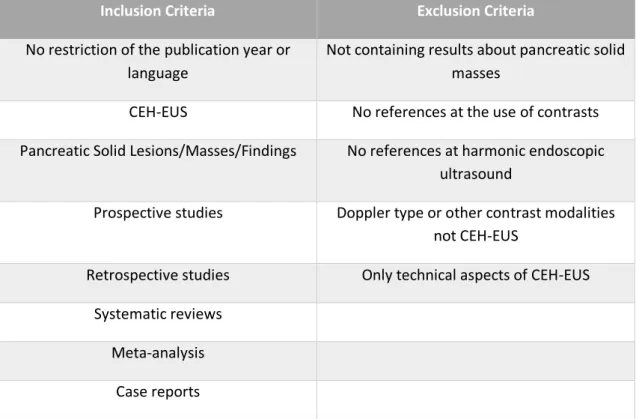
account including prospective and retrospective studies, systematic reviews, meta-analysis and case reports. The articles which did not contain results about pancreatic solid masses, no references at the use of contrasts or harmonic endoscopic ultrasound, and that concerned the Doppler type were excluded. Articles only about the technical aspects of CEH-EUS were also excluded. The inclusion and exclusion criteria used in this review are summarized on Table I.
RESULTS
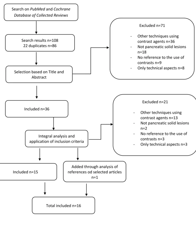
After the search 108 articles were found, and 22 were excluded for being duplicates. The abstracts of 86 papers were analyzed and after applying the exclusion criteria 36 articles were fully read. Upon reading, only 15 were considered valid according the criteria applied. 3 papers were included after scrutinizing the references of selected articles, but only 1 was used due to the other 2 not fitting on the inclusion criteria.
A lower intensity of enhancement comparing to the surrounding pancreatic tissue was considered as a hypoenhancement. Hyperenhancement is considered when a higher intensity of enhancement relative to the surrounding pancreatic tissue was shown. Isoenhancement was defined as an identical intensity of enhancement relative to the surrounding pancreatic tissue.(20)
The use of Sonazoid (Daiichi-Sankyo, Tokyo, Japan) and SonoVue (Bracco International BV, Amsterdam, The Netherlands) was reported for diagnosing solid pancreatic lesions (SPLs).(4)
According to Matsubara et all, pancreatic cancer has a hypovascular structure and the mean vascular density of pancreatic adenocarcinoma is low and often inferior comparing to the normal pancreatic parenchyma, thus considering that the explanation for pancreatic cancer appearing with an hypoenhancement pattern on CEH-EUS. (21) Concerning pathological examination, the
adenocarcinoma’s hard consistency is justified thanks to the presence of marked desmoplasia.(8)
On the other hand, neuroendocrine tumors appearance as hypervascular lesions using CEH-EUS with a sensitivity of 79%, described by Kitano et al. (22), is probably due to their abundant arterial
vascularization(17, 23) , being its appearance later confirmed on the research made by Park JS et
al., where 2 of 3 NETs were hyperenhanced and the other one was isoenhanced, being considered a possible explanation for the isoenhanced appearance as that the mass was misinterpreted as an isoenhancement pattern since the size of the tumor was too large (3.9 cm × 3.5 cm) to be accompanied by necrosis and then vascular structure was not visible on CEH-EUS. (20)
5 It was described that the differences between CEH-EUS using Sonazoid and SonoVue were that the two ultrasound contrast agents differ in terms of intensities and durations of signaling after infusion. (22) CEH-EUS images obtained after infusion of Sonazoid showed that parenchymal
perfusion can be observed throughout the pancreas only for 90 s (22) which limits the duration
of observation. However, with SonoVue it can last 120 s. (17) Therefore, an improving of the
observation of the pancreas can be achieved using SonoVue in CEH-EUS. Concerning the diagnostic accuracy for the pancreatic tumors by the two ultrasound contrast agents, it seems that the differences are related to differences in their signaling intensities and durations. (22)
The use of EUS-guided fine-needle aspiration (EUS-FNA) in patients subjected to CEH-EUS was reported to be more efficient comparing to the use of EUS-FNA in patients subjected only to EUS, as some studies have shown that the false-negatives according to EUS-FNA were detected as hypoenhancing tumors on CEH-EUS, thus lowering the number of biopsies that were not really pancreatic cancers . (24)
❖ SONOVUE
The use of SonoVue was reported in 10 of the 16 articles present in this study.
Its use on CEH-EUS was first reported in 2008 by Kitano et al. In this study, it was observed that hypovascular and heterogeneous images were seen in 4 of the 5 of malignant pancreatic lesions (80%) and isovascular images were observed in 100% of mass-forming pancreatitis lesions (3/3).(8) The first published study that tried to check if the use of CEH-EUS was able to
differentiate between pancreatic adenocarcinoma and mass-forming pancreatitis was made by A.Seicean et al. In that study, they used SonoVue and a low mechanical index of 0,3 to 0,4, followed by a diagnosis confirmation with EUS-FNA. The results were that a hypoenhanced pattern was seen in 14 patients with adenocarcinoma (15 in total) and in 10 cases of mass-forming pancreatitis (12 in total). The index of the contrast uptake ratio was significantly lower in adenocarcinoma than in mass-forming chronic pancreatitis. They concluded that visual discrimination between both cases of pancreatic adenocarcinoma and chronic pancreatitis was not possible, although the assessment of the index of the contrast uptake ratio can aid differentiation between malignant and benign masses but cannot replace EUS-FNA.(16)
In 2014, Park et al. found that hypoenhanced images were produced in 57 out of 62 pancreatic adenocarcinomas using SonoVue, which had a sensitivity of 92%, a specificity of 68% and an accuracy of 82% on that study. Those patients who had pancreatic adenocarcinoma showed a hypoenhanced pattern on CEH-EUS, being later confirmed via EUS-FNA or surgical resection. There were 3 cases which EUS-FNA was not able to get enough samples and 2 which were
6
negative for malignancy, but the 5 of them were reported as being hypoenhanced. (20) According
to Dietrich et al., considering hypovascularity as a sign of malignancy yields a diagnostic sensitivity of 92% and a specificity of 100%.(25)
In a retrospective study made by Leem G. et al., 207 patients with pancreatic solid masses were submitted to CEH-EUS with SonoVue in order to measure the sensitivity and specificity of this method to distinguish between ductal adenocarcinoma and neuroendocrine tumor as hypo or hyperenhanced on CEH-EUS. The sensitivity and specificity for ductal adenocarcinoma were 82.0% and 87.9%, respectively, and for neuroendocrine tumor were 81.1% and 90.9%, respectively. They described both ductal adenocarcinoma and neuroendocrine tumors as they were mostly hypoechoic in conventional EUS, but with the use of CEH-EUS it was possible to differentiate between them as ductal adenocarcinoma appeared with an hypoenhanced and inhomogeneous pattern whereas neuroendocrine tumors appeared with a hyperenhanced and homogeneous pattern, raising the sensitivity and specificity when compared to conventional EUS.(26)
In a study made in the Digestive Disease Center of the Department of Internal Medicine at Konkuk University School of Medicine in Seoul, Korea, the results found in 37 patients with pancreatic solid masses that were submitted to CEH-EUS with SonoVue were similar to the ones found in previous studies. Among pancreatic carcinomas, 28 out of 30 lesions (93%) had persistent hypovascular signals in the early and late phase, which indicates a sensitivity and diagnostic accuracy of 93% and 92%, respectively for the diagnosis of pancreatic cancer. Focusing on the speed of mass enhancement compared to surrounding pancreatic tissue, 26 out of the 30 pancreatic carcinomas (87%) had a slower enhancement. 94% of pancreatic carcinomas had a fast washout and had an inhomogeneous pattern. Concerning to neuroendocrine tumors, all of the 4 diagnosed showed hyperintense signals and a fast enhancement, but only 3 of them kept hyperintense signals in a late phase and a slower washout pattern and homogeneous enhancement pattern.(27)
The behavior of metastasis on CEH-EUS was reported by Dietrich et al., having 61% appeared as hyperenhanced and 28% as hypoenhanced. The etiology of those metastasis consisted mainly in renal cell carcinoma, followed by bronchial carcinoma, melanoma, breast carcinoma, ovarian carcinoma, anal carcinoma and thyroid carcinoma, in decreasing order of frequency, although it was not clarified which ones showed a hypoenhanced or hyperenhanced pattern. (23)
An isoenhanced pattern using CEH-EUS in a patient with autoimmune pancreatitis was described in a study reported by Iglesias-Garcia et al. (2) Some interesting results shown in the same study
made by Iglesias-Garcia et al. suggested that the use of CEH-EUS can be very helpful differentiating SPTs. However, on the NET’s case it can be hard to differentiate between the
7 benign and the malignant ones as there was a benign and a malignant NET that showed a hyperenhanced pattern and a benign NET that had a hypoenhanced pattern.(2)
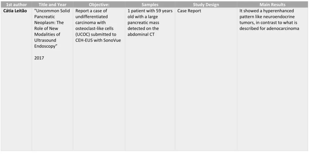
One case report made by Leitão described a patient with undifferentiated carcinoma with osteoclast-like cells submitted to CEH-EUS, showing a hyperenhanced pattern like neuroendocrine tumors, in contrast to what is described for adenocarcinoma.(28)
❖ SONAZOID
The use of Sonazoid was reported in 5 of the 16 articles present in this study.
CEH-EUS images of pancreatic cancers were described as having lower intensity of enhancement relative to the surrounding pancreatic tissue, and also a hypovascular pattern on all of the 5 articles analyzed. Studies using the Sonazoid contrast diagnosed pancreatic cancer with a sensitivity and specificity of 95% and 89% (22). In a different study, the presence of an
inflammatory mass depicted as an inflammatory pancreatic pseudotumor in patients with autoimmune pancreatitis had an isovascular pattern in 8 of 9 cases (89%), having the other one an hypovascular pattern. Neuroendocrine tumors were shown to be hypervascular. (29)
During contrast uptake and washout it is possible to evaluate in real time the pancreatic morphology, pancreatic circulation, tumor size and any invasion of an adjacent major vessel.(15)
In patients with pancreatic cancer, it is observed as hypoenhanced heterogeneous tumors on CEH-EUS, whereas pancreatic neuroendocrine tumor is observed as hyperenhanced tumors. Unenhanced areas in pancreatic lesions shown on CEH-EUS are believed to represent fibrosis or necrosis. Therefore, it would be difficult to obtain sufficient biopsy samples through unenhanced areas of SPLs. In the study made by Sugimoto et al., CEH-EUS was used to visualize SPLs and targeted the enhanced areas of lesions during FNA biopsy in order to minimize the false negatives. They hypothesized that, using CEH-EUS-guided FNA (CEH-EUS-FNA), fibrotic and necrotic areas could be avoided, improving the sampling rate and diagnostic ability, while reducing the number of needle passes required to obtain sufficient biopsy samples. It was also compared the diagnostic efficacy of CEH-EUS-FNA with that of conventional EUS-FNA.(4)
In some studies, there was used a multiphase study in order to compare the time-intensity of the contrast used with the types of pancreatic solid lesions. In a study made by T. Ishikawa et al. with 210 patients, there was recorded a movie for 60s continuously and the times of 20s, 40s and 60s were used for evaluation. The EI reduction was also used in the study in order to see if pancreatic ductal adenocarcinoma, pancreatic neuroendocrine neoplasm, solid pseudopapillary neoplasm and mass-forming pancreatitis had any differences that would help to diagnose these pancreatic lesions. PDAC lesions have shown a hypoenhanced pattern at each time (20s, 40s and
8
60s) in 102 of the 142 patients with PDAC lesions leading to an 83,1% of sensitivity, 86,8% of specificity and an 84,3% of accuracy. In PNEN and SPN lesions, prolonged hypervascular patterns were observed in all the times considered in 41% of the patients and in 61,3% that pattern was seen at either 20s or 40s, giving a 71,0% of sensitivity, 92,7% of specificity and an 89,5% of accuracy. In the MFP group, the lesions showed various enhancement patterns, leading to a non-relation between a pattern and MFP, although there was a high prevalence of an isovascular pattern, achieving 70,8% of sensitivity, 67,7% of specificity and a 68,1% of accuracy. Regarding EI reduction, CEH-EUS images of PDAC showed early reduction compared to the other types of lesions (35% at 1 minute) (14)
According to a study made in 2012 by Kitano et al. using CEH-EUS with Sonazoid, most of the diagnosed pancreatic carcinomas showed a hypoenhancement pattern, with a heterogeneous distribution and a lower intensity of enhancement when compared to the surrounding pancreatic tissue, with a sensitivity of 95 %, and the specificity of 89 %
❖ DEFINITY
Only one study made by
Romagnuolo
et al. was found reporting the use of Definity on CEH-EUS, allowing the depiction of malignant lesions as a hypoenhanced pattern and benign lesions as a hyperenhanced pattern, being later confirmed by FNA. However, it was described esophageal, pancreatic and liver tumors as a whole and specificity and sensitivity only for pancreatic solid masses was not available.(30)DISCUSSION AND CONCLUSIONS
The use of ultrasound contrast agents for diagnosing pancreatic solid lesions on CEH-EUS has been widely described with similar results. The appearance of the lesions was not changed between the use of SonoVue, Sonazoid or Definity, with pancreatic adenocarcinoma appearing in most cases as an hypoenhanced pattern, neuroendocrine tumors as an hyperenhanced pattern, and mass-forming pancreatitis as an isoenhanced pattern. CEH-EUS can be useful for the discrimination of hypoenhancing ductal adenocarcinoma of the pancreas from other hyper- or isoenhancing lesions and discrimination of mass-forming chronic pancreatitis from ductal adenocarcinoma in patients with chronic pancreatitis. (17) Patients with chronic pancreatitis have
9 impossible to distinguish between mass-forming pancreatitis and pancreatic adenocarcinoma, thus CEH-EUS has a huge importance on the ability to differentiate them in order to provide a best and faster approach.
The behavior of metastasis on CEH-EUS was described as either hyperenhanced or hypoenhanced, making it difficult to access their presence with the use of CEH-EUS. An isoenhanced pattern was seen in most cases of autoimmune pancreatitis, which mean that it can be misunderstood with mass-forming pancreatitis. Although they are both considered benign disorders, there is the need to, before anything else, access the clinical history and background of the patient, so the approach of each case can be decided carefully. Also, the B-mode of EUS used in patients with suspected mass-forming pancreatitis can provide very useful information regarding the presence of calcifications, an hyperechogenic duct or a lobular pattern.(32) In a study using SonoVue(16), mass-forming pancreatitis was described as having an
hypoenhanced pattern. This is probably due to the presence of a long inflammatory process which can led to decreasing vascularization caused by fibrosis.
The largest series of 277 patients with a solid pancreatic lesion who underwent CEH-EUS was reported by Kitano et al. in 2012, resulting in a 95% of sensitivity and 89% of specificity in detecting pancreatic cancer.(22)
Overall, the studies used in this review describing the results using SonoVue showed a variable sensitivity between 80% and 97%, a specificity of 67,85% to 100%, an accuracy of 85,5% to 92%, a positive predictive value ranging between 86,36% and 100% and a negative predictive value of 62,2% to 90% concerning the diagnosis of pancreatic adenocarcinoma. There was no information regarding NETs on these statistical topics. On the other hand, the studies concerning the diagnosis of pancreatic adenocarcinoma using Sonazoid described a sensitivity between 83,1% and 95,1%, a specificity of 71% to 89% and an accuracy of 84,3%, and there was no information regarding positive predictive value and negative predictive value. On the NETs case, a sensitivity between 78,9% and 81,1%, and a specificity ranging between 90,9% and 98,7% was described. According to these values, it seems that through the use of SonoVue is possible to achieve higher values of sensitivity and specificity, and endosonographers can say with more certainty if a lesion is definitely benign or malignant, although it can only be confirmed through histological analysis. However, neuroendocrine tumors were too few to analyze in this study. Further studies with more patients are therefore required to investigate the characteristic features of neuroendocrine tumors using CEH-EUS. As there were no comparative studies and the characteristics and the number of patients used in each study was different, as were the endoscopists, it is not possible to do a correct comparison between sensitivity and specificity of the contrasts used in CEH-EUS for diagnosing pancreatic solid lesions.
10
Most of the patients described in the studies used in this review were reported to have pancreatic adenocarcinoma. This could be happening as the major number of patients referred to CEH-EUS are the ones who have signs of malignancy in CEUS or in contrast enhanced power Doppler. (17)
It seems that the index of the contrast uptake ratio and echo intensity reduction is a good marker as it can aid differentiation between malignant and benign masses but still cannot replace EUS-FNA. More studies should be made concerning de application of this marker int the diagnosis of pancreatic solid lesions so it can be used in combination with the patterns’ analysis. The use of CEH-EUS can be helpful in reducing the number of needle passes required to obtain enough biopsy samples by EUS-FNA, thus improving the diagnostic efficacy and sampling rate of FNA. (4)
Deciding between follow-up or surgical resection in patients whose EUS-FNA findings are negative can be very difficult, as false -negatives EUS-FNA results cannot be excluded. Using CEH-EUS before EUS-FNA seems to be a good approach to help making decisions about the next treatment approach, as it identifies most false-negatives as hypoenhanced leading to a pathological re-evaluation or surgical resection of the tumor.(22) It improves the sensitivity with
which pancreatic adenocarcinomas are identified by EUS-FNA alone from 92,2% to 100%. (22)
It is not possible to suspect of any type of lesion without accessing correctly to the patient clinical history and background, as it is a major source of information and can be extremely helpful to decide how to act next.
The fact that an hypovascular pattern corresponded with heterogeneous tumor cells with fibrotic tissue, necrotic tissue and few vessels, and the isovascular pattern corresponded with homogeneous tumor cells with abundant vessels and no necrotic or fibrous tissues, can be relevant for the noninvasive evaluation of biological features of pancreatic cancer.(33) There is a
chance that using EUS-FNA in a lesion with a hypovascular pattern it would be unsuccessful because the samples can be of benign tissue, not allowing the correct diagnosis if it is pancreatic cancer, and the patient would be submitted to an invasive procedure without a correct outcome. Metastatic lung tumors found in the pancreas can also be described as having an hyperenhanced pattern being difficult to distinguish only by CEH-EUS if it is a case of neuroendocrine tumor or a pancreatic metastasis.(4) In the study made by Dietrich et al. (23) there was the description of
the origin of the pancreatic metastasis by order of prevalence in the patients included in the study, but there was no description of which types appeared as isoenhanced or hypoenhanced. As small solid pancreatic lesions make up a large variety of different tumor entities, a faster accurate and meticulous analysis and characterization of small solid pancreatic lesions should be made, in order to improve patients survival rate by providing an early detection and
11 appropriate management, or to avoid unnecessary risk exposure by submitting patients with benign lesions to the morbidity and mortality associated with radical pancreatic surgery. (23)
Giving the very high diagnostic accuracy in distinguish between benign and malignant lesions by elastography, which evaluates mainly tissue stiffness, the use of CEH-EUS can be helpful in order to help to characterize the type of malignancy in a lesion with a malignant appearance in elastography. (2)
Although there were very good results in what concerns the differentiation between solid pancreatic masses using CEH-EUS, the gold standard of diagnosis continues to be the histological analysis, achieved with samples acquired either by aspiration cytology with EUS-FNA or surgically. The concomitant use of CEH-EUS with EUS-FNA seems to have good outcomes in overcoming the false-negative results of each individual techniques, increasing the overall accuracy of diagnosis. (34) On the study reported by Kitano et al. in 2012, the use of CEH-EUS with
EUS-FNA was shown to rise the sensitivity to 100%(22), thus confirming that the application of
CEH-EUS is very helpful.
There were several limitations reported in the studies used in this review. Five were retrospective (4, 14, 20, 26, 35), seven had a small number of patients (4, 11, 14-16, 20, 27), six were unicentric (2, 4, 15, 16, 20, 27), verification or selection bias cannot be excluded in four (20, 23, 30, 35), and five used
subjective analysis.(2, 14, 15, 26, 35)
About this review, there were also a few limitations. As there was used some restrictive key words in the articles search and the search was made by only one person, there could be some selection bias. A meta-analysis should also be performed in order to synthesize data from a set of identical studies used to make this review.
To keep proving the real utility of contrast harmonic enhancement in endoscopic ultrasound to differentiate between pancreatic solid lesions in combination with EUS-FNA, more studies should be performed. A suggestion in the future can be to access the use of CEH-EUS to predict chemotherapy response in patients with pancreatic cancer by doing a correlation between the findings in the CEH-EUS, histological differentiation of tumors and clinical characteristics of pancreatic cancer. More multicenter trials should be made and more endosonographers with different degrees of experience should participate in those studies in order to validate these findings even more. By making a uniform dedicated software that allows the interplatform comparison and the decrease of interobserver and intraobserver variability through the use of more objective parameters could be an advantage in future studies.
12
APPENDIX I - FIGURES
Figure 1 - Fluxogram of the articles searched Search on PubMed and Cochrane
Database of Collected Reviews
Search results n=108 22 duplicates n=86
Selection based on Title and Abstract
Included n=36
Integral analysis and application of inclusion criteria
Included n=15
Added through analysis of references od selected articles
n=1
Total included n=16
Excluded n=71
- Other techniques using contrast agents n=36 - Not pancreatic solid lesions
n=18
- No reference to the use of contrasts n=9
- Only technical aspects n=8
Excluded n=21
- Other techniques using contrast agents n=13 - Not pancreatic solid lesions
n=2
- No reference to the use of contrasts n=3
13
APPENDIX II – TABLES
Table I - Inclusion and exclusion criteria
Inclusion Criteria Exclusion Criteria
No restriction of the publication year or language
Not containing results about pancreatic solid masses
CEH-EUS No references at the use of contrasts
Pancreatic Solid Lesions/Masses/Findings No references at harmonic endoscopic ultrasound
Prospective studies Doppler type or other contrast modalities
not CEH-EUS
Retrospective studies Only technical aspects of CEH-EUS
Systematic reviews Meta-analysis
14
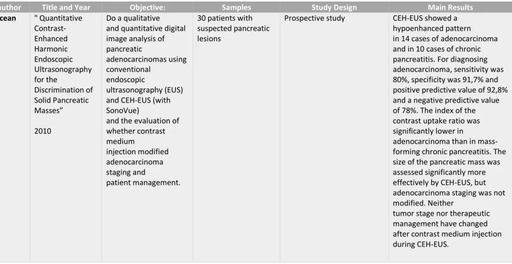
Table II - Seicean et al." Quantitative Contrast-Enhanced Harmonic Endoscopic Ultrasonography for the Discrimination of Solid Pancreatic Masses” (16)
1st author Title and Year Objective: Samples Study Design Main Results
A. Seicean " Quantitative Contrast-Enhanced Harmonic Endoscopic Ultrasonography for the Discrimination of Solid Pancreatic Masses” 2010 Do a qualitative and quantitative digital image analysis of pancreatic adenocarcinomas using conventional endoscopic ultrasonography (EUS) and CEH-EUS (with SonoVue)
and the evaluation of whether contrast medium injection modified adenocarcinoma staging and patient management. 30 patients with suspected pancreatic lesions
Prospective study CEH-EUS showed a
hypoenhanced pattern
in 14 cases of adenocarcinoma and in 10 cases of chronic pancreatitis. For diagnosing adenocarcinoma, sensitivity was 80%, specificity was 91,7% and positive predictive value of 92,8% and a negative predictive value of 78%. The index of the contrast uptake ratio was significantly lower in
adenocarcinoma than in mass-forming chronic pancreatitis. The size of the pancreatic mass was assessed significantly more effectively by CEH-EUS, but adenocarcinoma staging was not modified. Neither
tumor stage nor therapeutic management have changed after contrast medium injection during CEH-EUS.
15 Table III - Romagnuolo et al."Accuracy of contrast-enhanced harmonic EUS with a second-generation perflutren lipid microsphere contrast agent (with video)" (30)
1st author Title and Year Objective: Samples Study Design Main Results
Joseph Romagnuolo "Accuracy of contrast-enhanced harmonic EUS with a second-generation perflutren lipid microsphere contrast agent (with video)" 2011 To determine optimal settings and preliminary accuracy of CEH-EUS by using a second generation perflutren lipid microsphere contrast agent (Definity) and a prototype linear echoendoscope. Patients with esophageal/pancr eatic/liver tumor or adenopathy.
Contrast agent was injected (10 µL/kg intravenously in 1-2 doses), and the mechanical index was
optimized over 5 cases (0.3). Intermittent/continuous imaging was used with extended pure harmonic detection.
Before-contrast and after-contrast predictions of neoplasia (5-point Likert scale). The reference standard was positive tissue or 6-month follow-up. Perfusion factors (sequence, pattern,
washout) were noted, and phases were video recorded (arterial, venous, and postvenous).
30 sites (7 nodes and 16 pancreatic and 7 nonpancreatic masses) were imaged in 21 patients;
21 of 30 had FNA, and 5 had surgery; 4 cases (13.3%) were rated as undecided/indeterminate with EUS (vs 1 [3.3%] with CEH-EUS;
24 cases with confirmed diagnoses (12 malignant and 12 benign) were used for test performance:
positive/negative predictive values for CEH-EUS were 80.0% and 100% (with sensitivity of 100% and specificity of 72,7%) versus 84.6% and 100% of positive /negative predictive values (excluding the 3 undecided cases) and 100% and 80% of sensitivity and specificity for EUS. Accuracies, counting “undecided” (1 in CEH-EUS and 4 in EUS) as incorrect, were 83.3% and 79.2%. In 2 cases,
management would change significantly.
16
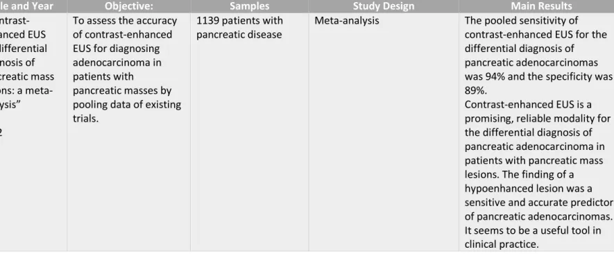
Table IV - Gong et al." Contrast-enhanced EUS for differential diagnosis of pancreatic mass lesions: a meta-analysis”(9)
1st author Title and Year Objective: Samples Study Design Main Results
Ting-ting Gong " Contrast-enhanced EUS for differential diagnosis of pancreatic mass lesions: a meta-analysis” 2012
To assess the accuracy of contrast-enhanced EUS for diagnosing adenocarcinoma in patients with
pancreatic masses by pooling data of existing trials.
1139 patients with pancreatic disease
Meta-analysis The pooled sensitivity of
contrast-enhanced EUS for the differential diagnosis of pancreatic adenocarcinomas was 94% and the specificity was 89%.
Contrast-enhanced EUS is a promising, reliable modality for the differential diagnosis of pancreatic adenocarcinoma in patients with pancreatic mass lesions. The finding of a hypoenhanced lesion was a sensitive and accurate predictor of pancreatic adenocarcinomas. It seems to be a useful tool in clinical practice.
17
Table V - Kitano et al." Characterization of Small Solid Tumors in the Pancreas: The Value of Contrast-Enhanced Harmonic Endoscopic Ultrasonography”(22)
1st author Title and Year Objective: Samples Study Design Main Results
Masayuki Kitano " Characterization of Small Solid Tumors in the Pancreas: The Value of Contrast-Enhanced Harmonic Endoscopic Ultrasonography” 2012 To evaluate how accurately CEH-EUS with Sonazoid characterizes
pancreatic lesions and compared its diagnostic ability with that of contrast-enhanced multidetector-row computed tomography (MDCT) and endoscopic ultrasonography-guided fi ne needle aspiration (EUS-FNA) 277 patients with pancreatic solid lesions
Prospective study CEH-EUS-depicted
hypoenhancement diagnosed ductal carcinomas with a sensitivity and specificity of 95.1% and 89.0 % respectively. For diagnosing small carcinomas by CEH-EUS, the sensitivity and specificity were 91.2 % and 94.4 % respectively. CEH-EUS-depicted hypervascular enhancement diagnosed neuroendocrine tumors with a sensitivity and specificity of 78.9 % and 98.7 % respectively. CEH-EUS was superior to MDCT in diagnosing small ( ≤ 2 cm)
carcinomas. In 12 neoplasms that MDCT failed to detect, 7 ductal carcinomas and 2
neuroendocrine tumors had hypoenhancement and
hyperenhancement on CEH-EUS, respectively. Combining CEH-EUS with EUS-FNA, the sensitivity of EUS-FNA increased from 92.2 to 100 % .
18
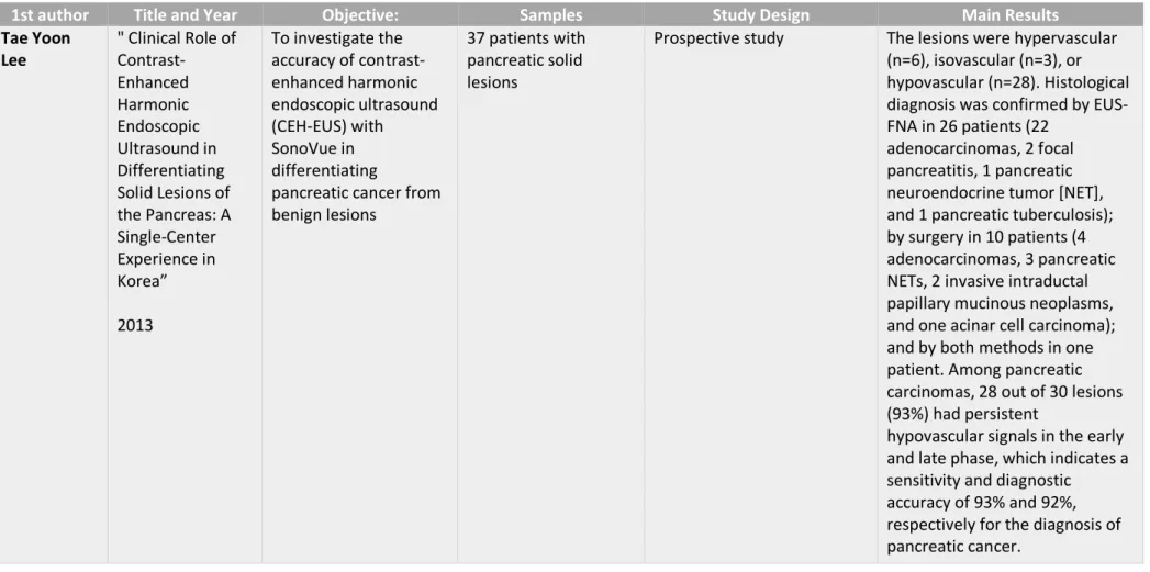
Table VI - Lee et al." Clinical Role of Contrast-Enhanced Harmonic Endoscopic Ultrasound in Differentiating Solid Lesions of the Pancreas: A Single-Center Experience in Korea” (27)
1st author Title and Year Objective: Samples Study Design Main Results
Tae Yoon Lee " Clinical Role of Contrast-Enhanced Harmonic Endoscopic Ultrasound in Differentiating Solid Lesions of the Pancreas: A Single-Center Experience in Korea” 2013 To investigate the accuracy of contrast-enhanced harmonic endoscopic ultrasound (CEH-EUS) with SonoVue in differentiating
pancreatic cancer from benign lesions
37 patients with pancreatic solid lesions
Prospective study The lesions were hypervascular
(n=6), isovascular (n=3), or hypovascular (n=28). Histological diagnosis was confirmed by EUS-FNA in 26 patients (22
adenocarcinomas, 2 focal pancreatitis, 1 pancreatic neuroendocrine tumor [NET], and 1 pancreatic tuberculosis); by surgery in 10 patients (4 adenocarcinomas, 3 pancreatic NETs, 2 invasive intraductal papillary mucinous neoplasms, and one acinar cell carcinoma); and by both methods in one patient. Among pancreatic carcinomas, 28 out of 30 lesions (93%) had persistent
hypovascular signals in the early and late phase, which indicates a sensitivity and diagnostic
accuracy of 93% and 92%, respectively for the diagnosis of pancreatic cancer.
19
Table VII - Park et al." Effectiveness of contrast-enhanced harmonic endoscopic ultrasound for the evaluation of solid pancreatic masses”(20)
1st author Title and Year Objective: Samples Study Design Main Results
Jin-Seok Park " Effectiveness of contrast-enhanced harmonic endoscopic ultrasound for the evaluation of solid pancreatic masses” 2014 To evaluate the usefulness of contrast-enhanced harmonic endoscopic ultrasound (CEH-EUS) with Sonovue in differentiating between pancreatic adenocarcinomas and other pancreatic disease
90 patients with solid pancreatic masses on EUS
Retrospective study 57 of a total of 62 patients that had pancreatic adenocarcinoma showed a hypoenhanced pattern on CEH-EUS, being later
confirmed via EUS-fine needle aspiration (EUS-FNA) or surgical ressection. There were 3 cases which EUS-FNA wasn’t able to get enough samples and 2 which were negative for malignancy, but the 5 of them were reported as being hypoenhanced. The 3 neuroendocrine tumors present appeared as hypervascular lesions. For diagnosing
adenocarcinoma, sensitivity was 91,93%, specificity was 67,85% and positive predictive value of 86,36% and a negative predictive value of 79,16%.
20
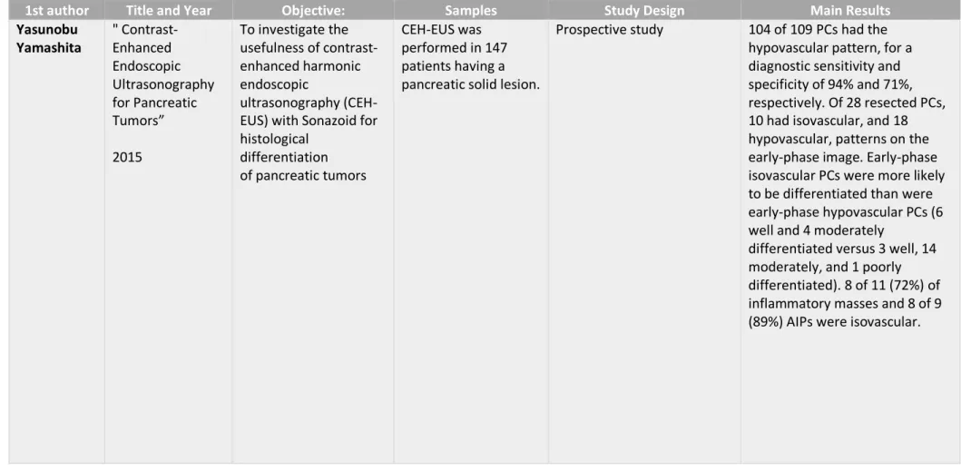
Table VIII - Yamashita et al." Contrast-Enhanced Endoscopic Ultrasonography for Pancreatic Tumors” (33)
1st author Title and Year Objective: Samples Study Design Main Results
Yasunobu Yamashita " Contrast-Enhanced Endoscopic Ultrasonography for Pancreatic Tumors” 2015 To investigate the usefulness of contrast-enhanced harmonic endoscopic ultrasonography (CEH-EUS) with Sonazoid for histological differentiation of pancreatic tumors CEH-EUS was performed in 147 patients having a pancreatic solid lesion.
Prospective study 104 of 109 PCs had the
hypovascular pattern, for a diagnostic sensitivity and specificity of 94% and 71%, respectively. Of 28 resected PCs, 10 had isovascular, and 18 hypovascular, patterns on the early-phase image. Early-phase isovascular PCs were more likely to be differentiated than were early-phase hypovascular PCs (6 well and 4 moderately
differentiated versus 3 well, 14 moderately, and 1 poorly differentiated). 8 of 11 (72%) of inflammatory masses and 8 of 9 (89%) AIPs were isovascular.
21 Table IX - Sugimoto et al." Conventional versus contrast-enhanced harmonic endoscopic ultrasonography-guided fine-needle aspiration for diagnosis of solid pancreatic lesions: A prospective randomized trial"(4)
1st author Title and Year Objective: Samples Study Design Main Results
Mitsuru Sugimoto " Conventional versus contrast-enhanced harmonic endoscopic ultrasonography-guided fine-needle aspiration for diagnosis of solid pancreatic lesions: A prospective randomized trial” 2015 To investigate the efficacy of CEH-EUS with Sonazoid guided fine-needle aspiration (CEH-EUS-FNA) compared with that of conventional EUS-FNA for the diagnosis of SPLs. 292 patients with pancreatic adenocarcinoma who were submitted to EUS-FNA and CEH-EUS
Retrospective study The overall sensitivity of EUS-FNA was 90.8% (265/292). The
sensitivities of EUS-FNA for lesions with and without an avascular area were 72.9% (35/48) and 94.3% (230/244), respectively, with the difference being statistically significant (P < .001).
EUS-FNA has lower sensitivity for pancreatic adenocarcinoma with avascular areas on CEH-EUS.
22
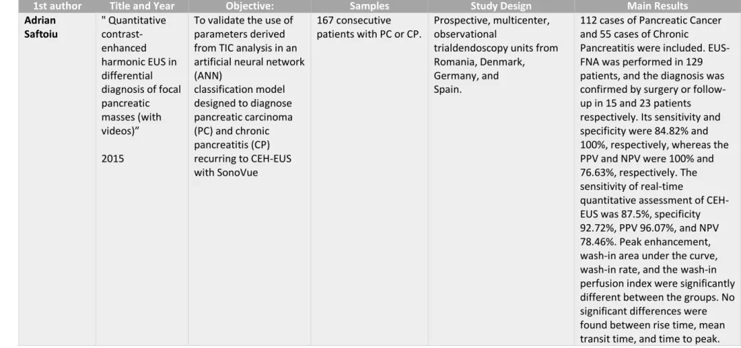
Table X - Saftoiu et al." Quantitative contrast-enhanced harmonic EUS in differential diagnosis of focal pancreatic masses (with videos"(11)
1st author Title and Year Objective: Samples Study Design Main Results
Adrian Saftoiu " Quantitative contrast-enhanced harmonic EUS in differential diagnosis of focal pancreatic masses (with videos)” 2015
To validate the use of parameters derived from TIC analysis in an artificial neural network (ANN) classification model designed to diagnose pancreatic carcinoma (PC) and chronic pancreatitis (CP) recurring to CEH-EUS with SonoVue 167 consecutive patients with PC or CP. Prospective, multicenter, observational
trialdendoscopy units from Romania, Denmark, Germany, and Spain.
112 cases of Pancreatic Cancer and 55 cases of Chronic
Pancreatitis were included. EUS-FNA was performed in 129 patients, and the diagnosis was confirmed by surgery or follow-up in 15 and 23 patients respectively. Its sensitivity and specificity were 84.82% and 100%, respectively, whereas the PPV and NPV were 100% and 76.63%, respectively. The sensitivity of real-time
quantitative assessment of CEH-EUS was 87.5%, specificity 92.72%, PPV 96.07%, and NPV 78.46%. Peak enhancement, wash-in area under the curve, wash-in rate, and the wash-in perfusion index were significantly different between the groups. No significant differences were found between rise time, mean transit time, and time to peak.
23 Table XI - Fusaroli et al." The clinical impact of ultrasound contrast agents in EUS: a systematic review according to the levels of evidence"(34)
1st author Title and Year Objective: Samples Study Design Main Results
Pietro Fusaroli " The clinical impact of ultrasound contrast agents in EUS: a systematic review according to the levels of evidence” 2016
To present the levels of evidence found in the literature about the use of CEH-EUS in routine clinical practice and its outcomes in the
appropriate perspective
210 articles about the use of CEH-EUS in pathologic conditions of the digestive tract
Systematic review For pancreatic solid neoplasms,
the pooled sensitivity and specificity in the diagnosis of pancreatic carcinoma were very high (LE 1); quantitative analysis and guidance of FNA were reported as investigational research (LE 2-3).
The differential diagnosis between benign and malignant lesions is the main field of application of CEH-EUS. Regarding to pancreatic solid neoplasms, the concomitant use of both CEH-EUS and EUS-FNA may have additive value in increasing the overall accuracy by overcoming the false-negative results associated with each individual technique. Other applications are promising but still investigational.
24
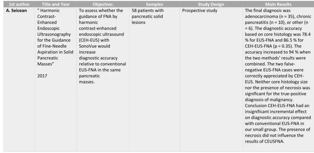
Table XII - Seicean et al."Harmonic Contrast-Enhanced Endoscopic Ultrasonography for the Guidance of Fine-Needle Aspiration in Solid Pancreatic Masses"(15)
1st author Title and Year Objective: Samples Study Design Main Results
A. Seicean " Harmonic Contrast-Enhanced Endoscopic Ultrasonography for the Guidance of Fine-Needle Aspiration in Solid Pancreatic
Masses” 2017
To assess whether the guidance of FNA by harmonic contrast-enhanced endoscopic ultrasound (CEH-EUS) with SonoVue would increase diagnostic accuracy relative to conventional EUS-FNA in the same pancreatic
masses.
58 patients with pancreatic solid lesions
Prospective study The final diagnosis was
adenocarcinoma (n = 35), chronic pancreatitis (n = 10), or other (n = 6). The diagnostic accuracy based on core histology was 78.4 % for EUS-FNA and 86.5 % for CEH-EUS-FNA (p = 0.35). The accuracy increased to 94 % when the two methods’ results were combined. The two false-negative EUS-FNA cases were correctly appreciated by CEH-EUS. Neither core histology size nor the presence of necrosis was significant for the true-positive diagnosis of malignancy.
Conclusion CEH-EUS-FNA had an insignificant incremental effect on diagnostic accuracy compared with conventional EUS-FNA in our small group. The presence of necrosis did not influence the results of CEUSFNA.
25 Table XIII - Garcia et al." Differential diagnosis of solid pancreatic masses: contrast-enhanced harmonic (CEH-EUS), quantitative-elastography (QE-EUS), or both?"(2)
1st author Title and Year Objective: Samples Study Design Main Results
Julio Iglesias-Garcia "Differential diagnosis of solid pancreatic masses: contrast-enhanced harmonic (CEH-EUS), quantitative-elastography (QE-EUS), or both?” 2017 To evaluate the diagnostic accuracy of CEH-EUS (with
SonoVue), QE-EUS, and the combination of both for the differential diagnosis of SPT (solid pancreatic tumors)
62 patients with pancreatic solid lesions
Prospective study Final diagnosis was pancreatic
adenocarcinoma (n=45), NET (n=3), inflammatory mass (n=10), pancreatic metastasis (n=2), autoimmune pancreatitis (n=1) and a mucinous
cystadenocarcinoma (n=1). Overall accuracies for determination of malignancy using: - QE-EUS: 98.4% (95% confidence interval (CI): 91.4–99.7) - CEH-EUS: 85.5% (95% CI: 74.7–92.2) - QE-EUS+ CEH-EUS: 91.9% (95% CI: 82.5–96.5)
- EUS-guided tissue acquisition: 91.5% (95% CI: 83.6–99.5) The combination of QE-EUS and CEH-EUS gives complementary information for the differential diagnosis of SPT. However, this combination does not
significantly increase the diagnostic accuracy of either of the techniques performed alone.
26
Table XIV - Leitão et al. "Uncommon Solid Pancreatic Neoplasm: The Role of New Modalities of Ultrasound Endoscopy”(28)
1st author Title and Year Objective: Samples Study Design Main Results
Cátia Leitão "Uncommon Solid Pancreatic Neoplasm: The Role of New Modalities of Ultrasound Endoscopy” 2017 Report a case of undifferentiated carcinoma with osteoclast-like cells (UCOC) submitted to CEH-EUS with SonoVue
1 patient with 59 years old with a large pancreatic mass detected on the abdominal CT
Case Report It showed a hyperenhanced
pattern like neuroendocrine tumors, in contrast to what is described for adenocarcinoma
27 Table XV - Leem et al. " Clinical Value of Contrast-Enhanced Harmonic Endoscopic Ultrasonography in the Differential Diagnosis of Pancreatic and
Gallbladder Masses"(26)
1st author Title and Year Objective: Samples Study Design Main Results
Galam Leem " Clinical Value of Contrast-Enhanced Harmonic Endoscopic Ultrasonography in the Differential Diagnosis of Pancreatic and Gallbladder Masses” 2017
To prove the clinical value of CEH-EUS in the differential diagnosis of pancreatic and
gallbladder masses by direct comparison with that of conventional EUS. 207 patients with pancreatic solid masses were submitted to CEH-EUS with SonoVue (Bracco)
Retrospective study The sensitivity and specificity for ductal adenocarcinoma were 82.0% and 87.9%, respectively, and for neuroendocrine tumor were 81.1% and 90.9%,
respectively. They described both ductal adenocarcinoma and neuroendocrine tumors as they were mostly hypoechoic in conventional EUS, but with the use of CEH-EUS it was possible to differentiate between them as ductal adenocarcinoma appeared with an hypoenhanced and inhomogeneous pattern whereas neuroendocrine tumors
appeared with a hyperenhanced and homogeneous pattern, raising the sensitivity and specificity when compared to conventional EUS.
28
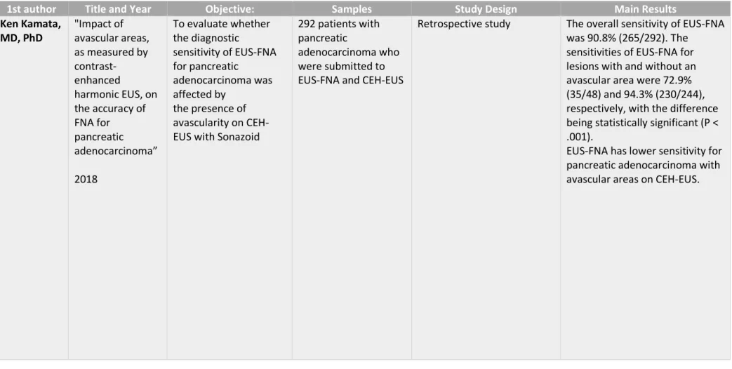
Table XVI - Kamata et al." Impact of avascular areas, as measured by contrast-enhanced harmonic EUS, on the accuracy of FNA for pancreatic adenocarcinoma"(35)
1st author Title and Year Objective: Samples Study Design Main Results
Ken Kamata, MD, PhD "Impact of avascular areas, as measured by contrast-enhanced harmonic EUS, on the accuracy of FNA for pancreatic adenocarcinoma” 2018 To evaluate whether the diagnostic sensitivity of EUS-FNA for pancreatic adenocarcinoma was affected by the presence of avascularity on CEH-EUS with Sonazoid
292 patients with pancreatic
adenocarcinoma who were submitted to EUS-FNA and CEH-EUS
Retrospective study The overall sensitivity of EUS-FNA was 90.8% (265/292). The
sensitivities of EUS-FNA for lesions with and without an avascular area were 72.9% (35/48) and 94.3% (230/244), respectively, with the difference being statistically significant (P < .001).
EUS-FNA has lower sensitivity for pancreatic adenocarcinoma with avascular areas on CEH-EUS.
29
Table XVII - Ishikawa et al. " Multiphase evaluation of contrast-enhanced endoscopic ultrasonography in the diagnosis of pancreatic solid lesions"(14)
1st author Title and Year Objective: Samples Study Design Main Results
Takuya Ishikawa " Multiphase evaluation of contrast-enhanced endoscopic ultrasonography in the diagnosis of pancreatic solid lesions” 2018 To propose a simplified method by evaluating multiple phases of CEH-EUS with Sonazoid in the diagnosis of
pancreatic solid lesions.
210 patients with pancreatic solid lesions including 142 with
pancreatic ductal cancer (PDAC), 31 with pancreatic
neuroendocrine neoplasm, 13 with solid pseudopapillary neoplasm and 24 with mass-forming
pancreatitis who underwent CE-EUS and achieved final diagnoses
Retrospective study The majority of the lesions in PDAC group showed
hypovascular pattern at 20 or 40 s after injection of contrast medium following early enhancement. The PDAC pattern in the differentiation of PDAC from other lesions resulted in a sensitivity of 83.1%,
specificity of 86.8% and accuracy of 84.3%. On histopathological analysis, significant differences were seen in histologic types, infiltration (INF), and neural invasion (ne) between those who showed PDAC pattern and those who didn't.
30
REFERENCES
1. Society AC. Cancer Facts & Figures 2012. American Cancer Society. 2012.
2. Iglesias-Garcia J, Lindkvist B, Lariño-Noia J, Abdulkader-Nallib I, Dominguez-Muñoz JE. Differential diagnosis of solid pancreatic masses: contrast-enhanced harmonic (CEH-EUS), quantitative-elastography (QE-EUS), or both? United European gastroenterology journal. 2017;5(2):236-46.
3. Gress F. Progress in Endoscopic Ultrasonography, An Issue of Gastrointestinal Endoscopy Clinics, E-Book: Elsevier Health Sciences; 2017.
4. Sugimoto M, Takagi T, Hikichi T, Suzuki R, Watanabe K, Nakamura J, et al. Conventional versus contrast-enhanced harmonic endoscopic ultrasonography-guided fine-needle aspiration for diagnosis of solid pancreatic lesions: A prospective randomized trial. Pancreatology.
2015;15(5):538-41.
5. Kato T TY, Naitoh Y, Hirooka Y, Furukawa T, Hayakawa T. Ultrasonographic and endoscopic ultrasonographic angiography in pancreatic mass lesions. Acta Radiol. 1995;36:381-7.
6. Kitano M, Kudo M, Sakamoto H, Komaki T. Endoscopic ultrasonography and contrast-enhanced endoscopic ultrasonography. Pancreatology. 2011;11 Suppl 2:28-33.
7. D’Onofrio M, Biagioli E, Gerardi C, Canestrini S, Rulli E, Crosara S, et al. Diagnostic Performance of Contrast-Enhanced Ultrasound (CEUS) and Contrast-Enhanced Endoscopic Ultrasound (ECEUS) for the Differentiation of Pancreatic Lesions: A Systematic Review and Meta-Analysis. Ultraschall in Med. 2014;35(06):515-21.
8. Kitano M, Sakamoto H, Matsui U, Ito Y, Maekawa K, von Schrenck T, et al. A novel perfusion imaging technique of the pancreas: contrast-enhanced harmonic EUS (with video). Gastrointest Endosc. 2008;67(1):141-50.
9. Gong T-t, Hu D-m, Zhu Q. Contrast-enhanced EUS for differential diagnosis of pancreatic mass lesions: a meta-analysis. Gastrointestinal Endoscopy. 2012;76(2):301-9. 10. Sugimoto M, Takagi T, Suzuki R, Konno N, Asama H, Watanabe K, et al. Contrast-enhanced harmonic endoscopic ultrasonography in gallbladder cancer and pancreatic cancer. Fukushima journal of medical science. 2017;63(2):39-45.
11. Săftoiu A, Vilmann P, Dietrich CF, Iglesias-Garcia J, Hocke M, Seicean A, et al. Quantitative contrast-enhanced harmonic EUS in differential diagnosis of focal pancreatic masses (with videos). Gastrointestinal Endoscopy. 2015;82(1):59-69.
12. Schneider M, Arditi M, Barrau MB, Brochot J, Broillet A, Ventrone R, et al. BR1: a new ultrasonographic contrast agent based on sulfur hexafluoride-filled microbubbles. Investigative radiology. 1995;30(8):451-7.
13. Sakamoto H, Kitano M, Matsui S, Kamata K, Komaki T, Imai H, et al. Estimation of malignant potential of GI stromal tumors by contrast-enhanced harmonic EUS (with videos). Gastrointest Endosc. 2011;73(2):227-37.
14. Ishikawa T, Hirooka Y, Kawashima H, Ohno E, Hashizume K, Funasaka K, et al. Multiphase evaluation of contrast-enhanced endoscopic ultrasonography in the diagnosis of pancreatic solid lesions. Pancreatology. 2018;18(3):291-7.
15. Seicean A, Badea R, Moldovan-Pop A, Vultur S, Botan EC, Zaharie T, et al. Harmonic Contrast-Enhanced Endoscopic Ultrasonography for the Guidance of Fine-Needle Aspiration in Solid Pancreatic Masses. Ultraschall in Med. 2017;38(02):174-82.
16. Seicean A, Badea R, Stan-Iuga R, Mocan T, Gulei I, Pascu O. Quantitative Contrast-Enhanced Harmonic Endoscopic Ultrasonography for the Discrimination of Solid Pancreatic Masses. Ultraschall in Med. 2010;31(06):571-6.
17. Piscaglia F, Nolsoe C, Dietrich CF, Cosgrove DO, Gilja OH, Bachmann Nielsen M, et al. The EFSUMB Guidelines and Recommendations on the Clinical Practice of Contrast Enhanced