MULTIPLE MYELOMA IN YOUNG WOMAN: CASE REPORT
Mieloma múltiplo em mulher jovem: relato de caso
Gabriela Arnoni Dias1
Nayara Lopes de Souza1
Evandro Barbosa dos Anjos1
José Alfreu Soares Junior1,2
Jéssica Aguiar das Virgens1
Jéssica Souza Rodrigues1
Antônio Guerra de Oliveira Neto3
Abstract: Multiple Myeloma is a hematological neoplasm that typically affects the elderly population. It
is rare in individuals under 40 years of age (2% of the total). The age group of the patient is fundamentally
important in order to define the most appropriate treatment. Multiple Myeloma is characterized by
dysregulated proliferation of plasma cells in the bone marrow, which produce and secrete monoclonal
immunoglobulin. Patients commonly present symptoms such as anemia, hypercalcemia, renal failure and
bone disease. The diagnosis is confirmed by detecting spinal cord plasma cytosis ≥ 10% and/or presence
of plasmacytoma and at least one of the following criteria: presence of target organ lesion associated with
Multiple Myeloma; presence of some biomarker. It is an incurable disease whose main goal of treatment is
to increase the patient’s survival and quality of life. Treatment is defined according to some parameters such
as age and presence of comorbidities. Young patients with no comorbidities should preferentially perform
high dose chemotherapy followed by autologous hematopoietic stem cell transplantation; while patients
older than 65-70 years with comorbidities are treated with chemotherapy alone. Multiple Myeloma presents
a heterogeneous evolution whose survival varies from a few months to more than a decade. The objective
of this study is to report the case of a young patient diagnosed with Multiple Myeloma.
Keywords: Multiple Myeloma; Neoplasms; Plasma Cells; Bone Marrow.
Autor para correspondência: Gabriela Arnoni Dias E-mail: [email protected]
1- Faculdades Unidas do Norte de Minas - FUNORTE. 2- Universidade Federal de Minas Gerais - UFMG.
Resumo: O Mieloma Múltiplo é uma neoplasia hematológica que afeta tipicamente a população idosa,
sendo raro em indivíduos com menos de 40 anos de idade (2% do total). A faixa etária do paciente é de
fun-damental importância para definição do tratamento mais adequado. O Mieloma Múltiplo caracteriza-se por
proliferação desregulada de plasmócitos na medula óssea, os quais produzem e secretam imunoglobulina
monoclonal. Os pacientes comumente se apresentam com sintomas de anemia, hipercalcemia, insuficiência
renal e doença óssea. O diagnóstico é firmado ao se detectar plasmocitose medular ≥ 10% e/ou presença de
plasmocitoma e no mínimo um dos critérios a seguir: presença de lesão de órgão alvo associado ao
Mielo-ma Múltiplo; presença de algum bioMielo-marcador. Trata-se de uMielo-ma doença incurável, cujo principal objetivo
do tratamento é aumentar a sobrevida e qualidade de vida do paciente. O tratamento é definido de acordo
com alguns parâmetros como idade e presença de comorbidades, sendo que os pacientes jovens e sem
co-morbidades devem realizar preferencialmente quimioterapia de altas doses seguida de transplante autólogo
de células-tronco hematopoiéticas; enquanto os pacientes com mais de 65-70 anos, com comorbidades, são
tratados somente com quimioterapia. O Mieloma Múltiplo apresenta evolução heterogênea cuja sobrevida
dos pacientes varia de alguns meses até mais de uma década. Este estudo objetivou relatar o caso de uma
paciente jovem diagnosticada com Mieloma Múltiplo.
Palavras-chave: Mieloma Múltiplo; Neoplasias; Plasmócitos; Medula Óssea.
REVISTA UNIMONTES CIENTÍFICA
INTRODUCTION
Multiple Myeloma (MM) corresponds to
1% of all malignant neoplasms but represents the
second most common hematological malignancy
1.
It is considered a disease of the elderly, with a
me-dian age at diagnosis between 65-74 years, with
ap-proximately 35% - 40% of the cases over the age
of 75 years. Of the patients diagnosed with MM,
less than 2% is below the age of 40 years, and the
cases in patients less than 30 years are extremely
rare
2. This age distribution has implications in the
population eligible for specific types of treatments,
such as high doses of chemotherapy and stem cells
transplantation
3. MM is two times more common
in African-caribbeans than white people, and the
incidence is higher in males
4. In recent years there
has been an increase in the incidence of MM, which
may be related to greater knowledge of the disease
natural history and its pathogenesis, the
improve-ment of laboratory resources, the increase in life
expectancy worldwide and to chronic exposure to
pollutants
1. It is a disease with a heterogeneous
de-velopment whose survival of patients is variable,
from a few months up to 10 years
5.
MM is a neoplasia arising from the lineage
of B lymphocytes, characterized by the
unregu-lated clonal plasmocytic proliferation in the bone
marrow, being that these plasma cells produce and
secrete monoclonal immunoglobulin or fragment
of this, the M protein
1. MM arises due to genetic
mutations that occur during the differentiation of
B lymphocytes into plasma cells. In about half of
cases there is a chromosomal translocation that puts
an oncogene in the gene of the heavy chain of
im-munoglobulin in chromosome 14. This results in
overexpression of the oncogene and unregulated
cell proliferation. The remaining cases are
charac-terized by trisomies of several odd chromosomes
(3, 5, 7, 9, 11, 15, 19 and 21). To the extent that the
multiple myeloma develops, other genetic events,
such as RAS mutations may occur
4.
Patients with MM commonly have
symp-toms of anemia (observed in 75% of patients at
di-agnosis), hypercalcemia (30%), renal insufficiency
(25%) and bone disease (70%). The skeletal
mani-festations may present as painful lytic lesions,
ver-tebral fractures or fractures of the long bones. High
values of monoclonal protein can cause symptoms
of hyperviscosity (headache, epistaxis, visual
tur-bidity and mental confusion), while the reduced
humoral immunity results in recurrent bacterial
in-fections
4.
For the diagnosis of MM clinical history and
detailed physical examination must be performed,
proceed initially with routine laboratory tests:
com-plete blood count, renal function, ions including
calcium, protein electrophoresis of serum and
uri-nary for quantification of monoclonal protein and
then the serum and/or urine immunofixation in
or-der to identify the subtype of monoclonal protein.
The examination of the bone marrow (myelogram
and/or biopsy with cytogenetic analysis) will help
to confirm the diagnosis
6. However, the criteria for
the diagnosis of MM were revised in 2014 with
the objective to allow an earlier recognition of the
disease. Therefore, biomarkers were incorporated
that in addition to elucidating the diagnosis can
also infer the biological behavior of the disease in
determining which patients will have a greater risk
of unfavorable evolution. Thus, for confirmatory
diagnosis of MM it is necessary, as a compulsory
criterion, the medullary plasma cytosis ≥ 10% and/
or presence of plasmacytoma confirmed by biopsy.
In addition, it is necessary to have at least one of the
following criteria: presence of any target-organ
in-jury attributed to MM (hypercalcemia, renal
insuf-ficiency, anemia and lytic bone lesion); presence of
a biomarker (medullary plasma cytosis ≥ 60%,
ra-tio of serum free light chains in the serum involved
and chains involved ≥ 100, more than 1 focal lesion
checked by MRI)
7. For all patients diagnosed with
MM a skeletal survey is indicated with plain
radio-graphs of the spine, skull, pelvis, chest, and upper
bones of the limbs to determine the extention of
the disease
4.
MM is considered an incurable disease.
Therefore, the main objective of treatment is to
in-crease the survival and quality of life of the patients
8.
The choice among the treatment options are based
on patient characteristics such as age and presence
of comorbidities, therapeutic options available and
service experience. In patients aged less than 65-70
years, without comorbidities and with normal renal
function the consolidation of treatment must be
per-formed with high doses of chemotherapy followed
by transplantation of autologous hematopoietic
stem cells (TCTHa). On the other hand, elderly
pa-tients over 65-70 years or with comorbidities and/
or with altered renal function usually are not
eligi-ble for transplantation, being then treated only with
chemotherapy
9. The traditional regien with
vincris-tine-doxorubicin-dexamethasone was replaced by
new therapeutic options and is currently in disuse.
Therefore, the regimens that contain bortezomibe
are suggested as first-line treatment associated with
other drugs such as dexamethasone, thalidomide,
doxorubicin and/or cyclophosphamide. The CTD
regimen
cyclophosphamide-thalidomide-dexa-methasone, is widely used in the United Kingdom
for those patients who are candidates for
transplan-tation
4. Whereas patients who are not candidates for
transplantation, the chemotherapy regimen
consid-ered commonly used is the combination of
mel-phalan, prednisone and thalidomide
6. The support
therapy for the control of the main clinical
mani-quality of life of patients with MM. Thus,
bisphos-phonates are used to reduce skeletal complications,
the erythropoietin is used for the control of anemia
and should be considered for prophylaxis for
op-portunistic infections
8. For the treatment of bone
pain, one should start with non-opioid analgesic
agents (for example, paracetamol and/or dipyrone).
The nonsteroid anti-Inflammatory drugs should be
avoided by the potential risk of worsening renal
function and the opiates should be introduced when
the no-opioid analgesic agents are ineffective
6. The
majority of patients respond to initial treatment by
acquiring a period of the disease stability, which
is usually associated with the relative improvement
of quality of life. Despite the impossibility of
cura-tive treatment, and relapse being inevitable, a large
proportion of patients respond to Chemotherapies
of second line with different drugs of the initial
regimen, or even similar. However, the subsequent
relapses become increasingly less sensitive to
treat-ment
4.
Thus, the present study aimed to report the
case of a young patient diagnosed with MM, besides
presenting the symptoms manifested at initial
diag-nosis, exposing the propaedeutic performed and the
initial therapy used. Because this is an extremely
rare disease in young adults it becomes necessary to
know MM, establish an investigation strategy and
more appropriate treatment to avoid an unfavorable
evolution in this age range.
The design of this study was evaluated and
approved by the Ethics and Research Committee
of Faculdades Unidade do Norte de Minas
(FU-NORTE), with the opinion number 2.526.747.
CASE REPORT
Carv-REVISTA UNIMONTES CIENTÍFICA
tal, complaining of pain in the body, arthralgia and
weakness four months ago. She had performed
he-mogram when anemia was identified, treated with
noripurum without improvement. Evolved with
diffuse abdominal pain, vomiting, episodes of
di-arrhea, loss of appetite, loss of weight (9 kg in 30
days), dry cough and fever in the afternoon with
chills. Patient had no other complaints or
comor-bidities.
Upon physical examination,
hemodynami-cally stable, vital data without changes, dehydrated
1+/4+, pale 2+/4+, regular general condition; the
abdomen was flat, diffusely painful to palpation,
splenomegaly; Blumberg, Rovsing and Murphy
signs were negative. The patient was hospitalized,
and laboratory exams were performed for
investi-gation of clinical signs (Table 1). Chest radiograph:
the pulmonary parenchyma without alterations.
Total abdominal ultrasonography: moderate
sple-nomegaly, kidneys increased dimensions (both of
textural aspect suggestive of acute nephropathy),
another abdominal organ studied were normal.
hypotheses pyelonephritis, intestinal parasitosis,
hemolytic anemia. She received treatment with
Ciprofloxacin and Albendazole. New examinations
were performed and showed a drop in hemoglobin,
hypercalcemia and increase in creatinine (Table 2).
Table 1 - Results of laboratory examinations of
a case study of MM, Montes Claros, MG, 2018.
Variable
Result
Hemoglobin
9.2 g/dL
Leukocytes
11.62/ mm3
Platelets
168,000/ mm3
AST
16 U/L
ALT
14 U/L
Amylase
39 U/L
Lipase
17 U/L
Gamma GT
22 U/L
PCR
8.6 mg/L
Creatinine
1.3 mg/dl
The patient evolved with dysuria and
per-sistence of other complaints from the beginning
of the clinical signs. It was arosen as diagnostic
Table 2: Results of laboratory examinations of
follow-up of case study of MM, Montes Claros,
MG, 2018.
Variable
Result
Hemoglobin
6.7 g/dL
Hematocrit
19.4%
Leukocytes
7,584/ mm3
Platelets
125,000
Serum iron
244 mcg/dL
Ferritin
745.2 ng/ml
Saturation Index of
transferrin
96.0%
Sodium
140 mEq/L
Albumin
3.3 g/dL
Globulin
1.7 g/dL
Rheumatoid Factor
Negative
Potassium
3.5 mEq/L
Ionic Calcium
1.45 mmol/L
Magnesium
2.5 mg/dL
Urea
38 mg/dL
Creatinine
1.57 mg/dL
PCR
36 mg/L
CPK
161 U/L
Total Proteins
5 g/dL
LDH
363 U/L
Direct Coombs
Negative
BAAR
Negative
EAS: bacterial flora slightly increased;
GRAM of gout: presence of gram-negative rods;
EPF: negative for protozoa and metazoan. Chest
CT: osteolytic lesions at the level of the thoracic
spine, ribs and sternum and scapula. Arose as
di-agnostic hypotheses MM and metastasis. Thus,
the following were requested: myelogram;
elec-trophoresis of serum and urinary protein and
x-rays.
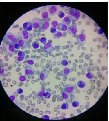
The myelogram showed the following
description: bone marrow infiltrated by 85% of
cells of Plasmacytic lineage (plasmocytes and
plasmablasts) (Figure 1). Bone marrow biopsy:
bone marrow hypercellularity.
diffusely, bone demineralization, and osteopenia;
reduction of articular spaces on coxofemorais,
incipient osteophytosis. The radiographs of the
left and right arms identified osteolytic lesions
and osteopenia, as well as the x-rays of the left
and right femur. Concerning the chest x-rays, the
results indicated a cardiothoracic index increased
bone structures with multiple osteolytic lesions. In
the radiography of the skull there was skullcap with
multiple osteolytic lesions and osteopenia.
After diagnosis of MM, the patient was
re-ferred to the onco-hematology service for treatment
and follow-up. Therapy performed: 6 cycles of
chemotherapy with CTD regimen
(Cyclophospha-mide, Thalidomide and Dexamethasone) associated
to Pamidronate with prediction of performing
TC-THa.
After initiation of chemotherapy, the
pa-tient showed significant improvement in all the
symptoms. New laboratory examinations were
per-formed after 6 cycles of chemotherapy (Table 3),
showing significant improvement, as well as new
electrophoresis of serum proteins (Table 4).
My-elogram performed after 4 cycles of chemotherapy
showed: bone marrow apparently normocelular and
4% of plasmocytes.
Figure 1 - Myelogram evidencing Plasmacytosis
of a case study of MM, Montes Claros, MG, 2018.
Regarding electrophoresis of serum proteins
the following results were observed: total proteins:
7.50 g/dL; Albumin: 4.20 g/dL (56.0%); Albumin/
Globulin ratio: 1.37; alpha 1 globulin: 0.40 g/dL
(5.3%); alpha 2 globulin: 0.89 g/dL (11.9%); beta
1 globulin: 0.44 g/dL (5.9%); beta 2 globulin: 0.39
g/dL (5.2%); gama globulin: 1.28 g/dL (17.1%).
Urinary protein electrophoresis: proteinuria 24
hours: 28.84g/24h; albumin 32.5%; alpha 1 globulin
10.5%; alpha 2 globulin 19,5%; beta globulin
11.5%; gama globulin 25.7%; urinary volume
1350ml; monoclonal bands were not observed.
The results of radiographs of hip left and
right femoral pointed multiple osteolytic lesions
Table 3 - Results of laboratory examinations
performed after chemotherapy of a case study
of MM, Montes Claros, MG, 2018.
Variable
Values
Hemoglobin
14.1 g/dL
Hematocrit
44.5%
Leukocytes
14,400/mm3
Platelets
312,000
Urea
28 mg/dL
Creatinine
0.85 mg/dL
LDH
17 U/L
REVISTA UNIMONTES CIENTÍFICA
DISCUSSION
MM is a malignant neoplasm originating
from B lymphocytes infiltrating the bone marrow
leading to bone destruction and bone marrow
insuf-ficiency
10, shows an incidence 50% greater in males
when compared to females
4, being that in Brazil the
average age at diagnosis is 61 years¹. In the case
reported there was a very atypical epidemiological
presentation, because in addition to the patient
be-ing female, she is in an age range below the usual.
MM has a heterogeneous presentation,
which may correlate to different cytogenetic
muta-tions that occur. However, the findings of anemia,
hypercalcemia, renal and bone pain are the most
common. For an initial evaluation complete blood
count must be requested with platelet count; urea;
serum creatinine and serum electrolytes; serum
calcium; albumin; lactate dehydrogenase;
beta-2-microglobulin and x-rays.
10In the present case,
the patient started the clinical sigs with symptoms
of anemia, arthralgia and asthenia. Among the
con-firmatory diagnostic criteria of the disease it is
ob-served that the patient had a myelogram with
in-tense plasmocitose (85%), presence of lytic lesions
(thoracic spine, ribs and sternum and scapula) and
hypercalcemia.
Despite the significant advances, MM is still
a disease considered incurable and the main
objec-tive of treatment is to achieve greater survival time
free from the disease. The time to relapse of the
dis-ease can vary from months to years depending on
the treatment regimen proposed
8. The patient’s age
and the presence or not of comorbidities directly
influences the choice of treatment and prognosis
10.
Thus, current therapy includes a phased approach,
often consisting of induction therapy,
consolida-tion and maintenance therapy. With a wider
pan-orama and more treatment options, the approach
of optimal therapy has become increasingly
com-plex. Especially in young patients aged less than
50 years, less than 15% of the cases, where there
are doubts whether more intense therapies could be
incorporated in this population, which may have a
greater tolerance to schemes of conditioning and,
consequently, a greater survival time free from
re-currence
11.
Upon defining the TCTHa as therapeutic
option one should consider the initial exposure to
chemotherapeutic agents that may affect the
col-lection of stem cells and also contribute to the
on-set of secondary neoplasms. Thus, among the main
schemes suggested for the first-line treatment of
the bortezomibe is more indicated. As examples,
the associations bortezomibe and dexamethasone;
bortezomibe, cyclophosphamide and
sone; bortezomibe, doxorubicin and
sone; bortezomibe, lenalidomide and
sone; bortezomibe, thalidomide and
dexametha-sone. However, bortezomibe is not available in
the Sistema Único de Saúde (SUS) of Brazil for
first-line regimens. For this reason, the CTD
regi-men was chosen, which has a reasonable response
rate of 65%
13, associated to pamidronate and then
Table 4 - Results of serum proteins
electrophoresis performed after chemotherapy
of a case study of MM, Montes Claros, MG,
2018.
Variable
Values
Albumin
56.4% (3.84g/dl)
Alpha 1 globulin
5.3% (0.36g/dL)
Alpha 2 globulin
14.2% (0.97g/dL)
Beta 1 globulin
5.9% (0.40g/dl)
Beta 2 globulin
4.4% (0.30g/dL)
Gamma globulin
13.8% (0.94g/dL)
Albumin/Globulin ratio
1.29
TCTHa is mandatory for consolidation of
treatment in eligible patients and has a mortality
rate relatively low, approximately 5%
11 12. Whereas
the allogeneic transplantation has a high mortality
rate, ranging between 30% and 50%, directly related
to the bacterial and fungal infections, interstitial
pneumonitis and graft versus host disease. Based on
this evidence, TCTHa was chosen for the patient.
In this modality of transplanting the stem cells are
removed from the peripheral blood from the own
patient after mobilisation by apheresis procedure,
high doses of chemotherapy are performed and
then the stem cells are reinfused into the patient.
Therefore, the fundamental point of TCTHa is
possibility of conducting an intense chemotherapy
for eradication of neoplastic clones.
The assessment of response to treatment
and follow-up of these patients must be done
through the quantification of serum and or urine
monoclonal protein, complete blood count with
platelet count, urea, creatinine, ionic calcium and
radiological study. According to the evolution and
clinical indication myelogram and bone marrow
biopsy, dosage of serum free light chains, MRI
and PET\CT might also be necessary
10. Until the
moment of conclusion of this work, the patient
had not performed the TCTHa, however, showed
significant improvement in new examinations and
on quality of life and is under propaedeutic for
the completion of treatment of consolidation with
TCTHa.
FINAL CONSIDERATIONS
The case report showed a young patient
diagnosed with MM through compatible clinical
history associated with bone marrow aspirate
showing 85% plasmocytes; hypercalcemia and
propaedeutic for completion of TCTHa.
MM is an uncommon neoplasm in young
people, which often can delay the initial diagnosis
and, therefore, affect the outcomes. It is concluded
that strategies for the early recognition in this
popu-lation should be defined and with the therapeutic
options currently generates a degree of uncertainty
about the best option for a more lasting complete
re-mission and control of the disease in the long term,
especially when access to new drugs is restricted.
The authors declare not having interest
confict.
REFERENCES
1. SILVA, R. O. P. et al. Mieloma múltiplo:
características clínicas e laboratoriais ao
diagnóstico e estudo prognóstico. Revista
Brasileira de Hematologia e Hemoterapia, São
Paulo, v. 31, n. 2, p. 63-68, abr. 2009.
2. DEVINE, H.; VERINA, D. Young Adults With
Multiple Myeloma. Seminars in Oncology
Nursing, v.33, n. 3, p. 316-331, ago. 2017.
3. SMITH, A.; WISLOFF, F.; SAMSON, D.
Guidelines on the diagnosis and management
of multiple myeloma 2005. British Journal
of Haematology, v. 132, n. 4, p. 410-451, fev.
2006.
4. SMITH, D.; YOUNG, K. Multiple Myeloma.
BMJ. v. 346, p. 30-35, jun. 2013.
5. FUNARI, M. F. A. et al. Mieloma Múltiplo:
50 casos diagnosticados por citometria de
fluxo. Revista Brasileira de Hematologia e
REVISTA UNIMONTES CIENTÍFICA