http://dx.doi.org/10.1590/jvb.2014.013
Transposition of cephalic vein to rescue hemodialysis
access arteriovenous istula and treat symptomatic
central venous obstruction
Transposição de veia cefálica para salvamento de fístula arteriovenosa
de hemodiálise e tratamento de obstrução venosa central sintomática
Felipe Jose Skupien1, Ricardo Zanetti Gomes1, Emerson Hideyoshi Shimada1, Rafael Inacio Brandao1, Suellen Vienscoski Skupien2
Abstract
It is known that stenosis or central venous obstruction afects 20 to 50% of patients who undergo placement of catheters in central veins. For patients who are given hemodialysis via upper limbs, this problem causes debilitating symptoms and increases the risk of loss of hemodialysis access. We report an atypical case of treatment of a dialysis patient with multiple comorbidities, severe swelling and pain in the right upper limb (RUL), few alternative sites for hemodialysis vascular access, a functioning brachiobasilic istula in the RUL and severe venous hypertension in the same limb, secondary to central vein occlusion of the internal jugular vein and right brachiocephalic trunk. he alternative surgical treatment chosen was to transpose the RUL cephalic vein, forming a venous necklace at the anterior cervical region, bypassing the site of venous occlusion. In order to achieve this, we dissected the cephalic vein in the right arm to its junction with the axillary vein, devalved the cephalic vein and anastomosed it to the contralateral external jugular vein, providing venous drainage to the RUL, alleviating symptoms of venous hypertension and preserving function of the brachiobasilic istula.
Keywords: arteriovenous istula; central venous obstruction; venous transposition; exotic vein graft; venous bypass.
Resumo
Sabemos que estenose ou obstrução venosa central ocorre em 20 a 50% dos pacientes que são submetidos à colocação de cateter em veias centrais. Nos pacientes que realizam hemodiálise pelos membros superiores, este problema causa sintomas debilitantes e um grande risco de perda do acesso para hemodiálise. Relatamos um caso atípico de tratamento em um paciente dialítico com múltiplas comorbidades, queixa de dor e edema severo do membro superior direito (MSD), escassas alternativas de acessos vasculares para hemodiálise e fístula braquiobasílica funcionante do MSD associada à severa hipertensão venosa deste membro, secundária à oclusão venosa central da veia jugular interna e do tronco braquiocefálico direito. O tratamento cirúrgico alternativo foi a transposição da veia cefálica do MSD, formando colar venoso na região cervical anterior, resultando em um bypass sobre o sítio venoso ocluído. Para isso, realizamos a dissecção da veia cefálica no braço direito até a sua junção com a veia axilar, devalvulamos e anastomosamos a veia cefálica na veia jugular externa contralateral, permitindo a drenagem venosa do MSD, aliviando os sintomas da hipertensão venosa e mantendo a fístula braquiobasílica funcionante.
Palavras-chave: fístula arteriovenosa; obstrução venosa central; transposição venosa; enxerto venoso exótico; bypass venoso.
1 Santa Casa de Misericórdia de Ponta Grossa – Ponta Grossa, PR, Brazil. 2 Universidade Estadual de Ponta Grossa – UEPG, Ponta Grossa, PR, Brazil.
Financial support: None.
Conlicts of interest: No conlicts of interest declared concerning the publication of this article. Submitted on: 09.15.13. Accepted on: 11.11.13.
he study was carried out at Santa Casa de Misericórdia de Ponta Grossa.
his study was presented as a poster at 40° Congresso Brasileiro de Angiologia e de Cirurgia Vascular, held in Florianópolis, Santa Catarina, Brazil.
63
J Vasc Bras. 2014 Jan.-Mar.; 13(1):63-66
Cephalic transposition for central venous obstruction
• Bypass of an occluded subclavian (with prosthesis) to the ipsilateral internal jugular, contralateral inter
-nal jugular, axillo-axillary;1,6
• Axillo-axillary, brachial-internal jugular, axillary-ipsilateral or contralateral internal jugular bypass; bypass of istula to jugular vein, of istula to con
-tralateral subclavian;1,7-10
• Bypass to veins of the lower limbs (axillo-iliac, ax
-illo-popliteal, axillo-femoral, axillo-saphenous);9,11
• Bypass with interposition of the contralateral inter
-nal jugular vein12,13 or transposition of the ipsilateral
jugular vein to the occluded segment;10,14
• Bypass to the right atrial appendage15,16 and to the
innominate vein;17
• Banding of the access to control istula low;18
• Arteriovenous axillary loop graft;19
• Anterior jugular-internal jugular bypass.20
Some authors consider that many stenoses or
occlusions become symptomatic in dialysis patients
because of extrinsic compression and recommend
that in such cases Thoracic Outlet Syndrome should
be considered.
21In some case series, surgical
treatment with resection of the irst rib or of the
clavicle, and liberation of external adherences to
the subclavian vein salvaged access and relieved
symptoms in up to 80% of patients.
22The patient described here had a brachiobasilic
AVF in the RUL that had been functioning
for approximately 6 months; but edema and
pain prompted a request for additional vascular
assessment.
Native accesses were exhausted in the left upper
limb (LUL) and the right forearm and the left lower
limb had been subjected to prior saphenectomy for
myocardial revascularization, and both lower limbs
had venous insuficiency (CEAP C 4). The patient
also had histories of ischemic claudication in lower
limbs, diabetes, hypertension, prior smoking habit
and myocardial infarction.
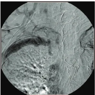
Investigation included Doppler venous ultrasound,
angiotomography of the thorax and phlebography,
which conirmed occlusion of the internal jugular
vein and right brachiocephalic trunk (Figures 1 and
2).
Venous Doppler ultrasound of the RUL showed
a mature brachiobasilic AVF and a cephalic
vein that was patent in the arm up to its junction
with the axillary vein, from where onwards it
exhibited pulsating reverse low– due to retrograde
transmission from the axillary vein (Figure 3).
Conservative treatment comprised elevation of
the limb, use of binding and elastic armbands, but
there was no signiicant improvement in symptoms.
We recommended endovascular intervention.
However, this would have entailed transferring the
OBJECTIVE
To describe an atypical case in which vascular
access for hemodialysis was preserved and central
venous obstruction treated.
METHOD
A patient with few remaining options for vascular
access presented with a functioning brachiobasilic
istula in the right upper limb (RUL), associated with
severe venous hypertension, and occlusion of the
internal jugular vein and the right brachiocephalic
trunk.
We decided to transpose the RUL cephalic vein,
forming a venous necklace in the anterior cervical
region, bypassing the site of venous occlusion.
In order to achieve this, we dissected the cephalic
vein in the right arm to its junction with the axillary
vein, devalved the cephalic vein and anastomosed it
to the contralateral external jugular vein, providing
venous drainage to the RUL, alleviating symptoms
of venous hypertension and preserving function of
the brachiobasilic istula.
RESULT
Reduction of edema, pain and venous congestion
in the RUL, in addition to preservation of an
arteriovenous istula (AVF) that was already mature
and functioning.
DISCUSSION
Stenosis or central venous obstruction affects 20 to
50% of patients who undergo placement of catheters
in the subclavian or internal jugular vein.
1,2Many
of these patients can remain symptomless for long
periods because of the rich network of collateral veins
to maintain venous drainage from the upper limb. In
patients with functioning arteriovenous istulae, this
can lead to venous hypertension, edema of the limb
and failure of vascular access.
3Percutaneous angioplasty, with or without
placement of stents or other endovascular devices
(HeRO, for example),
4,5has been evolving and is
becoming ever more common, taking its place in the
arsenal of options for treating such patients.
The simpler treatment options include rest and
elevation and elastic compression of the limb,
or deactivation of the AVF (although this option
involves sacriicing the access and subjects patients
to further central catheterization until the next AVF
matures).
The following surgical options are described in
isolated case reports and case series
Felipe Jose Skupien, Ricardo Zanetti Gomes et al.
patient to a different city and both the patient and her
family members refused.
We therefore chose an option designed to preserve
AVF function while reducing venous hypertension
in the RUL. To achieve this, under local anesthetic
(brachial plexus block) we dissected the cephalic vein
(patent in the distal third of the arm) up to its junction
with the axillary vein. Using a Mills valvulotome, we
achieved pulsating low in the entire cephalic vein.
Under supplementary local anesthetic to the
anterior cervical region, we dissected the left external
jugular vein, tunneled the devalved right cephalic
vein and created a terminal-lateral anastomosis with
the left external jugular vein.
At the end of surgery, we observed a thrill in the
transposed cephalic vein necklace and also at the
brachiobasilic AVF.
The procedure was accomplished without
intercurrent conditions and, after surgery, the patient
continued her normal hemodialysis program, using
the same access as prior to surgery (basilic vein of
right arm) and her right upper limb edema gradually
receded (Figure 4).
More than thirteen months after surgery, the
fistula was still functioning, the patient was on
hemodialysis three times a week and had no further
complaints caused by venous hypertension of her
right upper limb.
CONCLUSIONS
Our case bears out what is to be expected from
data described in the literature, including primary
patency rates of around 85% after twelve months,
3mean duration of access function of 9 months and
88% of cases with improvement in symptoms.
23Although rare, like other exotic grafts described
in the literature, the treatment described here is an
effective option for preserving dialysis vascular
access in patients with central venous occlusion.
Figure 1. Occlusion of right brachiocephalic trunk.Figure 2. Occlusion of internal jugular vein and right brachiocephalic trunk, and rich network of collateral veins.
Figure 3. Doppler ultrasound showing right cephalic vein with reversed, pulsating low.
Figure 4. Right upper limb twelve months after surgery.
65
Cephalic transposition for central venous obstruction
Occlusion, a New Approach. Ann Vasc Surg. 2009;23:465-8. PMid:19359137. http://dx.doi.org/10.1016/j.avsg.2009.01.001 17. Criado E, Marston WA, Jaques PF, Mauro MA, Keagy BA. Proximal
Venous Outlow Obstruction in Patients With Upper Extremity Arteriovenous Dialysis Access. Ann Vasc Surg. 1994;8:530-5. PMid:7865390. http://dx.doi.org/10.1007/BF02017408 18. Jennings WC, Miller GA, Coburn MZ, Howard CA, Lawless
MA. Vascular access low reduction for arteriovenous istula salvage in symptomatic patients with central venous occlusion. J Vasc Access. 2012;13(2):157-62. PMid:21983828. http://dx.doi. org/10.5301/jva.5000020
19. Frampton AE, Hossain M, Hamidian Jahromi A, Morsy M, Chemla ES. Rescue of an axillary-axillary arteriovenous graft not amenable to endovascular intervention by formation of an axillary loop: a case report. J Vasc Access. 2010;11(1):89.
20. Bacciu PP, Porcu P, Piredda F, Casu MA, Marongiu GM, Gherli T. Anterior jugular-internal jugular bypass to salvage a dialysis arteriovenous istula. J Mal Vasc. 2002; 27(3):165-9. PMid:12232533.
21. Illig KA. Management of Central Vein Stenoses and Occlusions: The Critical Importance of the Costoclavicular Junction. Semin Vasc Surg. 2011;24:113-8. PMid:21889100. http://dx.doi. org/10.1053/j.semvascsurg.2011.05.008
22. Glass C, Dugan M, Gillespie D, Doyle A, Illig K. Costoclavicular venous decompression in patients with threatened arteriovenous hemodialysis access. Ann Vasc Surg. 2011;25(5):640-5. PMid:21514107. http://dx.doi.org/10.1016/j.avsg.2010.12.020 23. Anaya-Ayala JE, Bellows PH, Ismail N, et al. Surgical Management
of Hemodialysis- Related Central Venous Occlusive Disease: A Treatment Algorithm. Ann Vasc Surg. 2011;25:1. PMid:21172586. http://dx.doi.org/10.1016/j.avsg.2010.11.002
Correspondence
Felipe Jose Skupien Felipe Jose Skupien Rua Santana, 200 - Centro CEP 84010-320 - Ponta Grossa (PR), Brasil Fone: (42) 3028-0033 Fax: (42) 3028.3033 E-mail: [email protected]
Author’s information
FJS é Cirurgião Vascular da Santa Casa de Misericórdia de Ponta Grossa (SCMPG). RZG é Chefe do Serviço de Cirurgia Vascular da Santa Casa de Misericórdia de Ponta Grossa (SCMPG). EHS é Médico Residente em Cirurgia Vascular da Santa Casa de Misericórdia de Ponta Grossa (SCMPG). RIB é Cirurgião Vascular da Santa Casa de Misericórdia de Ponta Grossa (SCMPG). SVS é Docente do Departamento de Enfermagem e Saúde Pública da Universidade Estadual de Ponta Grossa (UEPG); Mestranda em Tecnologia em Saúde pela Pontifícia Universidade Católica do Paraná (PUC-PR).
Author’s contributions
Conception and design: FJS Analysis and interpretation: FJS Data collection: FJS, EHS Writing the article: FJS, SVS Critical revision of the article: FJS, RZG, EHS, RIB, SVS Final approval of the article*: FJS, RZG, EHS, RIB, SVS Statistical analysis: N/A Overall responsibility: FJS Obtained funding: None.
*All authors have read and approved of the inal version of the article submitted to J Vasc Bras.
REFERENCES
1. Chandler NM, Mistry BM, Garvin PJ. Surgical Bypass for Subclavian Vein Occlusion in Hemodialysis Patients. J Am Coll Surg. 2002;194:416-21. http://dx.doi.org/10.1016/ S1072-7515(02)01127-4
2. Vanherweghem JL. hrombosis and stenosis of central venous access in hemodialysis. Nephrologie. 1994;15(2):117-21. PMid:8047195.
3. Jakimowicz T, Galazka Z, Grochowiecki T, Nazarewski S, Szmidt J. Vascular Access for Haemodialysis in Patients with Central Vein Thrombosis. Eur J Vasc Endovasc Surg. 2011;42:842-9. PMid:21852162. http://dx.doi.org/10.1016/j.ejvs.2011.07.022 4. Chen GJ, Anaya-Ayala JE, Ismail N, Smolock CJ, Davies MG.
Successful Use of the HeRO Device to Salvage a Functional Arteriovenous Fistula and Resolve Symptoms of Venous Hypertension. EJVES Extra. 2011;22:37-9. http://dx.doi. org/10.1016/j.ejvsextra.2011.06.008
5. Gage SM, Ahluwalia HS, Lawson JH. Salvaging vascular access and treatment of severe limb edema: case reports on the novel use of the hemodialysis reliable outlow vascular access device. Ann Vasc Surg. 2011;25(3):387.e1-5.
6. Bachleda P, Utikal P, Kalinova L, et al. Operating management of central venous hypertension complicating upper extremity dialysis access. Biomed Pap Med Fac Univ Palacky Olomouc Czech Repub. 2008;152(1):155-8. PMid:18795092. http://dx.doi. org/10.5507/bp.2008.025
7. Suliman A, Greenberg JI, Angle N. Surgical Bypass of Symptomatic Central Venous Obstruction for Arteriovenous Fistula Salvage in Hemodialysis Patients. Ann Vasc Surg. 2008;22:203-9. PMid:18346573. http://dx.doi.org/10.1016/j.avsg.2007.11.001 8. Montagnac R, Bourquelot P, Schillinger F. Arteriovenous
istula complicated by “fat arm” caused by proximal venous occlusion--salvage by axillo-jugular crossing bypass. Nephrologie. 1993;14(5):239-42. PMid:8159254.
9. Chemla ES, Korrakuti L, Makanjuola D, Chang RW. Vascular Access in Hemodialysis Patients with Central Venous Obstruction or Stenosis: One Center’s Experience. Ann Vasc Surg. 2005;19:692-8. PMid:16052387. http://dx.doi.org/10.1007/s10016-005-6624-z 10. Sottiurai VS, Lyon R, Ross C, Cooper M. Surgical Management
of Brachioaxillary-subclavian Vein Occlusion. Eur J Vasc Endovasc Surg. 1996;11:225-9. http://dx.doi.org/10.1016/ S1078-5884(96)80057-8
11. Kavallieratos N, Kokkinos A, Kalocheretis P. Axillary to saphenous vein bypass for treatment of central venous obstruction in patients receiving dialysis. J Vasc Surg. 2004;40:640-3. PMid:15472589. http://dx.doi.org/10.1016/j.jvs.2004.07.009
12. Hoballah JJ, Eid GE, Nazzal MM, Sharp WJ, Corson JD. Contralateral Internal Jugular Vein Interposition for Salvage of a Functioning Arteriovenous Fistula. Ann Vasc Surg. 2000; 14:679-82. PMid:11128468. http://dx.doi.org/10.1007/s100169910122 13. Tordoir JHM, Leunissen KLM. Jugular vein transposition of
the subclavian vein obstruction in haemodialysis patients. Eur J Vasc Surg. 1993;7:335-8. http://dx.doi.org/10.1016/ S0950-821X(05)80019-2
14. Puskas JD, Gertler JP. Internal jugular to axillary vein bypass for subclavian vein thrombosis in the setting of brachial arteriovenous istula. J Vasc Surg. 1994;19:939-42. http://dx.doi. org/10.1016/S0741-5214(94)70022-2
15. El-Sabrout RA, Duncan JM. Right atrial bypass grafting for central venous obstruction associated with dialysis access: Another treatment option. J Vasc Surg. 1999;29:472-8. http://dx.doi. org/10.1016/S0741-5214(99)70275-2
16. Glass C, Maevsky V, Massey T, Illig K. Subclavian Vein to Right Atrial Appendage Bypass without Sternotomy to Maintain Arteriovenous Access in Patients with Complete Central Vein