Joana Isabel Carvalho Pereira
Development of biomarkers for
the identification of pathogenic
Candida species
UMinho|20
12
Joana Isabel Car
valho P er eir a De velopment of biomark er s for t he identification of pat hogenic Candida species
Joana Isabel Carvalho Pereira
Dissertação de Mestrado
Mestrado em Genética Molecular
Development of biomarkers for
the identification of pathogenic
Candida species
Trabalho realizado sob a orientação da
Doutora Ana Paula Fernandes Monteiro
Sampaio Carvalho
e da
Nome: Joana Isabel Carvalho Pereira
Endereço Electrónico: [email protected] Telefone: (+351)911192326
Número do Bilhete de Identidade: 13347458 5ZY2
Título da Dissertação:
Development of biomarkers for the identification of pathogenic Candida species.
Desenvolvimento de biomarcadores para a identificação de espécies patogénicas de Candida.
Orientadores:
Doutora Ana Paula Fernandes Monteiro Sampaio Carvalho Doutora Célia Sacramento Santos Pais
Ano de Conclusão: 2012
Designação do Mestrado: Mestrado em Genética Molecular
É AUTORIZADA A REPRODUÇÃO PARCIAL DESTA TESE, APENAS PARA EFEITOS DE INVESTIGAÇÃO, MEDIANTE DECLARAÇÃO
ESCRITA DO INTERESSADO, QUE A TAL SE COMPROMETE.
Universidade do Minho, 31/10/2012
Agradecimentos
Ao longo do último ano diversas pessoas contribuíram, direta ou indiretamente, para o sucesso deste trabalho. A elas gostaria de expressar o meu mais sincero reconhecimento, por todas as criticas, sugestões e apoio manifestado.
Á Professora Doutora Célia Pais e Professora Doutora Ana Paula Sampaio pela inestimável orientação, conhecimentos transmitidos, criticas e sugestões, e sobretudo pela amizade e apoio em todas as situações.
A todos os responsáveis pelo Departamento de Biologia, e especialmente ao CBMA (Centro de Biologia Molecular e Ambiental) por me terem acolhido e por me proporcionarem as condições necessárias para a realização deste trabalho.
Á Catarina Carneiro, Catarina Vaz (o meu pinguim), Filipa Vale, Manoel Marques e Carina Silva por me terem ajudado e amparado nos primeiros passos como investigadora num verdadeiro ambiente de “família da Micro II”, e ao “nosso escravo” João Pacheco, pelas caixas de pontas enchidas, e pelos docinhos nos momentos de frustração.
A todos os docentes e funcionários do Departamento de Biologia, especialmente á Magda Graça, pela disponibilidade e ensinamentos sobre o sequenciador.
Á minha família e amigos, pelo apoio incondicional até nos momentos mais difíceis, especialmente á Cristina e ao Zé, por aturarem os meus devaneios e mau humor sem nunca se queixarem. Um agradecimento muito especial também aos meus “piolhinhos”, Hugo e Joaninha, pois embora a tenra idade sempre foram capazes de me animar nos piores momentos.
Finalmente, ás duas pessoas mais importantes da minha vida, os meus pais, José e Isabel, por acreditarem em mim, e lutarem sempre para que eu me torna-se na pessoa que sou.
A todos,
Development of biomarkers for the identification of pathogenic Candida species
Abstract
In the last decades the incidence of fungal infections has increased exponentially, due to the development of more aggressive therapeutic techniques, which increase the number of
immunocompromised and in risk individuals. The Candida species are the most common
etiological agents isolated from opportunistic fungal infections in these patients and Candida albicans appears to be the most common species isolated. However, infections caused by non-albicans species, such as C. parapsilosis, C. glabrata, C. tropicalis and C. krusei, are increasing alarmingly. It is thought that the susceptibility to antifungal drugs varies according to species, thus, the rapid and correct identification of infecting species are crucial. Microsatellite sequences have been largely used as molecular targets to differentiate and characterize
strains. However, no studies have been performed using microsatellite DNA for Candida
species identification. Therefore, the main objectives of this work were the evaluation of the potential of microsatellite markers for species differentiation and for identification of specific C. albicans lineages.
After an intensive search for described microsatellite markers for the main Candida species,
several markers were selected for C. albicans, C, glabrata, C. parapsilosis, C. tropicalis and C. krusei. These markers were combined into a multiplex strategy and this new developed system tested in 81 strains from 10 different species. All tested loci amplified correctly in single and multiplex conditions, except for the C. tropicalis selected locus that was unable to amplify in multiplex. After removal of the C. tropicalis primers, this system showed 100% specificity and 100% sensibility.
To test if the microsatellites were able to identify specific C. albicans lineages two microsatellite markers were used, CAI and CAVIII. These markers are located in two repeat-containing ORFs, CAI is located in the terminal-3’ of RLM1 gene and CAVIII in the terminal-3’ of SAP8 gene. CAI microsatellite has been previously described and CAVIII was described for the first time in this
study. CAVIII demonstrated to be highly specific for C. albicans strains and presented a
discriminatory power of 0.72. The two microsatellite markers were tested in 144 unrelated C. albicans strains isolated from different body locations, allowing the statistical differentiation of strains from oral cavity, vulvovaginal infections and from extra-mucosal (respiratory tract and
urine) infections. The number of extra-mucosal strains was increased to 224 and statistical analysis based in CAI genotypes, demonstrated significant differences between genotypes of strains isolated from superficial (oral and vagina) and invasive infections (respiratory tract, urine and blood).
The results obtained allowed to conclude that the microsatellite loci analysis can be used to
differentiate the most common Candida species, being an alternative in clinical diagnosis.
Moreover, it was also possible observe that analysis of repeat containing ORFs, such as RLM1 and SAP8 is able to differentiate lineages of C. albicans.
Desenvolvimento de biomarcadores para a identificação de espécies patogénicas de Candida
Resumo
Nas ultimas décadas a incidência das infecções provocadas por fungos têm aumentado exponencialmente. A principal razão proposta para esta mudança incide no desenvolvimento de métodos terapêuticos mais agressivos, responsáveis pelo aumento do número de
indivíduos imunocomprometidos. As espécies de Candida são os agentes etiológicos mais
frequentemente isolados de amostras de pacientes com infeções fúngicas oportunistas, sendo Candida albicans a espécie mais comum. Contudo, as infecções provocadas por outras espécies, nomeadamente C. parapsilosis, C. glabrata, C. tropicalis e C. krusei, têm aumentado em grande escala. Sabe-se que a susceptibilidade ás terapêuticas antifúngicas varia de acordo com a espécie causadora da infecção, assim, a correta identificação destes organismos é essencial. As sequências de DNA microssatélite têm sido frequentemente utilizadas como alvos para a diferenciação de estirpes. Contudo, não têm sido realizados estudos que utilizem
o DNA microssatélite na identificação das espécies patogénicas de Candida. Desta forma, o
principal objectivo deste trabalho consistiu na avaliação do potencial dos marcadores de microssatélites na diferenciação de espécies, assim como na diferenciação de linhagens de C. albicans.
Após uma intensa pesquisa por marcadores de microssatélites previamente descritos, cinco
marcadores foram selecionados para a identificação de C. albicans, C, glabrata, C.
parapsilosis, C. tropicalis e C. krusei. Estes marcadores foram combinados numa reação de PCR-multiplex e o sistema desenvolvido foi testado em 81 estirpes de 10 espécies diferentes. Todos os marcadores apresentaram uma amplificação específica em reação singleplex e multiplex, porém, o marcador selecionado para a identificação de C. tropicalis não foi capaz de amplificar em reação de multiplex. Após remover o marcador para a C. tropicalis do sistema de identificação foi obtida uma especificidade e sensibilidade de 100%.
Com o objectivo de verificar a utilidade da análise do DNA microssatélite na diferenciação de
linhagens de C. albicans foram utilizados dois marcadores, CAI e CAVIII, localizados no
terminal 3’ das regiões codificantes dos genes RLM1 e SAP8, respectivamente. O primeiro, CAI, já tinha sido previamente descrito apresentando elevada estabilidade e especificidade,
enquanto que o CAVIII foi descrito pela primeira vez neste trabalho. O microssatélite CAVIII demonstrou ter elevada especificidade para as estirpes de C. albicans e apresentou um poder discriminatório de 0.72. Ambos os microssatélites foram testados utilizando 144 estirpes de C. albicans isoladas a partir de diferentes locais, e a análise combinada dos genótipos obtidos com os dois microssatélites permitiram diferenciar as estirpes provenientes da cavidade oral, de infecções vulvovaginais e de infeções invasivas. Porém, o número de estirpes provenientes de infecções invasivas foi aumentado numa fase posterior do estudo, e a análise estatística foi realizada novamente utilizando apenas os genótipos obtidos com o marcador CAI. Esta análise demonstrou diferenças significativas entre as estirpes provenientes das infecções superficiais e estirpes provenientes das infecções invasivas, demonstrando a sua utilidade na diferenciação das linhagens de C. albicans.
Os resultados obtidos permitiram concluir que a análise de DNA microssatélite pode ser útil para diferenciar as espécies de Candida mais comuns, sendo uma excelente alternativa para o diagnóstico clínico. Para além disso, é também possível observar que a análise combinada com os marcadores CAI e CAVIII permite a diferenciação de linhagens de C. albicans.
Table of contents
Agradecimentos ... iii
Abstract ... v
Resumo ... vii
Table of contents ... ix
List of Abbreviations ... xiii
List of Figures ... xv
List of Tables ... xvii
General Introduction ... 1
1. Candida and candidiasis ... 2
1.1. Clinical manifestations of candidiasis ... 2
1.2. Epidemiology of candidiasis ... 5
1.3. Virulence factors of Candida species ... 7
1.3.1. Adhesion ... 8 1.3.2. Morphogenesis ... 8 1.3.3. Hydrolytic enzymes ... 9 1.3.4. Phenotypic switching ... 11 1.3.5. Biofilm formation ... 12 1.4. Treatment of candidiasis ... 13 1.4.1. Polyenes ... 13 1.4.2. Azoles ... 14 1.4.3. Echinocandins ... 15
1.4.4. Other antifungal agents ... 15
2. Identification of Candida species ... 16
2.1. Conventional methods ... 17
2.2. Serological methods ... 18
2.3.1. Restriction Fragment Length Polymorphism (RFLP) ... 19
2.3.2. Polymerase Chain Reaction (PCR) based methods ... 20
3. DNA microsatellite ... 22
3.1. Microsatellites described in Candida species ... 24
4. Objectives ... 26
New multiplex PCR based methodology to discriminate clinically important Candida species ... 3
1. Introduction ... 30
2. Materials and methods ... 31
2.1. Yeast Strains ... 31
2.2. Primers selection ... 33
2.3. Colony-PCR ... 33
2.3.1. PCR amplification conditions ... 34
2.4. DNA Sequence Analysis and Fragment Size Determination ... 35
2.5. Multiplex PCR optimization ... 35
3. Results and discussion ... 35
3.1. Microsatellite selection ... 35
3.2. Singleplex amplification ... 38
3.3. Multiplex amplification ... 39
3.4. Optimization of multiplex amplification conditions ... 41
3.4.1. Annealing temperature ... 41
3.4.2. Amplification cycles ... 42
3.4.3. Primers concentration ... 43
3.4.4. Magnesium Chloride (MgCl2) concentration ... 44
3.4.5. PCR additives ... 45
3.5. Multiplex amplification (without CT14 primers pair) ... 46
Genotypic differentiation of C. albicans lineages by microsatellite loci analysis . 51 1. Introduction ... 52
2. Materials and methods ... 53
2.1. Yeast Strains ... 53
2.3. Fragment Size Determination and DNA Sequence Analysis ... 54
2.4. Statistical analysis ... 55
3. Results and discussion ... 55
3.1. Microsatellite locus analysis ... 55
3.2. Use of the microsatellites CAVIII and CAI for strains differentiation ... 61
4. Conclusion and final remarks ... 77
Final Considerations ... 79
Bibliography ... 83
List of Abbreviations
5-FC 5-fluorocytosine Ab Antibody Ag AntigenAls Agglutinin-like sequence
ATCC American type culture collection
BC Blood culture
BSA Bovine Serum Albumin
CBS Centraalbureau voor Schimmelcultures CD4 Cluster of differentiation 4
DMSO Dimethyl sulfoxide DNA Deoxyribonucleic acid
dNTP Deoxynucleoside triphosphates
DP Discriminatory power
DTT Dithiothreitol
ECMM European Confederation of Medical Mycology EDTA Ethylenediamine tetraacetic acid
ELISA Enzyme-linked immunosorbent assay
Epa Epithelial adhesins
FAM 6-carboxyfluorescein HCl Hydrochloric acid HEX Hexachlorofluorescein
HIV Human Immunodeficiency Virus
ITS Internal transcribed spacer
KCl Potassium chloride
LA Latex agglutination
Lip Lipases
MgCl2 Magnesium chloride
NEMIS National Epidemiology of Mycosis Survey OPC Oropharyngeal candidiasis
PCR Polymerase chain reaction
Pga30 Glycophosphatidylinositol-anchored protein 30
Pl Phospholipase
PYCC Portuguese yeast culture collection RAPD Randomly Amplified Polymorphic DNA RFLP Restriction Fragment Length Polymorphism
RIA Radioimmunoassay
RNA Ribonucleic acid
RPLA Reverse passive latex agglutination
RT Respiratory tract
RVCC Recurrent vulvovaginal candidiasis Sap Secreted aspartyl proteinase SSR Simple sequence repeats STR Short tandem repeats
Ta Annealing temperature
TAMRA 6-carboxytetramethylrhodamine TET Ortetrachloro-6-carboxyfluorescein
UPGMA Unweighted pair group method with arithmetic mean
URT Upper respiratory tract
USA United States of America
VE Vaginal exudate
List of Figures
Figure 1.1 Different stages of disseminated candidiasis. Adapted from Naglik
et al. 2003. 4
Figure 1.2 Mechanisms of action of (1) polyenes, (2) azoles, (3)
echinocandins and (4) 5-FU. 13
Figure 1.3 Constitution of rRNA gene operon. 21
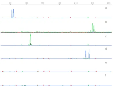
Figure 2.1 Gene Scan profiles obtained in a singleplex analysis using the
markers (a) CAI, (b) CAIII, (c) CAIV and (d) CAVIII. 37
Figure 2.2 GeneScan profile of PCR products amplified with different primer
pairs. 38
Figure 2.3 GeneScan profiles obtained with (a) CAIII, (b) Cp1, (c) 2bis, (d) CKTNR and (e) CT14 in strains S038 (C. albicans), 2257 (C. parapsilosis), 70V (C. glabrata), 109/RN0000.001 (C. krusei) and 2D (C. tropicalis) by singleplex PCR amplification.
39
Figure 2.4 GeneScan profiles obtained by multiplex amplification with (a) C. albicans, S040, (b) C. parapsilosis, 2252, (c) C. glabrata, M2, (d) C. krusei, H11, (e) C. tropicalis, 2D and (f) L. elongisporus strains, ISA 1421.
40
Figure 2.5 GeneScan profile of C. tropicalis strain (2D) amplified with CT14 at different annealing temperatures (a) 55ºC, (b) 58ºC, (c) 60ºC, (d) 62ºC and (e) 64ºC.
42
Figure 2.6 GeneScan profiles of C. krusei H11 strains obtained with
multiplex reaction using (a)2.0mM, (b) 2.5mM and (c) 3.0mM of
MgCl2.
44
Figure 2.7 GeneScan profiles of C. albicans, C. parapsilosis, C. glabrata and C. krusei, using the (a) initial concentrations and the (b) final concentrations.
47
Figure 3.1 GeneScan profile demonstrating a less intense stutter band ( ). 56
Figure 3.2 Genotypes and respective frequencies obtained in CAVIII analysis
of all C. albicans strains. 57
Figure 3.3 Genotypes and respective frequencies obtained in CAI analysis of
all C. albicans strains. 59
Figure 3.4 Genotypic frequencies based on CAI microsatellite analysis of Candida albicans strains from (a) oral group, (b) vaginal group and (c) extra-mucosal group.
67
Figure 3.5 Genotypic frequencies based on CAI microsatellite analysis of Candida albicans strains from (n) superficial group and (n) invasive group.
Figure 3.6 Specific genotypes and respective frequencies obtained with CAVI analysis of C. albicans strains from (a) Superficial group and (b) Invasive group.
73
Figure 3.7 UPGMA clustering of 244 C. albicans isolates based on the
genotypes, showing three phylogenetic groups (A, B and C). The percentage of strains with different origins in each group is represented by different shades: black, invasive infection; grey, superficial infections.
List of Tables
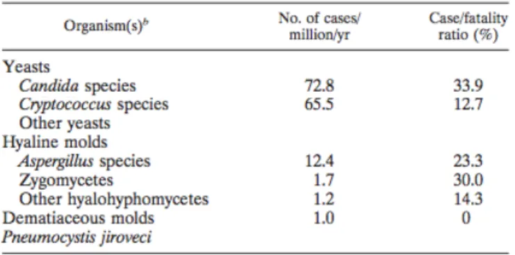
Table I.I Agents of opportunistic mycosis. Adapted from Pfaller et al.
2007. 5
Table I.II Species distribution of Candida bloodstream isolates. Adapted
from Tortorano et al. 2004. 6
Table I.III Mortality rates of Candida bloodstream infections. Adapted from
Tortorano et al. 2006. 7
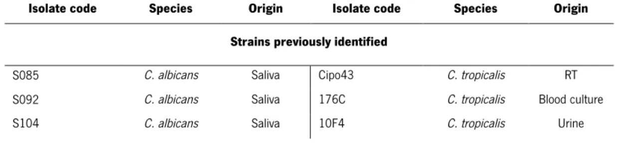
Table II.I Isolates used in the study and respective sources. 31
Table II.II Sequences and characteristics of the microsatellite loci selected. 33
Table II.III Primers concentrations tested in multiplex amplification. 43
Table II.IV Additives and combinations tested. 45
Table II.V Concentration of each primer pairs used in multiplex reaction. 46
Table II.VI Isolates tested with new multiplex mix, genotypes obtained e
respective identification. 47
Table III.I Alleles structure of CAVIII locus. The consensus sequence,
obtained from data base sequence for SC5314 strain is indicated and contain 10 repetitive units.
56
Table III.II Unbiased P-values of the probability test estimated by the Fisher
method and obtained for each population pair considering the combination of CAI and CAVIII microsatellite data.
61
Table III.III Significance of unbiased P-values of the probability test estimated by the Fisher method and obtained for each population pair considering microsatellite data. (+ when P<0.05 and - P>0.05). A- Results obtained with CAVIII. B- Results obtained with CAI.
62
Table III.IV C. albicans strains used and respective CAI and CAVIII
genotypes. 62
Table III.V Unbiased P-values of the probability test estimated by the Fisher
method and obtained for each population pair considering CAI microsatellite data.
CHAPTER I
General Introduction
1. Candida
and candidiasis
In the last two decades fungal infections have caused many difficulties in clinical practice. The main concerns about this problem are their prevalence, in which an alarming increase in number of cases, as well as the variety and complexity of the etiological agents involved (Guarro et al. 1999). Several reasons have been proposed to explain the increased incidence, including the increase lifespan in the populations of the developed world, the age related loss of immune-competence, as well as the use of more aggressive therapeutic methods, such as chemotherapeutic agents, bone marrow or solid-organ transplants, immunomodulatory agents, broad-spectrum antibiotics and more aggressive surgeries (Peres-Bota et al. 2004; Benjamin et al. 2010).
The most common etiological agents involved in fungal infections are Candida spp., Aspergillus spp. and Cryptococcus spp., although, other agents, such as Malasezzia spp., Fusarium spp.
or Trichosporon spp. may also be involved (Fridkin and Jarvis 1996). Several pathogenic
species have emerged in the last years but the ubiquitous Candida species remain the most
common cause of serious fungal infections (Fridkin and Jarvis 1996; Tortorano et al. 2004).
More than 200 species of Candida spp. have been isolated but only nearly 20 species have
been identified as being associated with human infections such as, C. albicans, C.
parapsilosis, C. glabrata, C. krusei, C. dubliniensis, C. tropicalis, C. guilliermondii, C. metapsilosis, C. bracarensis, C. kefyr among others (Guarro et al. 1999). Many of these pathogenic species are present in the commensal flora of genitourinary system, gastrointestinal tract, skin or oral cavity of healthy individuals (30-60% of humans), and only in disorders of the normal flora balance or when the immune defences are compromised, they may cause opportunistic infections, denominated candidiasis (Sanchez-Martinez and Perez-Martin 2001).
1.1. Clinical manifestations of candidiasis
Usually candidiasis is an endogenous infection, caused by prior colonization of mouth, gastrointestinal tract, vagina or skin. In these cases an unusual growth of normal flora occurs and the immune system is unable to react to this condition. However, the source of candidiasis may also be exogenous, and many species of Candida spp. have been isolated from hospital
environmental such as the floor, countertops, doorknobs, food and other inanimate surfaces (Perlroth et al. 2007).
Candida spp. is capable of causing a range of infections, from less-severe superficial lesions in mucosa and skin (including nails and hair) to life-threatening disseminated mycosis, characterized by the spread of fungi through the tissues and blood circulation (Fridkin and Jarvis 1996).
Superficial candidiasis affects mucosal epithelial tissues and is frequent in individuals with prior colonization when host physical barriers or immune system integrity are compromised. In the majority of these cases, the patient is symptom free and unaware of a problem, however, it can also cause a burning sensation, discomfort or pain (Jayatilake 2011). The most common clinical manifestations of superficial candidiasis are oropharyngeal candidiasis (OPC) and vulvovaginal candidiasis (VVC) although, infections in the urinary tract and skin are also observed (Jayatilake et al. 2009; Sobel et al. 2011).
Candida species are frequently associated with normal oral carriage in humans, occurring in the mouth of up to 80% of healthy individuals, but changes in the oral cavity environment can
enhance the Candida infection. Oropharyngeal candidiasis (OPC) is an acute condition often
affecting new-born babies due to the immature immune system and individuals infected with HIV (Samaranayake and Holmstrup 1989; Blignaut 2007). VVC is the most common vaginal infection and more than 75 % of women will have had at least one episode during their lives. It is known that about 40-50% of these women experience a recurrence, and up to 5% suffer more than four episodes during 1 year (recurrent vulvovaginal candidiasis – RVVC) (Buitron Garcia-Figueroa et al. 2009; Kalkanci et al. 2012). The presence of Candida species in urine is a common clinical finding, particularly in hospitalized patients, and several studies indicate that at least 10%–15% of hospital acquired urinary tract infections are caused by Candida species (Sobel et al. 2011).
Invasive candidiasis is verified only in severe cases of patient debilitation or immune compromisation and can involve the infection and spread of Candida cells via the bloodstream (candidaemia) to multiple organs, such as the brain, kidneys, heart, lungs and liver (Jayatilake 2011). This condition is more significant because of its associated high mortality rate (46-75%) and high morbidity in patients who survive the infection.
The route of bloodstream infection can occur through the “natural” way where yeast cells penetrate epithelial cells, the iatrogenic way through the use of medical devices (central venous catheters, peritoneal dialysis and cardiovascular devices) in which the formation of biofilms on its surface is important, or through the damage of defence barriers (polytrauma, surgery, drug treatment) (Figure 1.1) (Mavor et al. 2005).
Figure 1.1. Routes of entry into the bloodstream by Candida. Adapted from Mavor et al. 2005.
Numerous studies have identified common risk factors for patients developing candidiasis, and most of these causes are extremely common in hospitalized patients, increasing the possibility of development of nosocomial infections. These reasons include immunosuppression (chemotherapy, malnutrition, malignancy and neutropenia), prior colonization, disruption of normal skin barriers (intravascular catheters, extensive burns, invasive surgery, parenteral nutrition) and broad-spectrum antibiotics, since they disrupt the competition of bacterial flora (Peres-Bota et al. 2004; Benjamin et al. 2010). However, not all the predisposing factors equally favour superficial and invasive candidiasis, since immune protection of human host is site-specific. T- cell immune responses are important in protection against superficial candidiasis but resistance against systemic disease is more often associated with a functional phagocytic response (Calderone and Fonzi 2001). For example, HIV infected individuals suffer frequently from oral infections and onychomycosis due to a reduction in CD4+ cells counts, but rarely developed systemic infections (Mavor et al. 2005). Moreover, this feature is also supported by the fact that neutropenic patients are particularly susceptible to systemic infections (Koh et al. 2008).
1.2. Epidemiology of candidiasis
Fungal infections are an increasingly encountered threat among critically ill patients and are a significant cause of morbidity and mortality. Moreover, Candida species are the most common etiological agents of fungal infections, causing superficial or invasive candidiasis (Table I.I) (Pfaller and Diekema 2007).
Table I.I. Agents of opportunistic mycosis. Adapted from Pfaller et al. 2007.
Over the past decades several epidemiologic studies have been performed in European countries and in USA to evaluate the incidence of superficial and invasive candidiasis. These studies suggested that Candida infections are the third most common urinary tract infections with an incidence of ≈20%, and that the majority of episodes of Candida urinary tract infections occur in hospitalized patients with indwelling bladder catheters (Sobel et al. 2011). Moreover, Candida species are also responsible for 13.2% of all intra-abdominal infections, 70% of all onychomycosis (Jayatilake et al. 2009) and may colonize about 70% of women vagina (Sobel
2007). The incidence of invasive Candida infections have been studied by several
multi-institutional surveys, such as European Confederation of Medical Mycology (ECMM) (Tortorano et al. 2004) survey, National Epidemiology of Mycosis Survey (NEMIS) (Rangel-Frausto et al. 1999), among others. These studies concluded that the frequency of candidaemia among hospitalized patients has doubled during these two decades and candidaemia is now the third most common nosocomial blood-stream infection.
Data from ECMM indicate that C. albicans remains the most common species isolated from the blood of patients with invasive fungal infection (Tortorano et al. 2004). However, infections caused by non-albicans species are increasing (Lass-Florl 2009). This trend may be explained by the introduction of fluconazole in 1990 (Rodloff et al. 2011), since it was demonstrated that patients with candidaemia caused by non-albicans species received prophylactic antifungal
agents before the onset of their infections more frequently than patients with candidaemia by C. albicans (Hachem et al. 2008).
The most common non-albicans Candida species are C. glabrata, C. parapsilosis, C. tropicalis
and C. krusei, however, the incident rates of these organisms vary according to patient
population (Table I.II) (Tortorano et al. 2004).
Table I.II. Species distribution of Candida bloodstream isolates. Adapted from Tortorano et al. 2004.
C. parapsilosis has been associated with parental nutrition, neonatal population, intravenous catheters or contaminated prosthetic devices. The contamination may be caused by health
care workers since C. parapsilosis is the most common species isolated from the hands of
nurses (Pappas et al. 2003; Trofa et al. 2008). C. glabrata has a natural resistance to commonly used antifungals due to the constitutively expression of drug efflux pumps (Parkinson et al. 1995) and is commonly isolated from surgical patients, patients with urinary catheters, neutropenia and bone marrow transplant patients (Fidel et al. 1999). C. tropicalis is frequently responsible for invasive infections in patients with hematologic malignancies and neutropenia. Finally, C. krusei represents a significant challenge to clinicians due to the inherent resistance to azole drugs due to an altered target enzyme, and affects more frequently leukemic patients and bone marrow transplant recipients (Orozco et al. 1998).
Reported mortality rates from candidaemia range from 30 to 75% in European surveys, depending on species and geographic location studied (Table I.III) (Pappas et al. 2003; Tortorano et al. 2006; Lass-Florl 2009).
Table I.III. Mortality rates of Candida bloodstream infections. Adapted from Tortorano et al. 2006.
Small differences in the incidence of candidiasis in Europe and USA were found, probably due to differences in patient demographics or differences in medical practices (Pfaller and Diekema 2007). Candidaemia not only is associated with increased mortality and morbidity rates but also prolongs hospitalization and increases medical cares costs. Systemic Candida infections have been associated with an attributable intensive care unit cost of US $21,590 (Tortorano et al. 2004).
1.3. Virulence factors of Candida species
In order to establish an infection, an opportunistic pathogen have to colonise a host, penetrate the surface, survive and divide in the host environment, and avoid the immune response. Although some Candida species are commensal organisms of the normal flora, the ability to adapt to different environments, including changes in oxygen and carbohydrate levels, pH,
osmolality, availability of nutrients and temperature, improves the development of Candida
infections. The mechanisms required for the occurrence of these processes are designated as virulence factors (Mavor et al. 2005).
Candida species have developed an effective battery of putative virulence factors and specific strategies to assist in their ability to colonize host tissues, cause disease and overcome host
defences (Yang 2003). The Candida virulence factors most studied are adhesion capacity,
production of hydrolytic enzymes, hyphae formation and phenotypic switching. However, the virulence factors expressed may vary depending on the type, the site and the stage of infection, and the nature of the host response (Naglik et al. 2003).
1.3.1. Adhesion
The colonization and infection of Candida species are dependent on the ability to adhere to
host cells, tissues and medical devices in different stages of infection. However, the extent of adhesion is dependent on microbial, host and abiotic surface proprieties, such as cell-surface hydrophobicity and cell wall composition (Silva et al. 2011).
An important element that is correlated with the adhesion ability of Candida species is the
presence of specific cell-wall proteins, denominated adhesins. These proteins are defined as biomolecules that promote the adherence of Candida species to host cells or host cell ligands (Calderone and Fonzi 2001; Trofa et al. 2008; Silva et al. 2011). Mutants deficient in the genes encoding these adhesins exhibit decreased adherence to host substrates in vitro as well as a corresponding reduction in virulence in several experimental models of candidiasis (Sheppard et al. 2004).
Several genes encoding cell wall adhesins of Candida species have been identified. The most common adhesins studied are from the agglutinin-like sequence (Als) protein family, encoded
by eight ALS genes (ALS1-7 and ALS 9) (Yang 2003). Three domains characterize these
proteins and differences in N-terminal domain among distinct Als proteins are responsible for differences in their function. For example, Als1p has been shown to mediate binding to human vascular endothelial cells and epithelial cells in early stages of infection, whereas Als5p confers adherence to collagen, fibronectin, bovine serum albumin and laminin (Sheppard et al. 2004).
The ALS genes are differentially expressed depending on the growth conditions or on the
species analysed. Several strains of C. albicans express all eight ALS genes, however, in C. parapsilosis and C. tropicalis only five and three ALS genes were found, respectively (Silva et al. 2011).
Others adhesins have been identified, including the Epa (epithelial adhesin) family in C.
glabrata, the glycophosphatidylinositol-anchored protein 30 (Pga30) in C. parapsilosis or Hwp1 in C. albicans (Nobile et al. 2006; Silva et al. 2011).
1.3.2. Morphogenesis
Some Candida species are polymorphic yeasts that are able to undergo morphogenic switching from the unicellular budding yeast forms (blastopores) to the filamentous forms (hyphae or pseudohyphae). This transition is regulated by a complex network of signal transduction
pathways, which includes transcription factors such as Efg1, Cph1 and Tup1. The transcription factors are activated by morphogenetic stimuli such as the presence of serum or the interaction with innate immune cells (Heilmann et al. 2011). The yeast-to-hyphae transition is the most prominent morphological change in the Candida (especially C. albicans) life cycle and two important functions of hyphae formation have been suggested, including the ability to penetrate into tissue surfaces and the capacity to escape from host cells following internalization (Gow et al. 2002). In order to penetrate the epithelial tissue and to provide resistance to phagocytosis, the hyphae produce mechanical forces. The expression of adhesins, such as Hwp1p or Als3, for anchoring the Candida cells to host tissue is probably a prerequisite for hyphae invasion (Kumamoto and Vinces 2005). Another trend in hyphae penetration consists in the secretion of enzymes able to degrade proteins, lipids and other cellular components, facilitating the infiltration into solid substrates and tissues (Gow et al. 2002).
Although the hyphae formation is considered an important virulence factor in Candida
virulence, most lesions are populated by both morphological forms, suggesting that both have a role in the development and progression of disease (Calderone and Fonzi 2001). It has been suggested that yeast cells are better suited for dissemination while hyphae are important for tissue and organ invasion and for adaptation to different host niche conditions (Mavor et al. 2005; Lim et al. 2012).
This ability is observed in species such as C. albicans, C. parapsilosis or C. tropicalis and is considered to be crucial for virulence (Lim et al. 2012). C. glabrata is generally described as incapable to form hyphae and pseudohyphae. However, the ability of pseudohyphae formation was suggested in numerous studies, where this feature was observed in some strains (Odds et al. 1997; Csank and Haynes 2000; Lachke et al. 2002). Regarding C. krusei, no consistent filamentous studies have been performed.
1.3.3. Hydrolytic enzymes
The secretion of hydrolytic enzymes during the development of candidiasis may be required as a virulence attribute. This virulence factor may be involved in adhesion by degrading host cell surface molecules, invasion by digesting host cell membranes, resistance to host immunity by attacking the immune system, and nutrient acquisition. The three most significant extracellular
hydrolytic enzymes secreted by Candida species include secreted aspartyl proteinases (Sap), phospholipases and lipases (Mavor et al. 2005; Jayatilake 2011).
The secretion of secreted aspartyl proteinases (Sap) by Candida species is recognized as an important virulence factor since they facilitate invasion and colonization of host tissue. A family of 10 SAP genes encodes the Sap proteins and the virulence mechanism of Sap involves the disruption of host mucosal membranes and degradation of important immunological and structural defence proteins, such as immunoglobulin G heavy chains, C3 protein, collagen, fibronectin, albumin, haemoglobin, keratin among others (Yang 2003; Trofa et al. 2008). The
expression of SAP genes during infection has been studied by their disruption in several
models and differential expression profiles under various conditions it has been observed (Naglik et al. 2003; Naglik et al. 2008; Correia et al. 2010). Schaller and co-workers (Schaller et al. 2001) demonstrated that SAP genes family is differentially expressed in the yeast, hyphal and phenotypically switched states. SAP1-3 is predominantly expressed on cell walls and cytoplasm of blastopores, SAP4-6 is localized at the tips of hyphae and SAP1 and SAP3 are expressed by phenotypically switched cells. Moreover, Sap8 is predominantly detected in yeast cells grown at 25ºC and Sap9 is preferentially expressed in later growth phases (Yang 2003). Hereupon, the versatility of SAP genes expression may prove to be vital to the success of Candida as an opportunistic pathogen, by allowing the fungus to survive and cause infections on a variety of tissues (Naglik et al. 2003).
The secretion of Saps is recognized as an important virulence factor, however, the expression
of all ten SAP genes is only observed in C. albicans strains, whereas only four (SAP1-4) and
three (SAP1-3) genes have been identified in C. tropicalis and C. parapsilosis, respectively
(Trofa et al. 2008; Silva et al. 2011). Regard C. glabrata and C. krusei, some proteinase activity was detected, however, the number of these proteinases have not been well defined (Yang 2003). Although the expression of SAP genes has been recognized as an important virulence factor, Correia and co-workers (Correia et al. 2010) demonstrated that other factors must be the major contributors to invasion and cell damage in this model.
Phospholipases (PLs) are enzymes that hydrolyse phospholipids to fatty acids and glycerol. Depending on the different ester bonds cleaved, these enzymes have been classified into PLs A, B, C and D. However, only proteins encoded by the phospholipase B family (PLB1-5) seem
candidiasis (Calderone and Fonzi 2001; Silva et al. 2011). The presence of PLs during infection could contribute to host cell membrane damage and adherence of Candida species. Jayatilake and co-workers (Jayatilake et al. 2005) and Gahnnoum and co-workers (Ghannoum 2000) demonstrated that PLs are expressed at the tips of Candida hyphae and initial buds of C. albicans during invasion. These studies confirm that PLs of Candida are involved in the pathogenesis of candidiasis by facilitating the tissue penetration. Recent studies have indicated that C. tropicalis and C. parapsilosis are able to produce extracellular PLs, however, at much
lower levels than C. albicans. For C. glabrata and C. krusei very few studies were performed
and no clear PL activity was observed.
Lipases are involved in the hydrolysis and synthesis of triacylglycerols. These enzymes are
encoded by ten LIP genes (LIP1-10) differentially expressed at different stages and sites of
infection. In C. albicans and C. tropicalis ten LIP genes (LIP1-10) were detected. However, for C. parapsilosis, only two lipase genes, LIP1 and LIP2, have been reported (Trofa et al. 2008).
Moreover, no studies have been performed to investigate the expression of LIP genes in C.
glabrata and C. krusei (Silva et al. 2011). Gácser and co-workers (Gacser et al. 2007) demonstrated the significance of lipases, showing that the use of lipase inhibitors significantly reduce tissue damage during infection in reconstituted human tissue models.
1.3.4. Phenotypic switching
The colonies of Candida species can reversibly switch between different morphologies, and this process is known as phenotypic switching. The ability to undergo phenotypic switching is thought to aid survival in different microenvironments, and evasion from the host immune response. Moreover, phenotypic switching also affects adhesion, hyphal formation, sensitivity to neutrophils and increase the resistance to antifungals (Mavor et al. 2005). However, the basic mechanism of phenotypic switching and the involvement of this switching in the virulence are not clear (Calderone and Fonzi 2001).
The white-opaque switching in strain WO-1 of C. albicans is the most studied phenotypic
switching. In this case, the smooth and white colonies with round-ovoid cells can switch to flat and grey colonies with elongated or bean-shaped cells (Morschhauser 2010). The ultrastructural observations of white and opaque phenotypes have revealed differences in the cell shape, cell surface structures and germination at 37ºC, suggesting that phenotypic switching could affect the behaviour of the organism. For instance, opaque phase cells have
higher ability to colonize the skin whereas white cells are more virulent in a systemic animal model (Calderone and Fonzi 2001).
Although the phenotypic switching of C. albicans is the most studied, C. glabrata, C.
parapsilosis and C. tropicalis also present this capability. Laffey and co-workers (Laffey and Butler 2005) identified four core phenotypes in C. parapsilosis, including the crepe, concentric, smooth and crater phenotypes and demonstrated their relation with biofilms formation. Moreover, Lachke and co-workers (Lachke et al. 2002) identified four phenotypes in C. glabrata (White, Dark Brown, very Dark Brown and Light Brown) and França and co-workers (Franca et al. 2011) demonstrated the presence of four possible phenotypes in C. tropicalis
(Smooth, Rough, Ring, Semi-Smooth). The phenotypic switching of C. krusei has not been
studied.
1.3.5. Biofilm formation
The attachment of Candida cells to host or medical devices followed by cell division and
proliferation is called biofilm. Biofilms are complex and well organized microbial communities with fungal cells embedded within a mainly polysaccharide extracellular matrix (Lim et al. 2012). Biofilm formation is considered as an important virulence factor in the development of infection. The presence of biofilms confers significant tolerance to antifungal therapy and host immune responses, and causes the failure of indwelling medical devices (Trofa et al. 2008). Numerous Candida species produce biofilms, including C. albicans, C. tropicalis, C. glabrata
and C. parapsilosis, and their presence during infection has been linked to higher mortality
rates. However, the biofilm formation is dependent on several factors, such as the species, strains and environmental conditions (pH, medium composition and oxygen) (Silva et al. 2011).
Estivill and co-workers (Estivill et al. 2011) studied the biofilm formation by C. albicans, C. glabrata, C. parapsilosis, C. tropicalis and C. krusei on three clinical materials. This study
demonstrated that C. parapsilosis showed great biofilm formation capacity and its ability to
cause nosocomial infections can be related with this feature. Moreover, this study also demonstrated that the capacity of C. krusei to form biofilms is limited.
1.4. Treatment of candidiasis
The increasing incidence of fungal infections, including Candida infections, as well as the
increasing variety of pathogenic species have contributed significantly to the mortality in immunosuppressed patients. In order to reverse this condition several antimycotics agents have been developed, however, numerous species remain difficult to treat due to delayed diagnosis, drug toxicity, antifungal drug resistance, drug bioavailability and lack of oral or intravenous preparations. Recent epidemiological trends have confirmed the increasing importance of infections caused by resistant fungal species (Lass-Florl 2009). Thereby, it is crucial to understand the antifungal drug resistance and develop effective therapeutics.
The antifungal agents are classified into different groups according to the antifungal mechanism of action, namely polyenes, azoles, echinocandins and others antifungal agents (Figure 1.2) (Mathew and Nath 2009).
Figure 1.2. Mechanisms of action of (1) polyenes, (2) azoles, (3) echinocandins and (4) 5-FU. 1.4.1. Polyenes
Polyenes are the major class of antifungal agents and are isolated from Streptomyces species. The mechanism of action of polyenes is based on their interaction with ergosterol components of the fungal membrane. The complex polyene-esterol formed provides an aqueous pore and affect cell permeability, which causes cell leakage and cell death (Mathew and Nath 2009). The polyenes with therapeutic application are amphotericin B, nystatin, pimaricin and candicidin, however only the first two are commonly used. Amphotericin B has long been considered the gold standard for the treatment of fungal infections. This agent is active against
most fungal pathogens, namely Trichosporan beigelii, Aspergillus terreus, Pseudallesheria
Regarding candidiasis, this agent is active against most Candida species and can be used in the treatment of invasive or superficial candidiasis. However, the cytotoxicity associated with amphotericin B demanded the development of new formulations, which use liposomes or lipid complexes as delivery systems (Chen and Sorrell 2007).
The acquisition of polyenes resistance by C. albicans and other Candida species is unusual
however, numerous reports have demonstrated resistance to amphotericin B by C. albicans in patients previously treated with polyenes (Mokaddas et al. 2007). The molecular mechanisms involved in polyene resistance are the decrease in the total ergosterol content of the cell, replacement of some or all of the polyene-binding sterols, and reorientation or masking of existing ergosterol (Masia Canuto and Gutierrez Rodero 2002).
1.4.2. Azoles
Azoles are the second most studied antifungal agents and their mechanism of action is based in the inhibition of ergosterol biosynthesis. In more detail, exposure of fungal species to azoles inhibits the ergosterol enzymatic pathway, especially the enzyme cytochrome P450 sterol 14α-demethylase. This inhibition promotes the disruption of the structure of the membrane as well its functions in nutrient transport and chitin synthesis, reducing the fungal growth (Mathew and Nath 2009).
There are two azole groups in clinical use. The first azole compounds explored are the imidazole-based drugs, such as clorotrimazole, miconazole, ketoconazole and econazole. However, this group is only efficient in superficial treatment. Later, triazole-based drugs, including fluconazole, itraconazole, voriconazole, posaconazole and ravuconazole were developed, which are used as superficial and systemic fungicidal agents (Chen and Sorrell 2007). The differences in the structure of the different azoles are responsible for their variation on antifungal potency, bioavailability, drug interaction and toxicity (Mathew and Nath 2009). The introduction of fluconazole as antifungal agent of choice in the treatment of superficial candidiasis in the early 1990s triggered the appearance of azoles resistant strains. Moreover, this increasing azole resistance may be also explained by the appearance of species intrinsically resistant to fluconazole, such as C. glabrata or C. krusei (Chen and Sorrell 2007).
The mechanism of resistance to azoles in Candida species has been studied, and distinct
decreased accumulation of the drug from enhanced efflux interference of their action on the target enzyme, alterations in other enzymes of the biosynthetic pathway of ergosterol and decreased permeability of the fungal membrane to the drug (Masia Canuto and Gutierrez Rodero 2002).
1.4.3. Echinocandins
The increasing incidence of infection caused by fluconazole resistant species required the development of a therapeutic alternative and echinocandins have become an important group in the treatment of these infections. The echinocandins are lipopeptide molecules that act as inhibitors of the synthesis of β -1,3- D-glucan, which is an important component of the fungal cell wall, by blocking the action of a pathway enzyme, β -1,3- D-glucan synthase (Perlin 2007). The absence of β -1,3- D-glucan destabilizes the integrity of the fungal cell wall and promotes the osmotic instability and cell death (Kofla and Ruhnke 2011).
The echinocandins drugs used in antifungal treatment are caspofungin, micafungin and
anidulafungin. These agents have broad-spectrum antifungal activity against Candida and
Aspergillus species, however, are not active against C. neoformans and non-Aspergillus moulds (Perlin 2007). Moreover, echinocandins drugs are effective against azole-resistant species, since their target is the cell wall. Another vantage of these drugs is that the toxicity is infrequent since glucans are not found in mammalian cells (Chen and Sorrell 2007). The echinocandins resistance is unusual, however, some case reports have illustrated the potential for resistance development (Kofla and Ruhnke 2011).
1.4.4. Other antifungal agents
Although the polyenes, azoles and echinocandins are the three major classes of antifungal agents, other compounds, including allylamines, flucytosines, griseofluvins, sordarins, nikkomycins, ciclopiroxolamines, amog others, have been also used (Mathew and Nath 2009). The flucytosine (5-fluorocytosine or 5-FC) is one of the oldest antifungal agents and its mechanism of action is based in the conversion into 5-fluorouracil within target cells. Fluorouracil is incorporated into RNA, where it causes premature chain termination, and also inhibits DNA synthesis through effects on thymidylate synthase (Vermes et al. 2000). This drug
is selectively toxic to fungi because mammalian cells lack cytosine permease and do not convert flucytosine into 5-fluorouracil (Mathew and Nath 2009).
Although this agent shows antifungal activity against Candida species in cases of systemic
candidiasis, the development of resistance is frequent. In order to overcome the development of resistance, the use of monotherapy is not recommended and this agent must be combined with azoles. The mechanisms of resistance proposed are (1) the development of mutations that result in a deficiency in the enzymes necessary for cellular transports and uptake of 5-FC or for its metabolism, and (2) the increase in the synthesis of pyrimidines, which compete with the fluorinated antimetabolites of 5-FC and thus diminish its antimycotics activity (Vermes et al. 2000).
Although in the last years a number of antifungal agents have been developed, the selection of the most appropriate drug is imperative. As stated above, the susceptibility of the different species to the different antifungal agents varies considerable. Thus, the correct identification of infectious agents represents an important tool in reducing the mortality rate.
2. Identification of
Candida
species
The rapid and correct identification of infecting species is crucial for several reasons. The main
reason is the use of appropriate antifungal treatment, since Candida species differ in their
susceptibility to antifungal agents. For instance, C. krusei is intrinsically resistant to azoles and C. glabrata easily acquires resistance to fluconazole (Parkinson et al. 1995; Orozco et al. 1998). Moreover, species identification is also important for epidemiological purposes, for example, repeated identification of a particular species in a given hospital ward or locate may indicate a point source outbreak (Denning et al. 2003; Sabino et al. 2010). An additional reason to explain the significance of correct diagnosis is the fact that the risk of developing deep organ involvement, and the severity of clinical manifestations, differs depending on the infecting species (Rabkin et al. 2000).
Clinical microbiology laboratory methodologies for the identification of pathogenic fungal species are based on the morphological, physiological and biochemical tests. However, new serological and molecular tests have also been developed for the differential identification of the fungal species. These tests are classified as conventional, serological and molecular.
2.1. Conventional methods
The light microscopy analysis of biological products is the first methodology used in clinical laboratory practice, and is used to observe the presence, shape and size of blastopores as well as the hyphae/pseudohyphae formation. This method only allows the presumptive identification, since some species can present specific microscopic characteristics (Lee et al. 1999; Pinoni et al. 2007). For example, the presence of true hyphae in C. albicans or the
shape of the blastopores that in C. krusei is elongated, whereas in C. albicans or C.
parapsilosis is oval and spherical (Ellepola and Morrison 2005).
The growth and isolation of species present in clinical samples is an important method used in
microbiology laboratories. The media selected should sustain the growth of all the Candida
ssp., inhibit the growth of bacteria and should facilitate the identification of clinical specimens, however, should not interfere with the viability of the organisms (Sullivan et al. 1996; Alvarez-Perez et al. 2011). Several chromogenic media have been developed in order to distinguish Candida species. These culture media incorporates substrates linked to chemical dyes in a solid medium to differentiate Candida species by the colour and texture of the colonies (Okulicz et al. 2008; Ozcan et al. 2010). However, these media only allows the presumptive
identification of some Candida species, especially C. albicans, C. tropicalis and C. krusei
(Ghelardi et al. 2008; Okulicz et al. 2008). Examples of commercial chromogenic media are
ChromIDCandida (BioMerieux®), CandiSelect4 (BioRad®) or CHROMagar Candida (BD®) (Sendid
et al. 2007; Guzel et al. 2011).
For Candida species differentiation the physiological and biochemical methods are the most
commonly used. The biochemical identification consists in carbohydrate and nitrogen assimilation, such as glucose, xylose, urease, trehalose, saccharose, nitrates, among others; fermentation tests and enzymes detection (Lopez et al. 2001; Ellepola and Khan 2012). However, these tests can have a number of problems associated with the results interpretation. The results obtained may be inconsistent since different isolates from the same species could yield different profiles or genetically diverse species can yield similar profiles (Campbell et al. 1999; Cardenes-Perera et al. 2004). For example, C. parapsilosis, C. metapsilosis and C. orthopsilosis or C. albicans and C. dubliniensis have similar biochemical
APICandida (BioMerieux®), API20CAux (BioMerieux®) or Vitek Yeast Biochemical Card
(BioMerieux®) are examples of commercial kits for biochemical Candida spp. Identification.
Although conventional methods are the most commonly used in clinical microbiology laboratories, there are several limitations, such as inaccuracy, high cost and the long time required for identification (Ellepola and Morrison 2005). Therefore, the application of alternative methodologies is needed in order to overcome these limitations.
2.2. Serological methods
The species identification based in serological methods consists in the detection of specific antigens, antibodies or metabolites (such as D-anabinitol) in clinical samples. For this purpose several methods are used, such as radioimmunoassay (RIA), enzyme-linked immunosorbent assay (ELISA), latex agglutination (LA) or reverse passive latex agglutination (RPLA) (Ellepola and Morrison 2005).
Numerous antigens have been used as potential targets for the diagnosis of disseminated candidiasis, including secreted aspartyl proteinases, 1,3-β-D-glucans and mannans. Mannan is
an abundant cell wall polysaccharide of Candida spp. and is the most used and studied
antigen (Guery et al. 2009). However, the detection of mannan in clinical samples depends on the frequency of sampling, the underlying disease, the degree of immunosuppression, the Candida species involved, the specificity and titer of the capture antibodies and the method used. Another important limitation of this method is the rapid clearance of the antigen from the patient sera (Poulain et al. 1997; Ellepola and Morrison 2005).
A number of Candida antigens are highly immunogenic for humans and the detection of
antibodies against them in clinical samples may represent an important diagnostic method for invasive candidiasis (Quindos et al. 2004). The detection of anti-mannan antibodies is the most common used, however, the detection of antibodies against antigens with enzymatic activity (enolase or aspartyl proteinase) and antibodies against proteins of C. albicans germ tubes are also options (Quindos et al. 1987; Ponton et al. 1994). The limitations of this technique are the possibility of false-negative results in immunocompromised patients that produce low levels of antibody, false-positive results in patients with superficial colonization and the fact that antibody production may occur only at an advanced stage of disease. Nevertheless, it is
possible to overcome these limitations since the specificity of the tests has been improved by selecting the appropriate antigens (purified molecules, recombinant antigens, among others) (Quindos et al. 2004; Ellepola and Morrison 2005).
Several commercial kits have been developed for the diagnosis of Candida spp. based on
detection of antigens or antibodies. For example, Fungiter-G MK® or Glucatell®, which detects
the presence of 1,3-β-D-glucans, PlateliaCandida Ag Plus (BioRad®), which detects the
presence of mannans in blood samples in ELISA format, or PlateliaCandida Antibody Plus
(BioRad®), which is an ELISA-based test for of anti-mannan antibodies (Sendid et al. 2003).
Recent studies have suggested that the combined detection of mannan and anti-mannan antibodies considerably improves the diagnosis of candidiasis (Alam et al. 2007).
2.3. Molecular methods
Molecular methodologies, especially based in the analysis of DNA sequences, are characterized by their high specificity, sensibility and reproducibility. To overcome the limitations of conventional methods several molecular approaches have been developed in molecular research laboratories for Candida species identification.
2.3.1. Restriction Fragment Length Polymorphism (RFLP)
Restriction fragment length polymorphism (RFLP) analysis consists in the digestion of total chromosomal or plasmid DNA as well as PCR products with one or more restriction endonucleases. The endonucleases selected (EcoR1 is the most frequent) recognize specific nucleotide sequences, breaking the DNA into small fragments. The fragments are finally separated by agarose gel electrophoresis and the number and sizes of the restriction fragments depend on recognition sequence of the enzyme as well as the composition of the DNA. The different RFLP patterns obtained allow the species or strains differentiation (Sullivan et al. 1996).
Several RFLP studies have been performed to differentiate individual Candida species or
Candida strains, especially Candida albicans strains (Xu et al. 1999; Isik et al. 2003). Williams et. al (Williams et al. 1995) demonstrated the possibility to distinguish eight medically
C.tropicalis, C. stellatoidea, C. parapsilosis, and C. krusei) using three restriction enzymes, BfaI, DdeI and HaeIII. Pinto et al. (Pinto et al. 2004) also demonstrated this possibility, using
several enzymes, and the identification of six Candida species using only one enzyme, MspI,
was confirmed by Mirhendi et al. (Mirhendi et al. 2006).
The RFLP analysis presents several advantages, including high reproducibility and accuracy. However, this is a time-consuming technique, the RFLP patterns obtained from Candida spp. can contain a limited number of bands hampering the interpretation, and the same species can present different patterns (Sullivan et al. 1996).
2.3.2. Polymerase Chain Reaction (PCR) based methods
The polymerase chain reaction (PCR) based methodologies are sensitive, specific and rapid assays that have been accepted as the standard method for detecting nucleic acids from a number of microorganisms in clinical samples, including Streptococcus agalactiae (de Zoysa et al. 2012), Treponema pallidum spp. (Leslie et al. 2007), Aspergillus species (Walsh et al. 2011) or Candida species (Correia et al. 2004). The conventional PCR methodology was developed by Kary Mullis in 1983 (Mullis et al. 1986) to amplify target DNA sequences derived from dead or living cells by thermostable DNA polymerase-mediated extension of specific oligonucleotide primers. The PCR amplification is followed by PCR products detection or analysis, and the most common methods are the agarose or polyacrylamide gel electrophoresis, sequencing or pyrosequencing.
The design of specific oligonucleotide primers, complementary to DNA sequences unique to the organisms, is important since it can provide identification of an organism to the species level. For this purpose specific sequences need to be selected as DNA targets. For Candida species identification the most commonly used target is the ribosomal DNA (rDNA), which
encodes three subunits, 18S, 5.8S and 28S, and is largely distributed in Candida genome
(Sullivan et al. 1996; Ramos et al. 2006). Although the rRNA genes are highly conserved, the internal transcribed spacer (ITS) is variable and species specific. The ITS region is located between the 18S and 26S rRNA genes and is subdivided into the ITS1 region, between the 18S and 5.8S rRNA genes, and the ITS2 region, between the 5.8S and 26S rRNA genes (Figure 1.3). PCR with specific primers (ITS1 and ITS4), targeting the conserved sequences of 5.8S and 28S rDNAs, results in the amplification of the species-specific ITS1 and ITS2 regions,
which vary in amplicon length and sequence according to species (Ellepola et al. 2003; Coignard et al. 2004).
Figure 1.3.Constitution of rRNA gene operon.
The tRNA genes can also be used and DNA target to species identification into a specific genus (Baele et al. 2000). Although these genes are highly conserved, the lengths of tRNA intergenic spacers vary considerably and primers design in the highly conserved flanking tRNA genes can be used to amplify the polymorphic region in any organism that is sufficiently closely related. For inter-tRNA gene amplification several primers pairs have been developed and the length of the resultant PCR products, rather than its presence or absence, is characteristic of the species. The amplification of this region was firstly described by Welsh and McClelland (Welsh and McClelland 1991) in Staphylococcus strains, demonstrating the potential of investigation
of tRNA gene intergenic lengh polymorphism in species differentiation. For Candida species
identification, T3B primer pair, previously described in the identification of Staphylococcus
species (McClelland et al. 1992), has been successfully used in species differentiation (Correia et al. 2004).
In research laboratories, the most commonly used PCR based methodology for species or strains identification is RAPD (Randomly Amplified Polymorphic DNA) (Novak et al. 2004; Valerio et al. 2006). This method uses short primers, typically 9 to 10 nucleotides in length, which anneal at multiple genomic loci since it does not depend on prior knowledge of species-specific sequences. Following the amplification, the PCR products obtained are analysed by agarose gel electrophoresis and visualized after specific staining. The RAPD patterns obtained allow the species or strains differentiation. This methodology has been successfully used to identify Candida species, including C. albicans, C. tropicalis, C. parapsilosis, C. glabrata and C. krusei (Bautista-Munoz et al. 2003; Valerio et al. 2006). The RAPD-PCR has a high discriminatory power, its easy to perform, does not require radiolabelled probes and it is applicable to several microorganisms. However, presents some limitations, such as the necessity of fastidious conditions for reproducible PCR and the inter-laboratory reproducibility is very low (Tang et al. 1997).
In clinical laboratories, only Real Time PCR is performed using a variety of commercial kits,
including Septifast (Roche Diagnostics®) or Quantifast Pathogen (Quiagen®). The commercial kit
Septifast (Roche Diagnostics®) is the most used and allows the identification of twenty-five
clinically important microorganisms, including Candida species, directly from blood samples
(Vince et al. 2008). This methodology uses the internal transcribed spacer (ITS) region as the target region for fungal (18S–5.8S) species identification and the diagnosis is based in melting curves differences (Ellepola and Morrison 2005; Wellinghausen et al. 2009). Although Real
Time PCR using the commercial kit Septifast (Roche Diagnostics®) is an alternative to
conventional and serological methods, these techniques has several disadvantages, namely the use of nonspecific targets that can increase the appearance of nonspecific signals from environmental microorganisms from laboratory contamination, the presence of large amounts of host nucleic acid in blood samples that can interfere with primer hybridization and amplification or the presence of inhibitors of Taq DNA polymerase (Bravo et al. 2011). These limitations can be overcome by the prior DNA extraction and purification, which is not always simple due to the difficulty to lyse the complex fungal cell wall. Regarding to Candida species
identification, this methodology has low analytical sensitivity in C. glabrata identification,
probably due to the reduced efficiency of the amplification reaction owing to the larger genomic target flanked by the primers designed for the ITS region in this specific organism (Lehmann et al. 2008).
3. DNA microsatellite
The genomic DNA of all living organisms, including eukaryotes and prokaryotes, demonstrates a considerable number of repetitive sequences, namely transposons, which move around the genome and satellite DNAs. Satellite DNAs are tandemly repeated sequences, which can be subdivided into two classes according to the size of the repetitive motif, namely minisatellite DNA and microsatellite DNA (Richard and Paques 2000). Therefore, minisatellite DNAs are tandem arrays of longer units (10-100 bp), while microsatellite DNAs, also designated as simple sequence repeats (SSRs) or short tandem repeats (STRs), are tandem arrays of short units (1-6 bp). The STRs are stably inherited being unique to an individual and the same in all cells from the same individual. However, the special interest of these repetitive sequences is