Recebido em: 09 de maio de 2017.
Occurrence of splenic leiomyosarcoma in dog: anatomopathological findings
[Ocorrência de leiomiossarcoma esplênico em cão: Achados anatomopatológicos]
“Relato de Caso/
Case Report”
Thaís de Almeida Moreira
1*, Marcelo Coelho Lopes
2, Leilane Sousa Santos
3,
Rafael Rocha de Souza
1, Júnior Artur dos Reis
4, José Maurício da Rocha Júnior
5,
Márcio de Barros Bandarra
11Departamento de Patologia Animal, Universidade Federal de Uberlândia (UFU), Uberlândia-MG, Brasil 2Departamento de Patologia Animal, Universidade de Uberaba (UNIUBE), Uberaba-MG, Brasil.
3Setor de Clínica de Pequenos Animas, Universidade Federal de Uberlândia (UFU), Uberlândia-MG, Brasil.
4Faculdade de Medicina Veterinária, Centro Universitário de Patos de Minas (UNIPAM), Patos de Minas-MG, Brasil. 5Departamento de Patologia Animal, Centro Universitário de Patos de Minas (UNIPAM), Patos de Minas-MG, Brasil. *Autor para correspondência/Corresponding author: E-mail: thais-vet@outlook.com
Abstract
Leiomyosarcoma is a malignant neoplasm with origin in smooth muscles. It can originate in the vessel wall and interstitium of the spleen smooth muscle, which is considered rare. The objective of this study was to describe clinical and anatomopathological findings of a splenic leiomyosarcoma in an eight-year-old bitch of the Fila breed. This splenic leiomyosarcoma weighed 9.2 kg, and had 30×27×14 cm in dimension, rounded shape, irregular outline, reddish color alternating with pale areas, friable and hemorrhagic tissues, and necrotic center. After conventional histopathological examination, the suspected diagnosis (leiomyosarcoma) was confirmed by histochemical analysis using Masson's trichrome staining (which showed the reactivity of the muscle tissue) and by immunohistochemical analysis, showed positivity for smooth muscle actin, vimentin, and desmin. This study emphasizes the importance of associating histopathological evaluation with histochemical and immunohistochemical techniques for a diagnosis of this neoplasm. The occurrence of leiomyosarcoma reported here denotes the importance of considering it for a differential diagnosis in cases of splenic tumors.
Keywords: canine; spleen; neoplasm; histochemical.
Resumo
O leiomiossarcoma é uma neoplasia maligna com origem na musculatura lisa. No baço, pode se originar do músculo liso presente tanto na parede dos vasos quanto no interstício, sendo considerado raro. Objetivou-se com este estudo descrever os achados clínicos e anatomopatológicos de um leiomiossarcoma esplênico em uma cadela da raça Fila, de oito anos de idade. A neoformação pesou 9,200 quilogramas, com dimensões 30 x 27 x 14 centímetros, era arredondada, de contorno irregular, coloração avermelhada alternando com áreas claras, friável, hemorrágica e de centro necrótico. Após exame histopatológico convencional, a suspeita de leiomiossarcoma foi confirmada com análise histoquímica pela coloração por Tricrômio de Masson (TM), demonstrando reatividade para tecido muscular, e a análise imunohistoquímica demonstrou positividade para actina de músculo liso, vimentina e desmina. Nesse estudo ressalta-se a importância da associação da avaliação histopatológica com técnicas histoquímicas e imunohistoquímicas para diagnóstico dessa neoplasia. A ocorrência do leiomiossarcoma aqui relatado remete à importância de considerá-la como diagnóstico diferencial em casos de neoformações esplênicas.
Introduction
The spleen trabeculae and capsules have smooth muscles, which allows the spleen to expand and contract (Zachary and McGavin, 2013). The development of neoplasms, called leiomyoma (benign) or leiomyosarcoma (malignant) can occur in this smooth muscle. Leiomyosarcoma is more frequent in the uterus and gastrointestinal tract, and rare in other sites (Liptak and Forrest, 2013).
Macroscopically, leiomyosarcoma presents as sole or multiple, circumscribed masses with firm consistency, and necrotic and hemorrhagic areas (Liptak and Forrest, 2013; Comazzi and MacNeill, 2017). Leiomyosarcoma is histologically characterized by bundles of smooth muscle fibers arranged in different directions, with fusiform aspect and eosinophilic cytoplasm. These cells have intense anisokaryosis, bizarre nuclei, high mitotic index and dispersed chromatin due to their malignant character (Valli et al., 2017).
Histological characterization leaves doubt about animals with suspected leiomyosarcoma, thus, histochemical and immunohistochemical techniques can be employed for its diagnosis. However, in some cases of well-differentiated cells in neoplasms predominantly consisting of muscle cells, histochemical analysis by the Masson's trichrome staining (MTS) may be sufficient to confirm the diagnosis (Serin et al., 2010).
The occurrence of primary leiomyosarcoma in the spleen is considered rare (Comazzi and MacNeill, 2017), thus, reports on splenic neoplasia in dogs are extremely scarce. In this context, the objective of this study was to describe clinical and anatomopathological findings related to a giant splenic leiomyosarcoma in an eight-year-old bitch of the Fila breed, with diagnosis confirmed through histochemical evaluation with MTS and immunohistochemical expression of smooth muscle actin, vimentin and desmin.
Case report
A castrated 8-year-old bitch of the Fila breed was seen at the Veterinary Hospital of the Federal University of Uberlândia (HV-UFU). According to its history, it showed weight gain in the last four months and exercise intolerance.
The animal showed moderate apathy to physical examination. Its abdomen was distended, with painful sensitivity and intense stiffness on palpation. Tests were performed, and a mild regenerative normocytic normochromic anemia with a left shift was observed in the hemogram.
Biochemical analysis showed a slight increase in alkaline phosphatase (93.4 UI L-1), with creatinine,
calcium, and phosphorus within the normal range. An abdominal ultrasonography showed a large homogeneous hyperechoic mass, with imprecise limits, indicative of neoplasia. However, it was not possible to determine its origin due to its large dimensions.
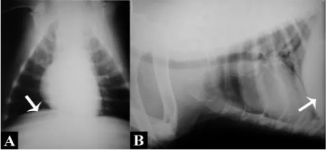
Thoracic radiographs were performed for pre-surgical evaluation, which showed absence of pulmonary alterations and diaphragm compression by a radiopaque rounded structure (Figure 1).
Figure 1. Female dog, Brazilian Fila breed, eight years old.
Thoracic radiograph in ventrodorsal (A) and left lateral (B) positions. A large, rounded and radiopaque structure (arrows) is observed dislocating the diaphragm cranially.
In exploratory abdominal laparotomy was observed a mass originating from the medial portion of the spleen, presenting strong adhesion to the omentum. Moreover, a whitish nodular structure with defined limits was found adjacent to this mass, in the caudal splenic region. A total splenectomy was performed and the surgically-removed fresh material was sent for analysis to the laboratory of animal pathology.
The bitch was maintained without water or food, with an intravenous saline solution, antibiotics, analgesic, and wound dressing. Postoperative chemotherapy treatment was recommended, but the owner refused.
A macroscopic evaluation showed a tumoral mass which weighed 9.2 kg, with dimensions of 30×27×14 cm, firm, encapsulated, and parts of the omentum adhered to the surface. A cut showed a reddish coloration with some pale areas, slightly yellowish, with a great amount of dark red blood flowing, friable tissues, and necrotic center. Adjacent to this mass, a whitish nodule with a clear regular outline, soft consistency, measuring 5.3×5.0×2.0 cm was found in the caudal splenic region. A cut showed that this smaller nodule was homogeneous and had a moderate amount of a
slightly yellowish translucent fluid flowing. The spleen was enlarged and had firm areas to the touch (Figure 2).
Figure 2. Macroscopic appearance of a giant splenic
leiomyosarcoma in a female dog. A) Intraoperative image of the splenic tumor. There is a whitish minor nodule with clear outline in the caudal splenic region (arrow). B) Surgical specimen after splenectomy: red tumor with some pale areas, irregular surface and attached to omentum. C) Cut of minor nodule attached to the splenic parenchyma, showing light colored interior (arrow). D) Interior of large mass showing red color with central pale area. Fragments of the larger tumor mass, minor nodule, and spleen were collected and fixed in buffered formalin (10%) for histopathological analysis. These samples were placed in paraffin blocks, cut in 5-µm thick slices and stained by Hematoxylin-Eosin.
Microscopically, the larger mass showed spindle cells, with intense anisocytosis and anisokaryosis, arranged in multidirectional bundles. An average mitotic count of 2/400x High Power Field (HPF), within a large amount of red blood cells and conjunctive stroma, which indicate a diagnosis of leiomyosarcoma. Necrotic areas and large numbers of vessels were also found, but the vascular endothelium was not altered. The smaller nodule (located in the caudal splenic region) consisted of red blood cells and corrugated structures, with variable thickness and indefinite length, compatible with collagen fibers. The spleen showed hemorrhagic areas and loss of architecture.
New slices were cut and stained with Masson's trichrome staining (MTS). The test was positive for muscle tissue, with red stained cytoplasm of spindle cells. The smaller nodule, adjacent to the tumor, showed fibers stained in blue
by the MTS, confirming to be collagen fibers. This nodule showed no neoplastic cells (Figure 3).
Figure 3. Photomicrography of splenic leiomyosarcoma: A)
Histopathological analysis of a large mass with hematoxylin and eosin stain (microscope objective10x) showing spindle cells with high pleomorphism forming multidirectional beams and atypical mitosis (B) (arrows). C and D) Masson´s trichrome stain (40x - C) and (100x – D) showing tumor cells staining red indicating muscle differentiation, admixed with collagen fibers (blue) and red blood cells (yellow). Photomicrography of small nodule: E) Collagen fibers characterized corrugated structures of varying thickness and indefinite length, which are colored by eosin (10x) and aniline blue, this one used in Masson's trichrome (F) (10x).
Immunohistochemistry analysis was performed for a definitive diagnosis of the histogenesis of the tumor. Tissue sections were placed on slides previously silanized. The antigenic recovery by the moist heat method was carried out in a steamer for 20 min. Primary antibodies incubation was performed by overnight at 4oC and
the revelation was using Advance system. Staining was performed with 3.3 diaminobenzidine and counterstained with hematoxylin. External and/or internal controls were used to validate the reaction. The antibodies tested were alpha smooth muscle actin (1A4, Dako, 1:250), vimentin (V9, Dako, 1:100), desmin (D33, Dako, 1:100), cytokeratin (Pan - Z0622, Dako, 1:200), CD34 (M7165, Dako, 1:100) and S100 protein (Z0311, Dako, 1:800).
Positive expression was found for alpha smooth muscle actin, vimentin and desmin (Figure
4) whereas cytokeratin, CD34 and S100 protein were not immunoreactive.
Figure 4. Immunohistochemical staining of splenic
leiomyosarcoma. More than 80% of the tumor cells are strongly positive for: A) Smooth muscle actin (original magnification: 40x), B) Vimentin (40x) and C) Desmin (40x). Hematoxylin counterstaining.
The postoperative hemogram, five days after surgery, showed a mild normocytic normochromic anemia, which was corrected with nutritional support. There was no clinical follow-up after this period. According to information provided by the owner, 20 days after the last evaluation, the bitch began to show apathy and inappetence, and died. Survival time after surgical excision was 32 days.
Discussion
Splenic leiomyosarcoma is very rare and thus, little described in the veterinary scientific literature. Most reports about this neoplasm focus on its occurrence in humans (Daudia et al., 2001; Piovanello et al., 2007; Patrono et al., 2014). Giant leiomyosarcoma has been reported in humans in different tissues, such as kidney (Öziş, et al., 2014), uterus (Senol et al., 2016), subcutaneous tissues (Kumar et al., 2016), and rectum (Anzai, 2017). However, no reports of giant leiomyosarcoma in canine species have been reported.
Splenic tumors in dogs are generally more frequent in middle-aged or elderly animals, and are more common in dogs of medium to large breeds (Spangler et al., 1994; Fossum and Caplan, 2015; Valli et al., 2017). These neoplasms grow silently; most dogs are asymptomatic, developing nonspecific clinical signs due to tumor mass growth, such as apathy, hyporexia, and abdominal distension (Morais, Argyle and O´Brien, 2010; Zachary and McGavin, 2013; Valli et al., 2017). Moreover, anemia has been most significantly found in dogs with malignant rather than benign splenic neoplasms (Sherwood et al., 2016).
Abdominal ultrasonography is essential in suspected splenic neoplasia. Leiomyosarcomas show homogeneous architecture on ultrasound, as well as hyperechogenicity and cavitary areas due to necrosis (Morrison, 2002). However, in the present
case, the origin of the leiomyosarcoma was not found and its detailed aspects were not well-defined via ultrasound due to its large mass, which occupied the entire abdominal cavity. Thus, ultrasound is more precise for detecting masses of smaller diameters (Fossum and Caplan, 2015).
Spleen rupture with consequent hemoperitoneum is one of the possible complications of malignant splenic neoplasms. However, in this case, despite the large mass dimensions, this complication did not occur. Studies indicate that hemangiosarcoma is more susceptible to rupture and bleeding prior to diagnosis when compared with other malignant splenic neoplasms (Spangler et al., 1994; Cleveland and Casale, 2016; Sherwood et al., 2016).
According to Morrison (2002), splenic leiomyosarcoma or leiomyoma result in splenomegaly, as observed in the present study during surgical resection. In addition, considering the other clinicopathological aspects of this tumor, although the occurrence of metastasis is common due to the sinusoidal structure of this organ (Morrison, 2002; Zachary and McGavin, 2013; Valli et al., 2017), it was not found here.
Total splenectomy is the choice treatment for this neoplasia (Fossum and Caplan, 2015). However, although this treatment was employed, the survival time in this case was short. According to Liptak and Forrest (2013), the average survival time for dogs with splenic leiomyosarcoma is eight months, and for Brearley and Murphy (2008), four months. This lower survival may be correlated with not performing post-surgical chemotherapy (Fossum and Caplan, 2015).
Splenic neoplasms are locally extensive and above the capsular surface, but usually delimited by the capsule, especially on the cut surface (Zachary and McGavin, 2013; Comazzi and MacNeill, 2017), as that found here. The occurrence of necrotic areas, hemorrhage, pale areas (Morais, Argyle and O´Brien, 2010; Comazzi and MacNeill, 2017), and involvement of the omentum with the tumor mass (Fossum and Caplan, 2015) found here are compatible with cases reported in the literature. Moreover, leiomyosarcoma may assume large dimensions in a short time (Senol et al., 2016; Kumar et al., 2016). The microscopic characteristics found in this case are similar to those described by Liptak and Forrest (2013) and Vaill et al. (2017). These authors emphasized the presence of anaplastic
multidirectional spindle-shaped cells, eosinophilic cytoplasm and large numbers of mitoses in this neoplasia. According to Zachary and McGavin, (2013), tumors must be vascularized to increase in diameter. Tumor angiogenesis promotes nutrient and oxygen supply to the neoplasia and stimulates the growth of adjacent tumor cells by the secretion of substances that are growth factors (Zachary and McGavin, 2013; Vaill et al., 2017). Thus, the large amount of red blood cells and vessels found in the histological sections is probably due to the tumor angiogenesis, which is necessary for the neoplasm growth.
Considering malignant mesenchymal tumors that may occur primarily in the spleen, the main differential diagnoses for the neoplasia reported in this case include hemangiosarcoma and fibrosarcoma (Liptak and Forrest, 2013; Zachary and McGavin, 2013). The histochemistry by Masson's trichrome staining (MTS) contributed significantly to the diagnosis, staining in red the cytoplasm of muscle cells, as indicated in the literature for well-differentiated splenic sarcomas (Morais, Argyle and O´Brien, 2010; Liptak and Forrest, 2013; Valli et al., 2017). Considering the microscopic characteristics by the Hematoxylin-Eosin (HE), MTS and the fibrosarcoma stained in blue by the MTS, as also found by Yang et al. (2014) and Park et al. (2015).
The immunohistochemical analysis observed in this study is in agreement with other studies on leiomyosarcoma (Kumar et al., 2016; Sazaki et al., 2016; Anzai et al., 2017), allowing the diagnostic conclusion.
According to the microscopic analysis, the small nodule adjacent to the leiomyosarcoma was compatible with collagen fibers. These fibers resemble reticulin fibers of the spleen, however, reticulin fibers of the spleen are not identifiable in paraffin sections via HE staining, but with PAS (Periodic acid-Schiff) reaction or argentic impregnation techniques (Tolosa, 2003). Thus, this smaller mass consisting of collagen fibers and red blood cells was probably due to the intense growth of the neoplasm. Nevertheless, additional studies are necessary to compare the characteristics presented in this case.
Conclusion
Canine splenic leiomyosarcoma may have large dimensions. Diagnosis of different types of sarcomas is considered difficult, making it necessary to use the histochemical and
immunohistochemical techniques for a diagnostic conclusion. The occurrence of leiomyosarcoma reported here denotes the importance of considering it a differential diagnosis in cases of splenic masses.
Conflito de Interesse
We have no conflict of interest to declare.
Agradecimentos
This study was supported by the Pathology department of the Federal University of Uberlândia.
References
Anzai, H.; Nozawa, H.; Tanaka, J.; Yasuda, K.; Otani, K.; Nishikawa, T.; Toshiaki, T.; Kiyomatsu, T.; Hata, K.; Kawai, K.; Ushiku, T.; Ishihara, S.; Takano, T.; Fukayama, M.; Watanabe, T. Giant leiomyosarcoma of the rectum with lymph node metastasis: a case report and review of the literature. International Journal of Surgery Case Reports, 34: 27-31, 2017.
Brearley, M.J; Murphy, S. Splenic tumors. In: Arlyge, D.J.; Brearley, M.J.; Turek, M.M. Decision making in small animal oncology. Iowa: Wiley-Blackwell, 2008. Cap.11, p. 211-216.
Cleveland, M.J.; Casale, S. Incidence of malignancy and outcomes for dogs undergoing splenectomy for incidentally detected nonruptured splenic nodules or masses: 105 cases (2009-2013). Journal of the American Veterinary Medical Association, 248(11): 1267-1273, 2016. Comazzi, S.; MacNeill, A.L. Cytology of lymphoid
tissues. In: Barger, A.M.; MacNeill, A.L. Small animal cytologic diagnosis. Boca Raton: CRC Press, 2017. Cap.8, p. 209-210. Daudia, A.T.; Walker, S.; Morgan, B.; Lloyd, D.M.
Leiomyosarcoma of the spleen. Surgery, 130, n.5, p.893-894, 2001.
Fossum, T.W.; Caplan, E.R. Cirurgias do sistema hemolinfático. In: Fossum, T.W. Cirurgia de pequenos animais. Rio de Janeiro: Elsevier, 2015. Cap.24, p.700-704.
Kumar, S.K.; Bhadani, P.P.; Sinha, R.; Shuchismita, Kumar, K. Giant subcutaneous leiomyosarcoma at knee joint: malignant tumour at rare site. International Journal of Medical and Dental Sciences, 5(2): 1279-1282, 2016.
Liptak, J.M.; Forrest, L.J. Soft tissue sarcomas. In: Withrow, S.J.; Page, R.; Vail, D.M. Small animal clinical oncology. St Louis: Elsevier Saunders, 2013. Cap. 21, p. 356-380.
Morais, H.A.; Argyle, D.J.; O´Brien, R.T. Diseases of the spleen. In: Ettinger, S.J.; Feldman, E.C. Textbooks of internal veterinary medicine: diseases of the dog and the cat. St Louis: Saunders Elsevier, 2010. Cap.206, p. 810-818. Piovanello, P.; Viola, V.; Costa, G.; Carletti, M.; Cecera, A.; Turchetta, F.; Iudicone, R.; Catalano, G.; Santucci, A.; Recchia, F.; Fiorillo, L.; Menichella, M.A.; Baiano, G. Locally advanced leiomyosarcoma of the spleen. A case report and review of the literature. World Journal of Surgical Oncology, 28(5): 135, 2007.
Morrison, W. B. Principles of cancer diagnosis. In:_____. Cancer in dogs and cats – medical and surgical management. Teton: New Media, 2002. Cap. 7, p-166-167.
Öziş, S.E.; Gülpinar, K.; Sahli, Z.; Konak, B.B.; Keskin, M.; Özdemir, S.; Ataoğlu, Ö. Recurrent renal giant leiomyosarcoma. Ulus Cerrahi Derg, 32(2): 145-148, 2014.
Park, H.; Jeong, C.; Kim, G.; Kim, H.; Do, S.; Park, H. Primary renal fibrosarcoma with local invasion into the mesenteric membrane of a mongrel dog. Korean Journal of Veterinary Research, 55(1): 65-69, 2015.
Sasaki, J.; Toyoshima, M.; Okamura, Y.; Goryo, M. Omental leiomyosarcoma with unusual giant cells in a beagle dog – short communication. Acta Veterinaria Hungarica, 64(2): 222–228, 2016.
Senol, T.; Kahramanoglu, I.; Muezzinoglu, B.; Yucesoy, I. Giant leiomyosarcoma: A case
report. International Journal of Surgery Case Reports, 19: 109–111, 2016.
Serin, G.; Aydogan, A.; Yaygingul, R.; Tunca, R. Uterine leiomyosarcoma in a dog: a case report. Veterinarni Medicina, 55(8): 405– 408, 2010.
Sherwood, J.M.; Haynes, A.M.; Klocke, E.; Higginbotham, M.L.; Thomson, E.M.; Weng, H.; Millard, H.A.T. Occurrence and clinicopathologic features of splenic neoplasia based on body weight: 325 Dogs (2003– 2013). Journal of the American Animal Hospital Association, 52(4): 220–226, 2016. Spangler, W.L., Culbertson, M.R., Kass, P.H.
Primary mesenchymal (nonangioma-tous/nomlymphomatous) neoplasms occurring in the canine spleen: anatomic classification, immunohistoche-mistry, and mitotic activity correlated with patient survival. Veterinary Pathology, 31: 37-47, 1994.
Tolosa, E.M., Rodrigues, C.J.; Behmer, O.A.; Freitas-Neto, A.G. Manual de técnicas para histologia normal e patológica. 2. ed. São Paulo: Manole, 2003. 331p.
Valli, V.E.; Bienzle, D.; Meuten, D.J.; Linder, K.E. Tumors of hemolymphatic system. In: Meuten, D. J. Tumors in domestic animals. Ames: Willey Backwell, 2017. Cap. 7, p. 203-321.
Zachary, J.F.; McGavin, M.D. Bases da patologia em veterinária. 5. ed. São Paulo: Elsevier, 2013, 1344p.
Yang, C.; Liao, J.; Yu, Y.; Kao, J. Case report: splenic fibrosarcoma in a campbell's hamster. Taiwan Veterinary Journal, 40(3): 145-149, 2014.