RELATIONSHIP BETWEEN CLINICAL, PLACENTARY, OBSTETRIC AND
NEONATAL ASPECTS AND INTRAUTERINE GROWTH IN HIGH RISK
PREGNANCIES
Ana Karina Marques Salge1, Renata Calciolari Rossi e Silva2, Janaína Valadares Guimarães3, Wilzianne Silva Ramalho4, Douglas Reis Abdalla5, George Kemil Abdalla6
1 Ph.D. in Health Sciences. Nursing Faculty and Post-graduate Program in Nursing Professor, Universidade Federal de Goiás (UFG). Goiânia, Goiás, Brazil. E-mail: [email protected]
2 Physiotherapist. Professor, Graduate Courses in Physiotherapy and Medicine, Universidade do Oeste Paulista. Presidente Prudente, São Paulo, Brazil. E-mail: [email protected]
3 Ph.D. in Health Sciences. Professor, Nursing Faculty and Post-graduate Program in Nursing, UFG. Goiânia, Goiás, Brazil. E-mail: [email protected]
4 M.Sc. in Nursing. Post-graduate Program in Nursing, UFG. Goiânia, Goiás, Brazil. E-mail: [email protected]
5 Ph.D. in Health and Medical Sciences. Professor, Faculdade de Telaentos Humanos (FACTHUS). Uberaba, Minas Gerais, Brazil, E-mail: [email protected]
6 Ph.D. in Health Sciences. Professor, FACTHUS. Uberaba, Minas Gerais, Brazil, E-mail: [email protected]
ABSTRACT
Objective: to analyze clinical, placental and obstetric aspects of women with and without high-risk pregnancy, and their relationship with intrauterine growth deviations and neonatal aspects.
Method: this is a cross-sectional descriptive study. Data collection was based on an analysis of the medical records of women with and without high-risk pregnancy and newborns, and anatomopathological characterization of the placenta.
Results: 265 placentas were analyzed, 130 (49.06%) women with high-risk pregnancy and newborns with intrauterine growth deviations. A higher occurrence of placental changes was found in high-risk pregnancy and uterine growth deviations in comparison to cases without high-risk pregnancy (p≤0.001). High-risk pregnancies with intrauterine growth deviations were associated with placental changes (p≤0.001). Intrauterine growth deviations was related to birth weight in cases of high-risk pregnancy compared to normal gestation (p=0.014). Conclusion: a higher occurrence of placental anatomopathological changes was found in maternal and fetal surfaces in cases of high-risk pregnancy and intrauterine growth deviations.
DESCRIPTORS: Fetal growth retardation. High risk pregnancy. Neonatology. Placenta. Newborn.
RELAÇÃO ENTRE OS ASPECTOS CLÍNICOS, PLACENTÁRIOS,
OBSTÉTRICOS E NEONATAIS E O CRESCIMENTO INTRAUTERINO NA
GESTAÇÃO DE ALTO RISCO
RESUMO
Objetivo: analisar aspectos clínicos, placentários e obstétricos de mulheres com e sem gestação de alto risco e sua relação com desvios de crescimento intrauterino e aspectos neonatais.
Método: trata-se de um estudo descritivo transversal. A coleta de dados baseou-se em análise dos prontuários das mulheres com e sem gestação de alto risco e dos recém-nascidos, e análise anatomopatológica placentária.
Resultados: foram estudadas 265 placentas, 130 (49,06%) de mulheres com gestação de alto risco e recém-nascidos com desvios de crescimento intrauterino. Houve maior ocorrência de alterações placentárias em gestação de alto risco e Desvios de crescimento uterino comparadas aos casos sem gestação de alto risco (p≤0,001). Gestação de alto risco com Desvios de crescimento intrauterino possui associação com alterações placentárias (p≤0,001). desvios de crescimento intrauterino está relacionado ao peso ao nascer em casos com gestação de alto risco, comparados com gestação normal (p=0,014).
Conclusão: existe maior ocorrência de alterações anatomopatológicas placentárias nas faces materna e fetal nos casos com gestação de alto risco e desvios de crescimento intrauterino.
DESCRITORES: Retardo do crescimento fetal. Gravidez de alto risco. Neonatologia. Placenta. Recém-nascido.
RELACIÓN ENTRE LOS ASPECTOS CLÍNICOS PLACENTARIOS,
OBSTÉTRICOS Y NEONATALES Y EL CRECIMIENTO INTRAUTERINO EN
LA GESTACIÓN DE ALTO RIESGO
RESUMEN
Objetivo: analizar aspectos clínicos, placentarios y obstétricos de mujeres con y sin gestación de alto riesgo y su relación con desvíos de crecimiento intrauterino y aspectos neonatales.
Métodos: investigación descriptivo transversal. La recolección de datos se basó en el análisis de los expedientes de las mujeres con y sin gestación de alto riesgo y de los recién nacidos, y análisis anatomopatológico placentario.
Resultados: fueron estudiadas 265 placentas, 130 (49,06%) de mujeres con gestación de alto riesgo y recién nacidos con desvíos de crecimiento intrauterino. Hubo mayor ocurrencia de alteraciones placentarias en gestación de alto riesgo y desvíos de crecimiento uterino comparados con los casos sin gestación de alto riesgo (p≤0,001). Gestación de alto riesgo con desvíos de crecimiento intrauterino posee asociación con alteraciones placentarias (p≤0,001). Desvíos de crecimiento intrauterino están relacionados al peso al nacer en casos con gestación de alto riesgo, comparados con gestación normal (p=0,014).
Conclusión: existe mayor ocurrencia de alteraciones anatomopatológicas placentarias tanto en la madre como en el feto en los casos con gestación de alto riesgo y desvíos de crecimiento intrauterino.
DESCRIPTORES: Retardo del crecimiento fetal. Embarazo de alto riesgo. Neonatología. Placenta. Recién nacido.
INTRODUCTION
Most pregnancies progress without any compli-cations, consisting of a necessary process of immune and systemic adaptation for the proper development of the fetus and the placenta.1 When the physiological
and balanced gestational period undergoes altera-tions that can generate risk for the mother-child bi-nomial, pregnancy is no longer considered a normal state, becoming high-risk pregnancy.
High-risk pregnancies are those in which the life or health of the mother and/or fetus/newborn are more likely to be threatened than the average population. A quantitative 10 to 20% of pregnant women have high-risk pregnancies, which is related to the occurrence of some disorders that represent risks to both the mothers and fetuses.2
Although the human placenta is a vital organ for maintaining pregnancy and promoting normal fetal development,3 a pattern of morphological
alteration found may indicate maternal and fetal clinical involvement related to gestational hyperten-sive syndromes,4 to intrauterine growth restriction
(IUGR),5 to Diabetes Mellitus (DM) or to placental
abruption (PA), with small for gestational age (SGA) newborns,5 decreased placental low (placental hy
-poxia) or miscarriage resulting as a consequence.6
Aggressions involving the uterine-placental unit may divert the fetus from its genetic growth potentia,7 resulting in a condition known as IUGR5
(Intrauterine Growth Restriction) and in complica-tions related to a higher incidence of fetal distress,
the presence of meconial amniotic luid, low Apgar
score, low birth weight (LBW) and perinatal death.3
Intrauterine growth deviations (IUGDs) are associated with perinatal morbidity and mortality,
and the risk of developing diseases in adult life such as DM and cardiovascular diseases. IUGR and macrosomia represent important clinical conditions associated with this condition.8 Thus, the IUGR
pathophysiology is related to changes in the avail-ability, transport and use of the substrate occurring in three different situations: reduced maternal-pla-cental supply; impaired fetal potential; or both being compromised. Placental and adnexa abnormalities are one of the main causes of IUGR.3 The incidence
of mortality in preterm newborns is higher if IUGR is also present, increasing the perinatal mortality percentages by two to ten times.9
Just as IUGR may result in adults with dis-eases, macrosomia in adulthood can also trigger obesity, dyslipidemia, hypertension and type 2 dia-betes.8 Fetal macrosomia is the term used to deine
fetuses or newborns considered abnormally large, weighing 4000 grams or more; large for gestational age (LGA) newborns are those who are above the 90th percentile for gestational age.10
DM is a known risk factor for excessive new-born weight. Macrosomia is observed among cases with poorly controlled diabetes, those with long evolution and those with vascular alterations.8
Understanding the relevance of IUGDs as a predisposing factor to the increased risk of fetal/ neonatal morbidity and mortality, and recogniz-ing deviations as well as their etiopathogenesis is extremely important,9 being a fundamental task in
quality prenatal care.11
problem due to the intercurrences that affect moth-ers and newborns at prenatal, delivery and in the puerperium periods, and they can be predictors of fetal changes in future pregnancies.
Thus, the objective of the present study was to relate obstetrical, fetal and anatomopathologi-cal aspects of maternal and fetal surfaces of the placentas of women with and without high-risk pregnancy, and their relationship with intrauterine growth deviations in a public maternity hospital in Goiânia-GO, Brazil.
METHOD
This is a descriptive cross-sectional study carried out from January 2010 to January 2015, in a public maternity hospital in Goiânia-GO, Brazil. The population consisted of 265 women presenting 130 with risk pregnancies and 135 without high-risk pregnancies, who had their medical records available for data collection with complete informa-tion, including newborn weight at birth, gestational age and placenta available for evaluation.
Data collection took place in two distinct stages: review of medical records and placen-tal macroscopic anatomopathological analy-sis, according to the protocols by Driscoll and Langston.5 The eligibility criteria considered in
the high-risk pregnancy and IUGD group were women with high-risk pregnancies who presented risk factors for fetuses/newborns with IUGD. All participants signed the Clear and Informed Consent Form (TCLE).
The group without high-risk pregnancy and without IUGDs included women without high-risk pregnancy, mothers of fetuses/newborns who did not present any disease considered risk factors for fetuses/newborn with IUGD during the gestation, those who did not present intercurrences during gestation and delivery evidenced by laboratory re-sults, as well as clinically normal fetuses/newborns who did not present IUGD according to the criteria established in 1963.3
All cases that did not meet the eligibility cri-teria were considered as exclusion cricri-teria; all cases in which the placenta was not available for analysis (not stored at a temperature of approximately 25°C in 0.9% saline solution, but in formaldehyde and was sent to the hospital’s pathology service) (four cases); all cases in which the medical record was not
available (ive cases), was incomplete or missing
information (11 cases) such as birth weight and/or gestational age; and also participant’s or the person
in charge refusing to sign the TCLE, or refusing to participate in the study (two cases).
For the macroscopic analysis, placentas stored in 0.9% saline solution for up to 48 hours postpartum were analyzed. All macroscopic changes found on maternal and/or fetal surfaces of the placenta and on the umbilical cord were evaluated, recorded and photographed.
The information source for this study was composed of the medical records of each patient, from where sociodemographic data (age, national-ity, education level, occupation, family income); baseline diseases, gestational age (determined by
the date of the last menstrual period, at the irst tri -mester ultrasound examination, and by the Capurro Method) and parity were collected and transcribed onto a separate form. Maternal-baseline diseases were grouped according to the criteria established by the 10th Revision of the International Classiica
-tion of Diseases (ICD-10).
The ethical principles of research involving human beings were respected, in accordance with Resolution 196/96.12 The research project was
sub-mitted and approved by the Human and Animal Medical Research of the Hospital das Clínicas of the
Universidade Federal de Goiás, under protocol number 101/2008. A CAAE number was not generated since the project was approved prior to the creation of the
Plataforma Brasil in 2012.
The information was analyzed through the SigmaStat ® version 2.0 electronic program. Quan-titative data were descriptively analyzed through frequency distribution, means and standard
devia-tion. Signiicance tests appropriate to the sample
size were applied to verify statistical differences between proportions. Proportions were compared
using the χ2 test, followed by the Yates’ correction
test. The differences found between the groups were
veriied using the Mann-Whitney test. Differences
where p was less than 5% (p<0.05) were considered
statistically signiicant.
RESULTS
Regarding maternal age, 107 (82.3%) women were between 19 and 35 years of age, eight women (6.15%) were under 18 years of age, and 15 women (11.55%) were in the age group above 35 years. Mean maternal age was 28.12 ± 6.2 years in the high-risk pregnancy group.
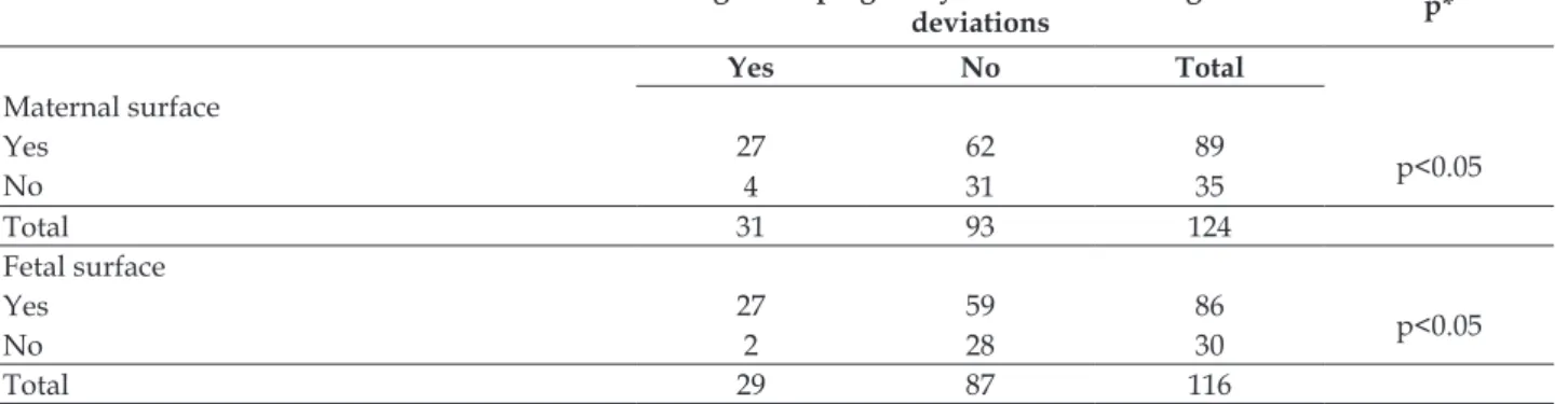
The comparison between the occurrence of macroscmpic anatomopathological changes on
maternal and fetal surfaces of the placenta in cases of high-risk pregnancy and newborns with IUGD, and cases of women without high-risk pregnancy and newborns without IUGD (control group) was
statistically signiicant (p<0.05, maternal surface,
and p<0.05, fetal surface). The occurrence of macro-scopic placental abnormalities was more prevalent in maternal and fetal surfaces in cases with high-risk pregnancy and IUGD (Table 1).
Table 1 - Comparison between the occurrence of macroscopic anatomopathological changes on the maternal and fetal surfaces of the placenta in women with and without high-risk pregnancy and their respective newborns with intrauterine growth deviation in a public maternity hospital in Goiânia-GO, Brazil, from January 2010 to January 2015
Placental anatomopathological changes High-risk pregnancy and intrauterine growth
deviations p*
Yes No Total
Maternal surface
Yes 27 62 89
p<0.05
No 4 31 35
Total 31 93 124
Fetal surface
Yes 27 59 86
p<0.05
No 2 28 30
Total 29 87 116
*χ2 test, followed by the Yates’ correction test.
Macroscopic anatomopathological changes in the maternal and fetal surfaces of the placentas of women with high-risk pregnancy are described
in Table 2. Calciphylaxis and ibrin were the most
prevalent changes in maternal and fetal surfaces (p<0.0001); calciphylaxis was more frequent on the
maternal surface (p<0.0001) and ibrin on the fetal
surface (p<0.0001).
Table 2 - Frequency of macroscopic anatomopathological changes in the maternal and fetal surfaces of the placentas of 130 women with high-risk pregnancy associated with intrauterine growth deviations in a public maternity hospital in Goiânia-GO, Brazil, from January 2010 to January 2015
Macroscopic anatomopathological changes Maternal surface n (%)
Fetal surface n (%)
Maternal surfasse vs Fetal surface
Calciphylaxis 75 (57.7)* 18 (13.84)† p<0.0001
Fibrin 31 (23.84)* 102 (78.5)† p<0.0001
Infarction 1 (0.76) 1 (0.76)
Detachment Area/abruption site 1 (0.76)
-Fibrosis 3 (2.3)
-Bruising - 3 (2.3)
Total changes 111 (85.4) 124 (95.4)
No changes 19 (14.6) 6 (4.6)
* p<0.0001 vs other Macroscopic anatomopathological changes on the maternal surface; † p<0.0001 vs other Macroscopic anatomopatholo-gical changes on the fetal surface. Mann-Whitney test. The proportions were compared by the χ2 test, followed by the Yates’ correction test.
Neonatal factors related to intrauterine growth deviations in women with high-risk pregnancy are
Table 3 - Neonatal factors related to intrauterine growth deviation in newborns of women with high-risk pregnancy in a public maternity hospital in Goiânia-GO, Brazil, from January 2010 to January 2015
Neonatal factors related to intrauterine growth deviation
Normal pregnancy n (%)*
High-risk pregnancy
n (%)* Odds Ratio (95%CI)† p
Cephalic perimeter
(<32 and >38 centimeters) 10 (8.0) 25 (23.8) 0.34 (0.15-0.73) 0.006
Thoracic perimeter
(<30 and >36 centimeters) 19 (16.4) 34 (35.4) 0.46 (0.25-0.86) 0.017
Abdominal perimeter (<28 and
>34 centimeters) 11 (8.8) 24 (22.6) 0.39 (0.18-0.84) 0.021
NB weight
(<2.500 and >4.000 grams) 11 (8.8) 29 (28.7) 0.31 (0.15-0.65) 0.002
Apgar at 1 (<8 points) 30 (28.6) 45 (52.9) 0.54 (0.31-0.93) 0.030
Apgar at 5 (<8 points) 3 (2.2) 4 (3.2) 0.72 (0.16-3.2) 0.718
*n: number of cases; † 95% CI: 95% of the conidence interval. The proportions were compared by the χ2 test, followed by the Yates’ cor-rection test.
According to the newborn classiication and
considering the relation between birth weight and gestational age (BW versus GA) according to the neonatal measurement curve3 related to the IUGD,
20 (15.38%) NBs were classiied as SGA, 7 (5.38%) as
LGA and 94 (72.31%) as appropriate for gestational age (AGA) (Table 4).
Table 4 – Classiication of newborns with intrauterine growth deviation using the birth weight and
gestational age ratio, according to a graph adapted from Lubchenco (1963), from high-risk pregnancy in a public maternity hospital in Goiânia-GO, Brazil, from January 2010 to January 2015
NB Classiication n (%)
Small for gestational age 20 (15.39)
Large for gestational age 7 (5.38)
Appropriate for gestational age 94 (72.31)
Birth weight not available 9 (6.92)
Total 130 (100)
Data on the association between newborn
classiication based on the criteria (BW versus GA)3
in high-risk pregnancy and normal gestation in 130
women with and without macroscopic placental changes, respectively, are described in Table 5.
Table 5 - Birth weight versus gestational related to intrauterine growth deviation, and associated to
high-risk pregnancy and anatomicopathological macroscopic placental changes in a public maternity hospital in Goiânia-GO, Brazil, from January 2010 to January 2015
Gestational agevs birth weight High-risk pregnancy (with placental change)
Normal pregnancy
(without placental change) p*
Small for gestational age 26 06 <0.05
Appropriate for gestational age 58 54 0.4583
Large for gestational age 20 - <0.05
* χ2 test, followed by the Yates’ correction test.
A statistically signiicant difference (p≤0.001)
was observed when comparing the occurrence of SGA and LGA (Intrauterine growth deviations)
LGA (NB births with IUGD) in cases of high-risk pregnancy (with macroscopic placental changes) was observed than in cases of normal pregnancy (without macroscopic placental changes). Cases
of gestational diabetes (33.07%) and preeclampsia (26.15%) can be highlighted among maternal predic-tors of high-risk pregnancy associated with IUGD (Table 6).
Table 6 - Maternal factors related to intrauterine growth deviation in women with high-risk pregnancy at a public maternity hospital in Goiânia-GO, Brazil, from January 2010 to January 2015
Maternal factors related to intrauterine growth deviation n (%)
Maternal age
≤ 18 years and ≥36 years 26 (20.00)
Gestational age
< 37 weeks and > 41 weeks and 6 days 29 (22.30)
Maternal Diseases
Preeclampsia 34 (26.15)
Eclampsia 3 (2.31)
Chronic hypertension 18 (13.85)
Gestational hypertension 11 (08.46)
HELLP syndrome 1 (0.77)
Gestational diabetes 43 (33.07)
Diabetes Mellitus I 15 (11.54)
Diabetes Mellitus II 5 (3.85)
DISCUSSION
This study evaluated 265 placentas, with 130 (49.06%) from women with high-risk pregnancy and fetuses/newborns with IUGD. The presence of macroscopic anatomopathological changes in the maternal and fetal surfaces of the placenta is asso-ciated with the occurrence of high-risk pregnancy IUGD in the investigated group.
Mean maternal age in the high-risk pregnancy
group was 28.12±6.2 years. A similar inding was
found in a study that investigated the sociodemo-graphic characteristics of women with high-risk pregnancy with newborns with IUGD. The mean maternal age was 30.1±1.4 years.13
A case-control study conducted in Chile with
22,227 newborns showed that there is a signiicant
relationship between the occurrence of IUGR and maternal age.14 In another study conducted in India
based on 36,674 births, a statistical association was found among births of SGA newborns with mater-nal age above 35 years. In addition, we found that maternal age below 19 years is a determinant of low birth weight (LBW).15 Maternal age extremes
can be considered a risk factor and inluence peri -natal mortality.
By comparing the occurrence of macroscopic anatomopathological changes found on the mater-nal and fetal surfaces of the placenta in women with
high-risk pregnancy and newborns with IUGD with the control group, it was possible to verify a higher occurrence in the former, as well as presenting
sta-tistical signiicance (p≤0.001).
Several placental changes were observed in a study carried out in Japan observing 53,650 placen-tae of pregnant women with high-risk pregnancy, mainly related to placental weight.16
A higher prevalence of calciphylaxis and ibrin
on both maternal and fetal surfaces was found in analyzing maternal and fetal surfaces of pregnant women with high-risk pregnancies regarding the presence and type/characteristic of macroscopic anatomopathological changes; however, a higher prevalence of calciphylaxis (57.7%) was found on
the maternal surface (p<0.0001), and ibrin on the
fetal surface (78.5%) (p<0.0001).
Divergent results were found in another sample with 518 placentas. The most frequent lesion was chorionic degenerative vascular injury (55.7%), followed by retroplacental hematoma (23.8%),
in-farction areas (10.9%) and ibrin deposits (9.2%).17
Fibrin deposits in the placenta relect impor -tant vascular changes, which can cause spontane-ous abortion, preterm birth, IUGD and fetal death. In a study carried out in Romania of 467 placentas examined macroscopically, around 188 (40%) had
a ibrin deposit. Alterations such as thrombosis and
recent infarcts were found on the fetal surface. On the maternal surface, important areas of placental infarction have been reported, which may contribute to the occurrence of IUGD by restricting the surface exchange area.18
Cases with no changes were found in both the maternal surface as well as the fetal surface analyzes, corresponding to a previously described study18
where placental morphological changes that were absent in 279 (60%) placentas were analyzed from a total of 467 placentas examined macroscopically.
A comparison between the cases of women with high-risk pregnancy and newborns with IUGD was carried out, with the cases of women without high-risk pregnancy showing
statisti-cal signiicance (p≤0.001). Cases with high-risk
pregnancy and IUGD have a higher statistical association with the occurrence of macroscopic anatomopathological changes.
Some high-risk pregnancies have been
associ-ated with both speciic macroscopic and microscopic
placental changes. In placentas from SGA newborns (an important type of IUGD) an increase in ischemic
lesions, infarctions and calcium and ibrin deposi -tion has been observed.19
In analyzing the data of fetuses/newborns regarding perimeters (cephalic - CP, thoracic - TP, and abdominal - AP, measured in centimeters), 25 (23.8%) among the 130 fetuses/newborns included in the study with case group criteria presented CP with IUGD at the time of investigation and data col-lection. Regarding TP, 34 (35.4%) cases with IUGD were included, and 24 (22.6%) cases for AP with IUGD were included. Regarding birth weight, 29 (28.7%) fetuses/newborns presenting IUGD birth weight were included. As for Apgar data at 1 min-ute, 45 (52.9%) cases with Apgar <8 points and 4 (3.2%) cases with Apgar <8 points in the 5th minute
were included.
There are many ways to diagnose perinatal hypoxia, and one of them is the Apgar score.
Sev-eral placental changes such as infarction relect fetal
distress diagnosed by Apgar indices below 7 at the 5th minute,20 suggesting the importance of studying
the relationship between fetal distress (evaluated through Apgar) and placental changes.
Mean cephalic circumference found in this study was 33.00±4.83 cm. This differs from a retro-spective cohort study performed in Zona da Mata of Pernambuco, Brazil, with 915 full-term children, in which the mean cephalic circumference was 34.5±1.16 for NBs without IUGD, 33.1±1.24 for LBW NBs, and 35.4±1.13 for macrosomic NBs.21
Cephalic perimeter can translate into patho-logical brain growth (hydrocephalus, microcephaly). Growth restriction of fetal segments in intrauterine life may be associated with the presence of maternal affections, such as hypertension due to placental hypoxia, placentation alteration, as well as oxygen and nutrient supply. This condition may alter bone formation and fetal organs, thus interfering with neonatal anthropometric measures such as head circumference and weight.22
When comparing the occurrence of SGA and LGA (IUGD) births with the occurrence of high-risk pregnancy (with placental changes) and normal pregnancy (no placental changes), a statistically
signiicant difference (p≤0.001) was found, evidenc -ing a greater number of SGA and LGA (NB births with IUGD) in cases of high - risk pregnancy (with macroscopic placental changes) than in cases of normal pregnancy (without macroscopic placental changes).
As for the comparison between the occurrence of high-risk pregnancy and macroscopic anatomo-pathological changes with neonatal clinical data (AI and BW) and maternal data (maternal age and GA),
a statistically signiicant difference was found only
when comparing the occurrence of macroscopic anatomopathological changes and neonatal clinical
data related to BW (≤ 2,500 g and > 4,000 g). Regard
-ing post-term pregnancy, no statistically signiicant
difference was found between groups.
Considering the maternal predictor “gesta-tional age”, the lower the maternal age, the greater the risk of preterm birth and the interruption of breastfeeding,23 especially before the age of 15 years.
Pregnant women of extreme ages (below 19 years and above 35 years) were also at higher risk for maternal and neonatal mortalities.24
The study presented some limitations in being a transversal descriptive study carried out with medical records and macroscopic placental analysis. Some information was often incomplete or inadequately recorded, with no standardization for the record. Thus, a great amount of informa-tion was lost, such as informainforma-tion about maternal disease history or newborn’s anthropometric data due to illegible handwriting and lack of records by the nursing team.
CONCLUSION
high-risk pregnancy. There was a greater occurrence of macroscopic anatomopathological changes in maternal and fetal surfaces of the placenta in cases of high-risk pregnancy and IUGD (p=0.029, mater-nal surface and p=0.007, fetal surface). Among the macroscopic anatomopathological changes,
calci-phylaxis and ibrin were the most prevalent changes
in maternal and fetal surfaces (p<0.0001), with calci-phylaxis being more frequent in the maternal surface
(p<0.0001) and ibrin in the fetal surface (p<0.0001).
Neonatal factors such as weight were
sta-tistically signiicant in NB with IUGD (p<0.05). A
higher number of SGA and LGA (NB with IUGD) was observed in cases of high-risk pregnancy (with macroscopic placental changes) than in cases of normal pregnancy (without macroscopic placental changes). Gestational diabetes (33.07%) and pre-ec-lampsia (26.15%) were among the maternal predic-tors of high-risk pregnancy associated with IUGD. Studying IUGDs related to obstetric, fetal and placental aspects of women with and without high-risk pregnancy deserves a vigilant and multiprofes-sional approach. Understanding the factors related to IUGD can positively contribute to health team
conduct, who should provide speciic and quality
care when carrying for newborns with IUGD in order to design care strategies that contemplate the newborn’s/child’s growth and development needs. Guidance can also be offered during women’s health and prenatal consultations, with the intention to inform about possible intercurrences that may be predictive of fetal changes in future pregnancies.
REFERENCES
1 Orczyk-Pawilowicz M, Jawien E, Deja S, Hirnle L, Zabek A, Mlynarz P. Metabolomics of human amniotic luid and maternal plasma during normal pregnancy. PLoS One [Internet]. 2016 Apr 12 [cited 2016 Apr 25];11(4):e0152740. Available from: http:// www.ncbi.nlm.nih.gov/pmc/articles/PMC4829258/ pdf/pone.0152740.pdf
2 Ministério da Saúde (BR). Secretaria de Atenção à Saúde. Departamento de Atenção Básica. Gestação de alto risco: manual técnico. Brasília (DF): MS; 2012. 3 Lubchenco LO, Hansman C, Dressler M, Boyd E. Intrauterine growth as estimated from liveborn birth-weight data at 24 to 42 weeks of gestation. Pediatrics. 1963;32:793-800.
4 Żyła MM, Wilczyński J, Nowakowska-Głąb A, Maniecka-Bryła I, Nowakowska D. pregnancy and delivery in women with uterine malformations. Adv Clin Exp Med [Internet]. 2015 Sep-Oct [cited 2016 Apr 25];24(5):873-9. Available from: http://www. advances.am.wroc.pl/pdf/2015/24/5/873.pdf
5 Driscoll SG, Langston C. Placental examination in a clinical setting. Arc Pathol Lab Med. 1991;115(7):668-71. 6 Daniel-Spiegel E, Mandel M, Nevo D, Ben-Chetrit
A, Shen O, Shalev E, et al. Fetal biometry in the Israeli population: new reference charts. Isr Med Assoc J [Internet]. 2016 Jan [cited 2016 Apr 25];18(1):40-4. Available from: http://www.ima.org. il/FilesUpload/IMAJ/0/183/91559.pdf
7 Amaral LM, Cunningham MW Jr, Cornelius DC, LaMarca B. Preeclampsia: long-term consequences for vascular health. Vasc Health Risk Manag [Internet]. 2015 Jul [cited 2016 Apr 25];11:403-15. Available from: http://www.ncbi.nlm.nih.gov/pmc/articles/ PMC4508084/pdf/vhrm-11-403.pdf
8 Juárez-Olguín H, Buendía-Soto E, Lares-Asseff I. Pharmacology for the fetus and the newborn]. Gac Med Mex [Internet]. 2015 May-Jun [cited 2016 Apr 25];151(3):387-95. Available from: http://www.anmm. org.mx/GMM/2015/n3/GMM_151_2015_3_387-395.pdf 9 Kamana Kc, Shakya S, Zhang H. Gestational diabetes mellitus and macrosomia: a literature review. Ann Nutr Metab [Internet]. 2015[cited 2016 Apr 25];66 Suppl 2:14-20. Available from: http://www.karger. com/Article/Pdf/371628
10 Kintiraki E, Papakatsika S, Kotronis G, Goulis DG, Kotsis V. Pregnancy-induced hypertension. Hormones (Athens) [Internet]. 2015 Apr-Jun [cited 2016 Apr 25];14(2):2112-23. Available from: http:// www.hormones.gr/8566/article/article.html 11 Zhang S, Regnault TR, Barker PL, Botting KJ, McMillen
IC, McMillan CM, et al. Placental adaptations in growth restriction. Nutrients [Internet]. 2015 Jan [cited 2016 Apr 25];7(1):360-89. Available from: http:// www.ncbi.nlm.nih.gov/pmc/articles/PMC4303845/ pdf/nutrients-07-00360.pdf
12 Ministério da Saúde (BR). Conselho Nacional de Saúde. Resolução 196 de 10 de outubro de 1996. Aprova as diretrizes e normas regulamentadoras da pesquisa envolvendo seres humanos. Brasília (DF): MS; 1996. Available from: http://conselho.saude.gov. br/Resolucoes/Reso196.doc
13 Albu AR, Horhoianu IA, Dumitrascu MC, Horhoianu V. Growth assessment in diagnosis of fetal growth Restriction. Review. J Med Life [Internet]. 2014 Jun [cited 2016 Apr 25];7(2):150-4. Available from: http:// www.ncbi.nlm.nih.gov/pmc/articles/PMC4197512/ pdf/JMedLife-07-165.pdf
14 Canals C A, Cavada C G, Nazer H J. Identiication of risk factors for congenital malformations. Rev Med Chil [Internet]. 2014 Nov [cited 2016 Apr 25];142(11):1431-9. Available from: http://www. scielo.cl/pdf/rmc/v142n11/art10.pdf
www.ncbi.nlm.nih.gov/pmc/articles/PMC4324804/ pdf/12884_2015_Article_440.pdf
16 Matsuda Y, Ogawa M, Nakai A, Hayashi M, Satoh S, Matsubara S. Fetal/Placental weight ratio in term Japanese pregnancy: its difference among gender, parity, and infant growth. Int J Med Sci [Internet]. 2015 Mar 25 [cited 2016 Apr 25];12(4):301-5. Available from: http://www.ncbi.nlm.nih.gov/pmc/articles/ PMC4402432/pdf/ijmsv12p0301.pdf
17 Folescu R, Motoc AG, Zamir CL, Ilie AC. Anatomical and histological considerations of placenta vascular diseases with implications in forensic medicine. Rom J Morphol Embryol [Internet]. 2014 [cited 2016 Apr 25];55(2 Suppl):579-83. Available from: http://www. rjme.ro/RJME/resources/iles/551214579583.pdf 18 Pinar H, Goldenberg RL, Koch MA, Heim-Hall J,
Hawkins HK, Shehata B, et al. Placental indings in singleton stillbirths. Obstet Gynecol [Internet]. 2014 Feb [cited 2016 Apr 25];123(2Pt1):325-36. Available from: http://www.ncbi.nlm.nih.gov/pmc/articles/ PMC3948332/pdf/nihms541867.pdf
19 Wise LA, Mikkelsen EM, Sørensen HT, Rothman KJ, Hahn KA, Riis AH, et al. Prospective study of time to pregnancy and adverse birth outcomes. Fertil Steril [Internet]. 2015 Apr [cited 2016 Apr 25];103(4):1065-73. Available from: http://www.ncbi.nlm.nih.gov/pmc/ articles/PMC4394049/pdf/nihms666521.pdf 20 Fadigas C, Saiid Y, Gonzalez R, Poon LC, Nicolaides
KH. Prediction of small-for-gestational-age neonates: screening by fetal biometry at 35-37 weeks. Ultrasound
Obstet Gynecol [Internet]. 2015 May [cited 2016 Apr 25];45(5):559-65. Available from: http://onlinelibrary. wiley.com/doi/10.1002/uog.14816/epdf
21 Gonçalves FC, Lira PI, Eickmann SH, Lima M de C. Weight/head circumference ratio at birth for assessing fetal growth. Cad Saude Publica [Internet]. 2015 Sep [cited 2016 Apr 25];31(9):1995-2004. Available from: http:// www.scielo.br/scielo.php?script=sci_arttext&pid= S0102311X2015000901995&lng=en&nrm=iso&tlng=en 22 Amare EB, Idsøe M, Wiksnes M, Moss T, Roelants
M, Shimelis D, et al. Reference ranges for head circumference in Ethiopian vhildren 0-2 years of age. World Neurosurg [Internet]. 2015 Dec [cited 2016 Apr 25];84(6):1566-71. Available from: http://www.worldneurosurgery.org/article/S1878-8750(15)01061-X/pdf
23 Abreu FCP, Marski BSL, Custódio NC, Carvalho SC, Wernet M. Breastfeeding preterm infants at home. Texto Contexto Enferm [Internet]. 2015 Out-Dez [cited 2016 Jun 13];24(4):968-75. Available from: http://www.scielo.br/pdf/tce/v24n4/pt_0104-0707-tce-201500000300014.pdf
24 Althabe F, Moore JL, Gibbons L, Berrueta M, Goudar SS, Chomba E, et al. Adverse maternal and perinatal outcomes in adolescent pregnancies: the global network’s maternal newborn health registry study. Reprod Health [Internet]. 2015 [cited 2016 Apr 25];12 Suppl 2:S8. Available from: http://www.ncbi.nlm. nih.gov/pmc/articles/PMC4464033/pdf/1742-4755-12-S2-S8.pdf
Correspondence: Ana Karina Marques Salge Faculdade de Enfermagem – UFG
Rua 227 Qd 68, S/N
74605-08 – Setor Leste Universitário, Goiânia, GO, Brasil E-mail: [email protected]