Universidade de Lisboa
Faculdade de Ciências
Departamento de Biologia Animal
Effect of amino acid substitutions in alpha-‐gliadin
peptides on their immunogenicity in coeliac disease
Beatriz Fernandes Côrte-‐Real
Dissertação
Mestrado em Biologia Humana e Ambiente
2015
Faculdade de Ciências
Departamento de Biologia Animal
Effect of amino acid substitutions in alpha-‐gliadin
peptides on their immunogenicity in coeliac disease
Beatriz Fernandes Côrte-‐Real
Dissertação
Mestrado em Biologia Humana e Ambiente
Tese orientada por: Professor Doutor Paul John Ciclitira Professora Doutora Deodália Dias
2015
F
OREWORD
The work created in this thesis was elaborated at the department of Gastroenterology at St. Thomas Hospital London in collaboration with Kings College London.
It was co-‐oriented by Doctor Tanja Suligoj and Master Nika Japelj.
Results obtained in this work, were presented at the 16th International Coeliac Disease
Symposium in Prague, held between 21 and 24 of June 2015, in a form of a poster. The poster was entitled ‘Abrogation of coeliac immunogenicity of gluten peptides by amino acid point
substitutions’ (Attachments I).
The Portuguese texts present in this document do not obey the new writing accord.
A
CKNOWLEDGEMENT
First of all I would like to thank Professor Paul Ciclitira for giving me the amazing opportunity of doing my thesis abroad. Thank you for let me be part of your great team, for let me stay in your house during this year without asking for anything and to allow me an experience that I will never forget.
To Tanja and Nika who taught me so much during this year. Thank you for all the patience, for all the support and for all the fun times. London would not have been the same without you.
To Janja and Melody. Even if I didn’t saw you or talked to you every day, you where always there to help me with everything, so thank you.
To Professor Deodália Dias that was always ready to help and guide me through my time as a master student and my time abroad.
To my girls that even miles away, I knew I could always count on them. They never made me feel that I was in a different place, always support me and encourage me to do better and bigger. Friends are the family we choose and I’m so happy I have you. Love you gals.
To Vanessa and Inês. One for being my person and, even if we didn’t speak every day it was like we did. Thank your for the support, confessions, laugh and tears. The other for making London an even funnier and more amazing place. For all the fun times, night outs, lunches and walks in the park. Thank you for all the conversations and for the major support during this experience, it would definitely not be the same without you.
To Ric for being who you are. Thank you for the unconditional support, thank you for believing in me and to never let me give up. You are one of the most important people in my life and even far away you kept showing how much you love me and support me. Thank you for being my best friend, thank you for all the rambling and all the serious conversations and
thank you for never look bored while I talked about my work. Distance is just a test to see how love can travel; I think we did it well. However far away, I will always love you. However long I stay I will always love you.
To my little brother and my little sister, who are not so little anymore. Thank you for believing in me, thank you for rooting for me and thank you for being just awesome. Even if sometimes I wish I could throw you of the window, life would not be the same without you. For all the laugh, jokes and non-‐sense, I thank you partners.
To my dog, that I have to admit was the one I missed the most.
To my family that were always rooting for me and supporting me to do my best and be my best. The funniest, craziest and most amazing family anyone could ask for.
And, most importantly, to my parents. Thank your for supporting me in this crazy and scary experience that was living on my own in a place that I had never visited before. Thank you for encouraging me to do it and to never give up. Thank you for being my biggest fans, for guiding me, for loving me always. Thank you for being the best role models and the best parents anyone can ask for. I know everyone says that his or her parents are the best in the world, but believe me; mine really are the best of the bests. Thank you mommy and daddy, I love you lots.
S
UMÁRIO
A doença celíaca, ou intolerância ao glúten, é uma enteropatia crónica do intestino delgado mediada pelo sistema imunitário, que é activada pela presença de glúten na dieta em indivíduos geneticamente predispostos a ela.
Na sua forma clássica, manifesta-‐se como uma inflamação na mucosa do intestino delgado que prejudica a arquitectura das vilosidades. Isto conduz à má absorção e, por sua vez, à perda de peso, diarreia e/ou deficiências de nutrientes, levando ao aparecimento de anemia e osteoporose. As complicações a longo prazo incluem aumento do risco de desenvolvimento de doenças autoimunes e malignidade.
A doença celíaca é tratada com uma dieta muito rigorosa isenta de glúten ao longo da vida do doente, o que envolve a eliminação de trigo, centeio e cevada da alimentação destes indivíduos. Alguns pacientes também são intolerantes à aveia. A prevalência é maior na Europa e na América do Norte, onde estima-‐se que afete 1% da população.
A maioria das pessoas que sofrem de intolerância ao glúten são positivas para o antígeno leucocitário humano (ALH) DQ2, enquanto que a minoria (5%) são ALH-‐DQ8 positivo. Moléculas ALH-‐DQ2 e DQ8 são um pré-‐requisito para a ligação e reconhecimento de peptídos de glúten. Estas moléculas favorecem resíduos de aminoácidos carregados negativamente em certas posições dos péptidos.
Proteínas de glúten que integram gliadinas e gluteninas são tóxicas para doentes celíacos. O tecido transglutaminase, uma molécula encontrada em vários tipos de tecidos e órgãos responsável por catalisar diferentes tipos de reacções, tem a capacidade de desaminar resíduos alvo de glutamina em péptidos de glúten, o que leva à conversão de glutamina neutra em ácido glutâmico de carga negativa. Péptidos desaminados de glúten são melhores imunogénicos para as células T que se ligam com uma maior afinidade a moléculas de ALH-‐ DQ2 e DQ8 no ligando peptídeo alterado. Por sua vez, estas moléculas apresentam os péptidos de glúten às células T.
Em resposta à apresentação do antigénio, células T especificas CD4+ começam a proliferar e a segregar o interferão-‐γ (IFN-‐γ). O último induz o aumento do número de células T citotóxicas na lâmina própria. A doença celíaca é considerada como sendo uma enteropatia mediada por células T específicas de glúten.
Celíacos não tratados têm um risco aumentado de desenvolverem doenças malignas e auto-‐imunes. Uma dieta rigorosa sem glúten, é então fundamental para a sua saúde.
a dieta é muito restritiva. O glúten está presente em muitos alimentos processados (tais como sopas, molhos, doces, batatas fritas e pão) e alimentos sem glúten são menos agradáveis ao palato e mais caros, resultando numa insatisfação e desvantagem social dos pacientes com este tipo de intolerância. Estes factores, por sua vez, levam a uma baixa adesão a este tipo de dieta, que é essencial para o manejo clínico da doença.
Com este trabalho tivemos o objectivo de testar a potencial toxicidade de dois péptidos de glúten (DQ2.5-‐glia α3 simbolizado por α2a e DQ8-‐glia αI simbolizado por α3I) encontrados no trigo e das suas versões modificadas (α2c e α3II, respectivamente) em pacientes diagnosticados com a doença celíaca.
Para tal recorreu-‐se ao uso de técnicas de cultura celular, nomeadamente de cultura de células T. Estas células foram isoladas a partir de biopsias do intestino de pacientes diagnosticados como celíacos, e foram mantidas em cultura durante várias semanas (entre duas e quatro semanas). Após estimuladas com glúten ao longo das semanas e do número de células por cultura atingir um valor satisfatório (≈ 100x104 células), estas foram submetidas a
um ensaio de proliferação na presença dos péptidos a testar e onde também se recorreu ao uso de radioactividade. A incorporação da radioactividade pelas células foi medida, os índices de estimulação (SI) calculados e as conclusões retiradas.
Biopsias recolhidas de vinte e quatro pacientes foram usadas para cultura de células T, submetidas a um ensaio de proliferação e os SI foram calculados. Destas vinte e quatro, apenas seis produziram resultados positivos e de possível conclusão. Das seis amostras apenas uma mostrou a potencial toxicidade de dois dos péptidos testados, o α2a e o α2c, apresentando valores de SI superiores a dois. Os restantes péptidos testados com esta cultura tiveram SI<2, o que representa falta de toxicidade. Para as restantes culturas de células, apenas uma apresentou SI>2 para o péptido α2a, apresentando SI<2 para os restantes péptidos. As outras quatro culturas tiveram sempre resultados negativos independentemente do péptido testado.
Os nossos resultados mostram que um dos péptidos testados, que resulta da substituição de vários aminoácidos de um péptido conhecido como tóxico, poderá também ele ser tóxico, levando à conclusão que estas substituições poderão não eliminar a 100% a toxicidade encontrada no péptido original. Apesar destes resultados, as conclusões tiradas estão longe de ser definitivas e novos testes e ensaios têm de ser realizados de modo a comprovar-‐se o que neste estudo se concluiu.
A
BSTRACT
Coeliac disease (CD) is a chronic small intestinal immune-‐mediated enteropathy triggered by dietary gluten in genetically predisposed individuals.
Most CD sufferers are positive for human leucocyte antigen (HLA) DQ2, whereas a minority (5%) are HLA-‐DQ8 positive. HLA-‐DQ2 and -‐DQ8 molecules are a prerequisite for binding of gluten peptides; majority of gluten proteins that comprise gliadins and glutenins are CD-‐toxic. Tissue transglutaminase (tTG) deamidates target glutamine residues in gluten peptides, which leads to conversion of neutral glutamine to negatively charged glutamic acid. Deamidated gluten peptides are better immunogens for T cells as they bind with higher affinity to HLA-‐DQ2 and -‐DQ8 molecules on antigen presenting cells (APC).
The aim of this study was to test the potential toxicity of two different wheat epitopes and their variants (labelled as α2a, α2c, α3I and α3II) for patients diagnosed with CD.
Peptides were tested using small intestinal gluten sensitive T cells. Biopsies from twenty-‐four CD patients were collected, T cells isolated and stimulation indices (SI) obtained. The cells were stimulated with gluten every week. After, they were submitted to proliferation assays using the different peptides. Radioactivity was injected to each assay and the amount incorporated in the cells measured and expressed as SI.
From the twenty-‐four samples, only six developed positive results. In one assay, α2a and α2c peptides resulted as CD-‐toxic. The other tested peptides had SI below 2, which indicate they were no immunogenic. For the other cell lines, one of them also responded with SI above 2 for the peptide α2a but not for the remaining peptides. All the other four cell lines responded with SI below two for every peptide tested.
Our results show that the substitutions made in the core of the epitope DQ2.5-‐glia α3 (α2a) induces toxicity in some patients, which contradicts results from previous studies. On the other hand, the substitutions made to epitope DQ8-‐glia α1 appeared to eliminate the CD-‐ toxicity of this epitope.
Despite these results, we can not take very strong conclusions and further tests and assays need to be made in order to completely ensure that our findings are correct.
Key words: Coeliac disease, gluten epitopes, toxicity, immunogenicity and therapeutic strategies
C
ONTENT
F
OREWORD... iii
A
CKNOWLEDGEMENT... iv
S
UMÁRIO... vi
A
BSTRACT... viii
C
ONTENT... ix
L
IST OFF
IGURES... xi
L
IST OFT
ABLES... xii
L
IST OFA
BBREVIATIONS... xiii
1.
I
NTRODUCTION... 1
1.1 Pathogenesis ... 2 1.1.1 Gluten ... 2 1.1.2 Immune responses ... 4 1.1.2.1 Adaptive response ... 4 1.1.2.2 Innate response ... 5 1.1.3 Genetic factors ... 6 1.1.3.1 HLA genes ... 6 1.1.3.2 Non-‐HLA genes ... 7 1.1.4 Peptide binding ... 81.1.4.1 HLA DQ2/8-‐peptide binding ... 8
1.1.4.2 TCR-‐peptide binding ... 9
1.1.5 Methods for investigating coeliac disease ... 10
1.1.5.1 In vitro methods ... 10
1.1.5.1.1 Organ Culture of intestinal tissue ... 10
1.1.5.1.2 T cell lines and clones ... 11
1.1.5.2 In vivo methods ... 11
1.1.5.2.1 Long-‐term Challenge ... 12
1.1.5.2.2 Short-‐term Challenge ... 12
1.2 Symptoms and Diagnosis ... 12
1.3 Treatment ... 13
2.
P
URPOSE OFT
HESIS... 15
3.
P
ATIENTS ANDM
ATERIALS... 17
3.1 Patients ... 18
3.2 Reagents, Chemicals, Solvents ... 19
3.3. Solutions ... 21
3.3.1. Autologous serium medium (ASM)-‐ 50mL ... 21
3.3.2. Organ culture medium (OCM)-‐ 10mL ... 21
3.3.4. Stock solution of IL-‐2 (1000U/mL) ... 22
3.3.5. Working concentration of IL-‐2 (100U/mL) ... 22
3.4. Materials and equipment ... 22
3.4.1. Plastics ... 22 3.4.2. Radioactivity work ... 23 3.4.3. Non-‐consumables ... 23
4.
E
XPERIMENTAL WORK... 25
4.1. Antigen preparation ... 26 4.2. tTG mix ... 26 4.2.1. Gluten solution ... 26 4.2.2. CaCl2 solution ... 26 4.2.3. tTG solution ... 264.3. Isolating Peripheral Blood Mononuclear Cells (PBMCs) from blood ... 27
4.4. Obtaining autologous plasma for cultured medium ... 28
4.5. Freezing PBMCs for liquid nitrogen storage ... 28
4.6. Small intestinal organ culture for T cell work ... 29
4.7. Isolating T cells from in vitro gluten-‐challenged duodenal biopsies ... 30
4.8. Thawing of PBMCs ... 31
4.9. Maintenance of Tcell lines in culture ... 31
4.10. T cell proliferation assay ... 31
5.
P
EPTIDES ANDE
PITOPES... 33
6.
R
ESULTS... 36
6.1. T cell work ... 37
7.
D
ISCUSSION... 39
7.1. Raising gluten-‐specific T cell lines ... 40
7.2. T cell epitopes ... 41
7.3. Effect of amino acid alteration in the toxic gluten epitopes and the impact on T cell proliferation ... 42
8.
C
ONCLUSIONS ANDF
UTURE PERSPECTIVES... 46
9.
R
EFERENCES... 48
10.
A
TTACHMENTS... 56
Attachment I ... 57
Poster presented at the 16th ICDS in Prague ... 57
Attachment II ... 58
Consent Form signed by all patients ... 58
Attachment III ... 59
Patient information sheet ... 59
L
IST OF
F
IGURES
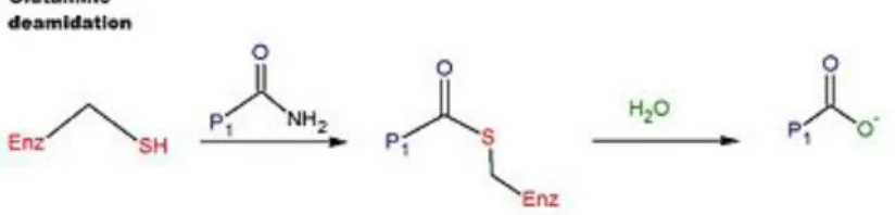
Figure 1. Deamidation reaction catalyzed by tTG ... 5
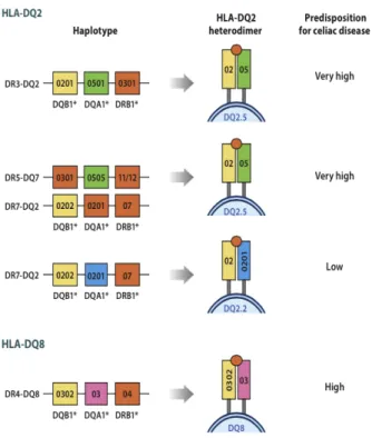
Figure 2. Human leucocyte antigen (HLA) associations in CD ... 7
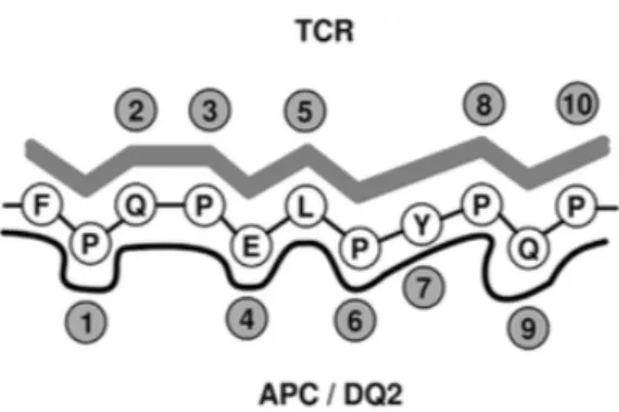
Figure 3. Binding sites for APC and TCR for an epitope of α2 gliadin ... 9
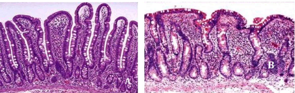
Figure 4. Small intestinal villous (A-‐ Normal; B-‐ Absence of villous due to gluten sensitivity) ... 13
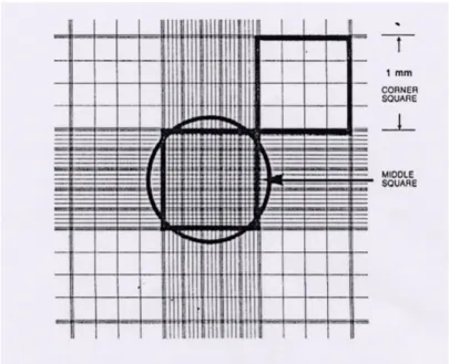
Figure 5. Standard Haemocytometer chamber to count PBMCs and T cells ... 27
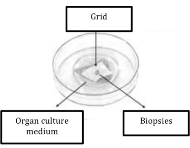
Figure 6. Schematic image of the organ culture dish with grid and biopsies ... 29
L
IST OF
T
ABLES
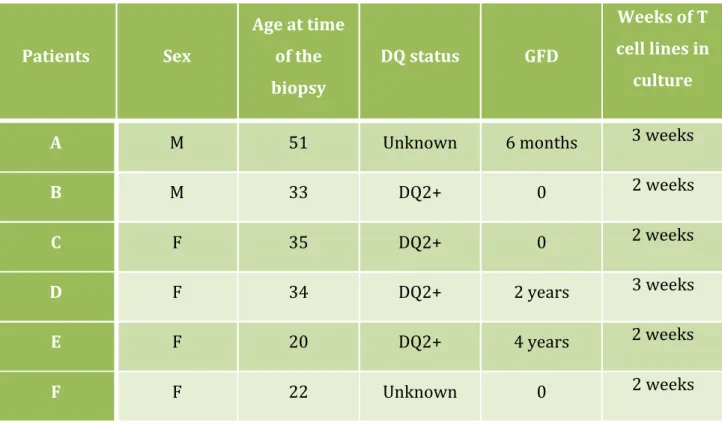
Table 1. General information about the patients used in T cell trial (Sex-‐ M for male and F for female; Age; Time on a GFD; DQ status; Time of T cells in culture) ... 19
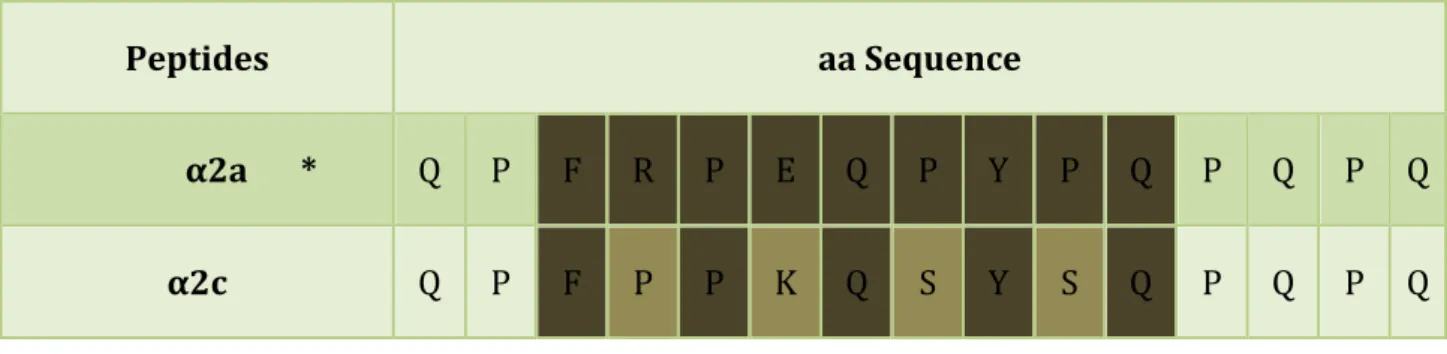
Table 2. Peptides’ labels and their amino acid (aa) sequences. Amino acid substitutions in known immunogenic peptides are marked in red ... 34
Table 3. Amino acid comparison sequence between α2a peptide and its altered version (α2c) and the epitope they harbour ... 35
Table 4. Amino acid comparison sequence between α3I peptide and its altered version (α3II) and the epitope they harbour ... 35
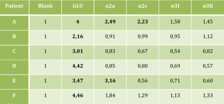
Table 5. Stimulation indexes values from the proliferation assays of 6 gluten-‐specific T cell lines (A, B, C, D, E and F) tested with blank (medium only), gluten (GLU) and four different α-‐gliadin peptides (α2a, α2c, α3I and α3II). Positive SI (SI≥2) are marked in bold. ... 38
L
IST OF
A
BBREVIATIONS
%-‐ Percentage > -‐ Higher < -‐ Lower ºC-‐ Celsius μCi-‐ Micro Curie μm-‐ Micrometre μL-‐ Microliter g-‐ Gram L-‐ Litter
mCi-‐ Milli Curie mL-‐ Millilitre mg-‐ Milligram M-‐ Molar α-‐ Alpha β-‐ Beta γ-‐ Gamma ω-‐ Omega δ-‐ Delta 3H-‐ Tritiated thymidine A
aa-‐ Amino acid ALH-‐Antigénio Leucocitário Humano APC-‐ Antigen Presenting Cells
APL-‐ Altered Peptide Ligand
ASM-‐ Autologous Serum Medium B BL-‐ Blank C
CaCl2-‐ Calcium Chloride
CD-‐ Coeliac Disease CO2-‐ Carbon Dioxide
Cpm-‐ Counts Per Minute CV-‐Coefficient of Variation D DMSO-‐Dimethyl Sulphoxide E
ECH-‐ Enterocyte Cell Height
EMA-‐ Anti-‐endomysial Antibodies
G
GFD-‐ Gluten Free Diet GLU-‐ Gluten
GWAS-‐ Genome wide association studies Gy-‐ Gray H
HLA-‐ Human Leucocyte Antigen HMWG-‐ High Molecular Weight Glutenin HCl-‐ Hydrogen Chloride I IEL-‐Intra-‐Epithelial Lymphocytes
INF γ-‐ Interferon Gamma IL-‐2-‐ Interleukin 2 L LMWG-‐ Low Molecular Weight Glutenin M MHC-‐Major Histocompatibility Complex N
NaOH-‐ Sodium Hidroxyde NK-‐ Natural Killer
O
O2-‐ Oxygen
OC-‐ Organ Culture
OCM-‐ Organ Culture Medium P PBMC-‐ Peripheral Blood Mononuclear Cell PBS-‐ Phosphate Buffered Saline
Psi-‐ Pound-‐force Per Square Inch
PT Gluten-‐ Peptic-‐Tryptic Digest of Industrial Gluten
R
rpm-‐ Revolutions per minute
RPMI-‐ Rosewell Park Memorial Institute S SD-‐ Standard Deviation
SI-‐ Stimulation Index SIL-‐ Small Intestinal Lymphocytes SNP-‐ Single nucleotide polymorphism T TCR-‐ T Cell Receptor tTG-‐Tissue Transglutaminase
1. I
NTRODUCTION
Coeliac disease (CD) is an inflammatory autoimmune disorder that affects at least 1% of the adult population in many countries. It is characterised by small intestinal damage with loss of absorptive villi and hyperplasia of the crypts, leading to malabsorption. In addition to nutrient deficiencies, prolonged CD is associated with an increased risk for malignancy, especially intestinal T cell lymphoma (Dieterich et al., 1997; Fraser & Ciclitira, 2001; Lichtwark et al., 2014; Nasr et al., 2012). The disease is also known as sprue, coeliac sprue, gluten sensitive enteropathy and gluten intolerance (Ludvigsson et al., 2013).
CD is precipitated and triggered by ingestion and exposure to gluten, a generic term to collectively describe all the cereal storage proteins in wheat, barley and rye, that are toxic for the individual, that is genetically predisposed to CD and that carries genes encoding HLA-‐DQ2 or HLA-‐DQ8 molecules (Dewar et al., 2012; Fraser & Ciclitira, 2001; Ludvigsson et al., 2013; Nasr et al., 2012). While the disease is primarily an intestinal disorder that is histologically characterised by a raised number of intraepithelial lymphocytosis, crypt hyperplasia and villous atrophy, there is an increasing support for a broader concept of the condition being a systemic inflammatory disease (Lichtwark et al., 2014).
1.1
Pathogenesis
1.1.1 Gluten
Some storage proteins found in wheat, barley and rye have been shown to cause intestinal inflammation in patients with CD. In some patients there were also found intolerance to oats, however it is only found in, approximately 50% of individuals with CD (Ciclitira, 2003; Dieterich et al., 1997; Fraser & Ciclitira, 2001; Molberg et al., 1998).
Wheat gluten proteins can be divided into two major groups: the glutenins and the prolamins (Fraser & Ciclitira, 2001). Prolamins, the aqueous alcohol soluble fraction of storage proteins, are one group responsible for triggering the disease (Wieser et al., 2014). Wheat, barley and rye, being closely related, all contain prolamins with a high composition of glutamine and proline, whereas the prolamins of oats and more distantly related cereals contain less glutamine and proline and more alanine and leucine (Fraser & Ciclitira, 2001). Glutenins comprise subunits, which are connected with intramolecular disulphide bonds, to form polymers. They are insoluble in neutral aqueous solution or saline solution but can be extracted with a mixture of aqueous alcohol (such as 60% ethanol or 50% propanol) plus reducing agents (such as dithiothreitol) and disaggregating compounds (such as urea)
(Wieser et al., 2014). The high levels of peptides from each of these groups in proline (10-‐ 15%) and glutamine (30-‐35%) residues contributes to their resistance against proteolysis in human gut, allowing them to reach the whole of the small intestine and crossing the epithelial barrier into the lamina propria, that lies bellow the epithelial barrier (Shan et al., 2002).
Depending on the cereal, prolamins may be called gliadins from wheat, hordeins from barley, secalins from rye and avenins from oats (Bai et al., 2013). The gliadins may be subdivided according to their relative electrophoretic mobility into α, β, γ and ω sub fractions; or according to their N-‐terminal amino acid (aa) sequence into α, β or ω sub fractions (Fraser & Ciclitira, 2001; Tighe & Ciclitira, 1995). CD toxicity has been shown to reside with all four classes of gliadins, although α-‐gliadins appear to have the greatest in vivo effects. Glutenins can be divided into two groups, high molecular weight (HMWG) and low molecular weight (LMWG). The HMWG contains proteins that are important for the baking quality of the flour (Bai et al., 2013). The toxicity of this fraction was, previously, very hard to evaluate since the extraction and purification of glutenins was hard to achieve (Dewar et al., 2006).
A total of thirty-‐four T cell epitopes derived from both of these protein groups have been identified and tested as toxic for CD patients, at this time. The majority of these epitopes are associated with binding to HLA-‐DQ2.5 followed by HLA-‐DQ8. The peptides tested in this work contain two different T cell epitopes previously determined to be CD toxic, the DQ2.5-‐ glia α3 (FRPEQPYPQ) and the DQ8-‐glia α1 (EGSFQPSQE) (Sollid et al., 2012). Vader et al., (2002) were the first research group to demonstrate that the epitope DQ2.5-‐glia α3 (ie Glia α20 according to the previous nomenclature) was toxic for CD patients. Their experiments were undertaken with children diagnosed with CD from whom gluten specific T cell lines and clones were generated. They were able to characterize different new and toxic T cell epitopes, and the DQ2.5-‐glia α3 was among them (Vader et al., 2002). Van de Wal et. al discovered the DQ8-‐glia α1 epitope earlier in 1998. In this case only one patient, that expressed both HLA-‐ DQ2 and DQ8, was used from whom gluten specific T cell clones were generated. With this study they were able to identify and demonstrate that gluten sensitive T cell epitopes are not located only in the N-‐terminal of gliadins but also in the C-‐terminal, since the epitopes discovered with this research are all located in that region (van de Wal et al., 1998)
Besides these two epitopes, point nucleotide alterations in the core of each of them were made in order to detect potential lack of CD toxicity. This approach is based in previous experiments that showed that alteration of certain aa at particular positions could decrease and even eliminate T cell responses (Ellis et al., 2003; Japelj, 2013; Mitea et al., 2010). For example, the study made by Japelj in 2013, showed that point substitutions of a proline to
serine residue at position 67 of α2-‐gliadin, decreased the T cell stimulation and, when more than one substitution was introduced to the core of that particular epitope, changing the key aas responsible for T cell receptor (TCR) binding, abrogated T cell responses.
1.1.2 Immune responses
Immune activation occurs after the ingestion of CD toxic cereals, when CD toxic peptides are presented, in connection with the major histocompatibility complex (MHC) class II molecules, HLA-‐DQ2/8 (Fraser & Ciclitira, 2001).
1.1.2.1 Adaptive response
The adaptive immune response plays a central role in the pathogenesis of CD. It provides a link between the main genetic factors and gluten (Wieser et al., 2014).
Tranglutaminases are one of the contributors for the aberrant adaptive immune response. They are enzymes that catalyse the acyl transfer of a glutamine side chain to a primary amine (Fig.1). Tissue transglutaminase (tTG), or transglutaminase 2, is a ubiquitous cytoplasmic enzyme, which is found mainly in respiratory and gut epithelial cells. It’s a monomeric protein comprise 687 aa composed by four different domains (Wieser et al., 2014).
tTG plays a critical role in biological processes and it is important in the prevention of tissue damage, by catalysing protein cross-‐linkage, causing formation of iso-‐peptide bonds between glutamine and lysine residues. If the pH is low (pH<7, can occur when there is an inflammation), or there are no primary lysine residues available, tTG catalyses deamidation of proteins with glutamine residues to glutamic acid, making it negatively charged (Fig. 1) (Fraser & Ciclitira, 2001; Koning et al., 2005; Molberg et al., 1998; Tjon et al., 2010).
The enzyme is not a requirement for T cell stimulation but deamidation of glutamine residues to glutamic acid (Fig. 1), with negatively charged aas, favours the binding to the DQ molecules (Fraser & Ciclitira, 2001; Wieser et al., 2014).
Activation of T cells leads to interferon-‐gamma (INF-‐γ) production and, consequently, higher presentation of gluten peptides to gluten sensitive T cells (Koning
et al., 2005; Tjon et al., 2010). The final result is a significant T cell response, with more
INF-‐γ inducing tissue damage and increasing release of tTG (Koning et al., 2005; Tjon
et al., 2010).
1.1.2.2 Innate response
The innate immune response collaborates with the adaptive response to induce a pro-‐inflammatory Th1 response, to increase the number of intra-‐epithelial lymphocytes (IELs). This favours a cytolytic attack on the epithelium. Certain gluten epitopes are not recognized by the adaptive immune system but are able to activate an innate response. However, the mechanisms involved remain unknown (Wieser et al., 2014).
An increase in IEls is one of the main features of CD. They represent an abundant and heterogeneous population of T cells that reside between the intestinal epithelial cells at the basolateral side of the epithelium. Their most important role is to promote immune protection by preventing the entry and spread of pathogens and avoiding excessive inflammatory reactions that can damage the intestinal epithelium (Wieser et al., 2014). They are composed of the antigen experience of memory effector T cell subtype CD8+ and natural killer (NK) cells (Wieser et al., 2014). Even though, IELs have regulatory functions that contribute to the repair and healing of the epithelium, they also contribute to inflammatory and tissue destructive reactions such as the ones occuring in CD. They are the main producer of INF-‐γ in active CD. They are enriched in cytolytic proteins where their expression is associated with increased epithelial apoptosis (Wieser et al., 2014). A count of 20-‐25 IELs per 100 enterocytes is
Figure 1. Deamidation reaction catalysed by tTG (Wikipédia. Tissue Transglutaminase)
estimated to be the borderline between normal and CD-‐damaged biopsies, with <20 being generally accepted as normal (Wieser et al., 2014).
1.1.3 Genetic factors
Susceptibility to gluten sensitivity appears to be, at least in part, genetically determined. It is known that HLA-‐DQ2/8 explains 40% of hereditability to CD; the 60% remaining is explained by an unknown number of non-‐HLA genes. The incidence of CD in first degree relatives of an affected individual, has been estimated at 10%-‐15%; while monozygotic twin data indicate a 75%-‐80% disease concordance, and 30%-‐50% concordance in HLA identified CD-‐ affected siblings (Wieser et al., 2014).
1.1.3.1 HLA genes
The influence of HLA genes have been well characterised both in family and population studies. The predisposition to the disease is closely associated with the inheritance of two alleles at the HLA-‐DQ loci that encode for the α and β chains of a specific HLA class II molecules. These alleles are found in >90% of individuals with CD (Tighe & Ciclitira, 1995). These HLA-‐DQ alleles are located at the loci within the MHC on chromosome 6 (Wieser et al., 2014). They are usually inherited together with alleles occurring at neighbouring loci on what is termed an extended haplotype. This common inheritance of several alleles has created a difficulty in the identification of the primary susceptibility alleles (Tighe & Ciclitira, 1995).
All patients with CD have been found to express either HLA-‐DQ2 or HLA-‐DQ8 class II molecules. HLA class II molecules are glycoproteins, with α and β chains, located on the surface of cells membranes of antigen presenting cells (APC). They are responsible for biding exogenous proteins and presenting them to CD4+ T cells (Bergseng, 2007; Ciclitira, 2003; Wieser et al., 2014). The large majority of CD patients are DQ2 positive (90-‐95%); the remainder are DQ8 positive. Two common DQ2 isoforms have been found: DQ2.5 and DQ2.2; most DQ2 patients have the DQ2.5 isoform which is encoded by DQA1*05 (α-‐chain) and DQB1*0501 (β-‐chain). The DQ2.2 heterodimer is encoded by the DQA1*0201 and the DQB1*0202 alleles. The DQ8
heterodimer is formed by α and β chains encoded by DQA1*03 and DQB1*0302, respectively (Kooy-‐Winkelaar et al., 2011).
The HLA-‐DQ2.5 genotype is associated with a high risk for CD, followed by DQ8 and DQ2.2. The different between the risk of DQ2.5 and DQ2.2 correlates with the different ability of these HLA molecules to form stable complexes with many gluten peptides (Fallang et al., 2009).
1.1.3.2 Non-‐HLA genes
Most individuals who express DQ2 or DQ8 never develop CD; therefore, even thought they are necessary they are insufficient for the development of CD. A number of new loci, including many immunological candidates have been identified. Fourty genomic regions harbouring more than sixty candidate genes have been described (Hunt et al., 2008; Tjon et al., 2010; Trynka et al., 2010). Most of these loci contain immune related genes (Gutierrez-‐Achury et al., 2011).
Most non-‐HLA genes related to CD are shared with other immune-‐related diseases such as type 1 diabetes and autoimmune thyroiditis. Genome-‐wide association studies (GWAS) have started to uncover genetic components contributing
Figure 2. Human leukocyte antigen (HLA) associations in CD (Wieser et al., 2014)
to CD but the challenge is to find the primary target of the genetic association to uncover the functional consequences of the true causal risk variant (Kumar et al., 2012; Wieser et al., 2014)
1.1.4 Peptide binding
1.1.4.1 HLA DQ2/8-‐peptide binding
HLA molecules have a characteristic-‐binding groove, which differs in size, shape and position between HLA class II alleles, and which can be used to predict the sequence of peptides needed to fit into it (Bergseng, 2007; Ciclitira, 2003; Fraser & Ciclitira, 2001). As mentioned before, HLA-‐DQ2 and HLA-‐DQ8 play a key role in the pathogenesis of the disease by presenting peptides to antigen specific T cells, which promulgate the observed inflammatory response (Ciclitira, 2003; Fraser et al., 2003; Tighe & Ciclitira, 1995).
The combined α and β chains bind immunogenic gluten epitopes in a ‘peptide-‐ groove’ and present them to T cells in the lamina propria. The N-‐terminal domains of the heterodimers combine to form the groove that has a five-‐turn α-‐helix from the α-‐ chain that runs parallel to a longer but kinked α-‐helix from the β-‐chain forming the side wall of the groove (Kim et al., 2004). In this groove, antigen peptide is bound for presentation to the TCR. Within this groove, pockets are sited to accept amino acid side chains of the antigen peptide to enable binding (Ciclitira, 2003). Alterations in the aa sequence of peptides by aa substitutions within this biding groove, predominantly at the location of these pockets, can affect binding affinity to a particular HLA molecule (Ciclitira, 2003; Tighe & Ciclitira, 1995).
The peptide binding of HLA-‐DQ2.5 and DQ2.2 are similar but DQ2.5 has the capacity to hold immunogenic peptides for much longer compared to DQ2.2. This fact also explains the higher disease risk associated with HLA-‐DQ2.5 (Fallang et al., 2009; van de Wal et al., 1997). As in HLA-‐DQ2, the HLA-‐DQ8 favours the binding of negatively charged peptides. In additon functional binding studies suggest anchor positions P1 and P9 for glutamic acid and P4 for hydrophobic residues. In contrast to DQ2, that requires only one glutamic acid residue, DQ8 requires two (Wieser et al., 2014). The glutamate introduced by tTG is usually in positions P4, P6 or P7 in HLA-‐DQ2.5 restricted epitopes and, as mentioned, at position P1 and/or P9 in HLA-‐DQ8 restricted
epitopes. These glutamatic acid residues serve as anchor residues important for binding of the peptides and both HLA-‐DQ2.5 and DQ8 prefer negatively charged residues at these anchors sites. This positioning of deamidated glutamine residues is strongly related to the positioning of proline residues, which is particularly strict in the case of DQ2.5 epitopes, as DQ2.5 only accepts proline at certain position in the peptide-‐ binding groove (Kim et al., 2004). This results in a dominant presence of proline at P1, P6 and P8 and leads to the modification by tTG of the glutamine residues at P4 and P6, respectively. Such positioning of proline residues is less strict in the case of the DQ8 epitopes (Sollid et al., 2012).
1.1.4.2 TCR-‐peptide binding
HLA-‐DQ2 and DQ8 heterodimers bind to gluten peptides in their peptide groove and then present them to T cells (Fig. 3) (Wieser et al., 2014). CD4+ T cells express a TCR, which is responsible for binding to the HLA-‐peptide complex. This receptor comprises of a α, β, γ or δ chains than contain three domains: one extracellular Ig-‐like, a trans membrane and a cytoplasmic tail (Bergseng, 2007). TCRs possess unique antigen specificities determined by the structures of the antigen-‐biding site formed by the α and β chains, and which are activated by contact with a peptide/MHC complex. APCs that have bound the antigens in the intestinal tissue can travel to the mesenteric lymph node, where antigen presentation and priming of naïve CD+ T cells take place (Qiao et al., 2012; Wieser et al., 2014).
Figure 3. Binding sites of APC and TCR for an epitope of α-‐2 gliadin (Wieser et al., 2012)
Following activation, gluten sensitive CD4+ T cells migrate to the lamina propria. In the tissue, primed CD4+ T cells are either reactivated by local resident APCs presenting gluten peptides or remain dormant as memory T cells (Wieser et al., 2014).
1.1.5 Methods for investigating coeliac disease
There are a variety of different methods, including in vivo and in vitro methodology, that help investigate the pathogenesis of CD.
1.1.5.1 In vitro methods
1.1.5.1.1 Organ Culture of intestinal tissue
The most reliable of the in vitro methods is the organ culture of small intestinal tissue obtained from CD patients. This tissue samples are taken from the small intestine and incubated with culture medium under special conditions at 37°C. With this method the biopsies survive up to 24 hours. It is used to evaluate peptide toxicity for CD. The biopsies are incubated with gluten and gluten fractions; then histological changes of the biopsies are evaluated after the incubation period (Browning & Trier, 1969; Ciclitira et al., 2005; Lindfors et
al., 2012).
One of the benefits of this method is the fact that various features characteristic for CD can be reproduced in biopsies from treated patients, thus allowing researchers to find mechanisms related to development of CD (Falchuk
et al., 1972). Because intestinal biopsies contain enterocytes and lamina
propria, this model is useful in determining both innate and adaptive responses. The main disadvantage of the method is that it is not a high-‐throughput method and the tissue lacks circulation, a nervous system and connection to lymphatic organs (Wieser et al., 2014).
1.1.5.1.2 T cell lines and clones
T cell lines and clones of the small intestinal mucosa have been used to measure immunogenic effects of proteins and peptides and to test potential novel treatments (Wieser et al., 2014). CD4+ T cells play the most important role in the adaptive immune response in CD pathology. To isolate the T cells, biopsies from the small intestinal are first incubated in the presence of an antigen toxic (gluten fractions or gluten peptides) for CD patients. Multiple restimulations are required to cultivate gluten sensitive T cells in vitro (Lindfors
et al., 2012; Wieser & Koehler, 2008).
Following activation with the antigen, interleukin 2 (IL-‐2) receptor (CD25) increases in number. If IL-‐2 is later added as stimulant, only lines that are specific for the antigen will grow. Irradiated peripheral blood mononuclear cells (PBMCs) are used as APC, helping gluten-‐sensitive T cells to proliferate (Lindfors et al., 2012; Wieser & Koehler, 2008). Proliferation of T cells in culture when stimulated with antigen can be determined in T cell proliferation assays. In this assay, T cells are incubated with the antigen, APCs and tritiated thymidine (3H). The 3H is incorporated into the nuclei of the cells that are
dividing and this amount is then measured by a scintillation counter (Lindfors
et al., 2012; Wieser & Koehler, 2008).
T cells tests have been used widely to compare the level of immunogenic effects and they are the first approach used when testing toxic epitopes in CD. However, immunogenicity in T cells do not always correspond to the CD toxicity demonstrated in vivo or in OC tests, that is why there is always the need to apply other methods, to confirm the results obtained with T cells (Hooper et al., 2012; Maynard et al., 2012).
1.1.5.2 In vivo methods
Small intestinal in vivo gluten challenge is used for purposes of evaluating CD toxicity and help diagnosing patients and it can be a short-‐term challenge or a long-‐term challenge. The problem with in vivo studies and methods is that they can be highly invasive and very time consuming (Ciclitira et al., 2005).