MARCO ANDRÉ MIRANDA GALÉSIO
NEW ANALYTICAL METHODOLOGIES FOR
DOPING CONTROL
DETECTION OF ANABOLIC ANDROGENIC STEROIDS IN HUMAN URINE
MARCO ANDRÉ MIRANDA GALÉSIO
NEW ANALYTICAL METHODOLOGIES FOR
DOPING CONTROL
DETECTION OF ANABOLIC ANDROGENIC STEROIDS IN HUMAN URINE
Dissertação apresentada para obtenção
do Grau de Doutor em
(Bioquímica-Biotecnologia) pela Universidade Nova de
Lisboa, Faculdade de Ciências e
Tecnologia.
Title: New Analytical Methodologies for Doping Control – Detection of Anabolic Androgenic Steroids in Human Urine
Author: Marco André Miranda Galésio
Dissertation submitted to Faculdade de Ciências e Tecnologia - Universidade Nova de Lisboa in fulfilment of the requirements for the degree of Doctor of Philosophy (Biochemistry - Biotechnology) by Marco André Miranda Galésio
Dissertation defended on February 9th, 2011.
The dissertation was approved by unanimity by the following members of the Final Oral Committee:
• Carlos Lodeiro; Professor at Faculdade de Ciências de Ourense - Vigo University, Spain • Florentino Riverola; Professor at Faculdade de Ciências de Ourense - Vigo University, Spain • Francesco Botrè; Scientific Director at WADA Anti-doping Laboratory of Rome, Italy • José Luís Capelo; Professor at Faculdade de Ciências de Ourense - Vigo University, Spain
• José Moura; Full Professor at Faculdade de Ciências e Tecnologia - Universidade Nova de Lisboa, Portugal
• Mário Diniz; Assistant Researcher at Faculdade de Ciências e Tecnologia - Universidade Nova de Lisboa, Portugal
• Xavier de la Torre; Vice-Scientific Director at WADA Anti-doping Laboratory of Rome, Italy
ISBN:
i
ACKNOWLEDGEMENTS
First and foremost I offer my sincerest appreciation to my supervisor, Prof. Dr. José Luis Capelo, who has supported me throughout my PhD. thesis with his knowledge, advices and friendship whilst allowing me to carry out my own scientific choices.
Many thanks to my co-supervisors, Prof. Dr. Jesus Símal and Prof. Dr. Raquel Rial, for all the support given during the last years. A special word to Raquel, whose indispensable help was vital to the success of the developed work.
I am also deeply thankful to Prof. Dr. Francesco Botrè and Prof. Dr. Xavier de la Torre that made my four months stay in Laboratorio Anti-doping di Roma, one of the greatest experiences of this journey. I thank you for all your support, for having shared your knowledge with me and for your friendship.
I acknowledge “Fundação para a Ciência e a Tecnologia”, for financial support through the grant SFRH/BD/31652/2006, and all the institutions involved in my PhD. thesis, in particular, Faculdade de Ciências from Universidade Nova de Lisboa, Universidad de Vigo and Laboratorio anti-doping di Roma.
I thank the SING research group, particularly Prof. Florentino Fernández Riverola, for all their support in the development of MLibrary software.
I am truly thankful to all people in Laboratorio anti-doping di Roma for all their sympathy and support. I am glad to think of most of them as my friends.
Thank you Simone and Alessandra for making me feel like home while in Rome. Thank you Daniele and Fabrizio for your great meals. Great Roberto, come sta il tuo ginocchio??
I thank all BIOCOPE GROUP collegues, in particular, Bruno, Elisabete, Gonçalo, Hugo, Luz and Ricardo. A special word to Gonçalo and Ricardo for their friendship and great fellowship that made this a unique journey, full of joy.
To Prof. Dr. Carlos Lodeiro for all his support and friendship.
ii
I thank to all my family and friends that are the best anyone could ask for. To my parents and brother for their unlimited care. To my parents-in-law and “little sister” for their support.
iii
ABSTRACT
The use of anabolic androgenic steroids (AAS) and other banned substances to enhance athletic performance has important health and social implications. The AAS are a major group included in the prohibited list of the world anti-doping agency (WADA) as well as of major sports authorities. This class of drugs, along with other anabolic agents, represent 64,9 % of all adverse analytical findings reported by WADA accredited laboratories, as stated in the WADA statistic report for 2009. The AAS are a class of hormones that include the natural male sex hormone, testosterone, and its many synthetic derivatives. They exert multiple actions affecting both the physiology of the human body and the individual behaviour. Under intensive training, the AAS induce the synthesis of proteins in muscle and bone causing an accelerated growth of these organs. Furthermore, during acute endurance workout, as well as during competition, androgen’s action seems to be critical to enhance the performance capacity, since they affect the production of red blood cells and increase neural conduction. In addition, after intense exercises, androgens are thought to prevent muscle catabolism and exhaustion and to speed up the recovery process.
In general, the normal proceeding for AAS determination includes chromatographic separation coupled to mass spectrometry detectors. The use of GC-MS methodologies is the most employed strategy for AAS control. However, over the last years, with the development of suitable LC-MS and LC-MS/MS systems, some AAS presenting poor chromatographic properties for GC-MS analysis, even after derivatisation, are being analysed by LC-MS(/MS) procedures.
The aim of the research programme presented in this thesis was, primarily, the development of a new screening method based on mass spectrometry (MS) using the soft ionisation technique matrix-assisted laser desorption/ionisation (MALDI). The major goals to be achieved were the development of an accurate, sensitive and robust methodology able to improve the screening of AAS for doping control in both analysis time and sample throughput. Additionally, the developed method should be capable to overcome the GC-MS limitation related to thermo-labile and polar AAS, so that the initial screening method could be extended to all AAS included in the prohibited list.
In parallel with the development of a screening procedure based on MALDI-MS(/MS) techniques, and applying the deep expertise of the research group on reaction enhancement by delivery energy based techniques, the improvement of the global sample preparation for the analysis of AAS by anti-doping control laboratories was also included in the research programme.
iv
Chapter II deals with the analysis of anabolic androgenic steroids by the analytical technique of MALDI-TOF-MS. In this study, the applicability of a variety of matrices for MALDI analysis was tested for 15 compounds. Nine organic and two inorganic matrices were evaluated in order to determine the best matrix for the identification of steroids in terms of ionisation yield and interference by characteristic matrix ions. The best results were achieved for the organic matrices 2-(4-hydroxyphenylazo) benzoic acid (HABA) and trans-3- indoleacrylic acid (IAA). Good signals were obtained for concentrations as low as 0.010 and 0.050 µg / mL on the MALDI sample plate for the HABA and IAA matrices, respectively.
For these two matrices, the sensitivity achieved by the technique of MALDI-TOF-MS is comparable to the sensitivity shown by GC-MS. Additionally, the accuracy and precision of the mass spectra obtained by MALDI-TOF-MS was very good, when compared to the theoretical masses of the molecular ions. The experimental methodology developed was applied to urine samples spiked with AASs. The results obtained present a good indicator concerning the use of MALDI strategies for the rapid screening of anabolic steroids in doping analysis in a near future. These results are particularly relevant to the analysis of exogenous steroids, in which its mere presence is a case of doping.
In Chapter III, a novel database search engine - Mlibrary - to assist the detection and identification of anabolic androgenic steroids, by the analytical technique of MALDI-TOF-TOF-MS, was developed in collaboration with the SING - Next Generation Computer Systems - group. The detection and identification of banned substances is achieved in two steps. In the first step, the developed software identifies the presence of a possible positive in the mass spectrum against a database that contains the theoretical mass of the molecular ions of AAS. In this operation the software includes possible changes that may have occurred to the AAS during the sample treatment. After the identification of possible positives and subsequent analysis of those ions by tandem mass spectrometry, in the second step, the mass spectrum obtained, after fragmentation of the molecular ion, is compared with a library of mass spectra contained in the database. The mass spectra library is easily constructed and introduced into the system by the user. With the help of the MLybrary software application, the use of MALDI techniques for doping control is simplified and the time required for data analysis is reduced. Moreover, by applying simple statistical tests, the software facilitates the interpretation of the results.
v
Chapter IV describes the development and optimization of a rapid sample treatment methodology for the analysis of anabolic androgenic steroids in urine by GC-MS. The new procedure makes use of ultrasonic energy to reduce the reaction times and increase the overall sensitivity of the current methodology used to analyse these compounds in WADA accredited laboratories. Ultrasonic energy was applied to the key steps of the methodology, which include the enzymatic hydrolysis with the enzyme β-glucuronidase from Escherichia coli K12 of the steroids excreted in urine as conjugated compounds, and subsequent derivatisation of the free compound to improve the analytical characteristics for analysis by GC-MS. The results indicate that, under an ultrasonic field, it was possible to reduce the hydrolysis time for 10 minutes, about six times lower than the reaction time normally required. After the enzymatic hydrolysis, the derivatisation procedure of the compounds with the trimethylsilyl (TMS) reagent, methyl-N-trimethylsilyltrifluoroacetamide (MSTFA)/NH4I/dithioerythritol (DTE) (1000:2:4, v/w/w) was also accelerated with the application of ultrasonic energy. The results demonstrated that after 3 min of ultrasonication, 19 of the 35 compounds studied showed similar reaction yield to those obtained with the classic procedure that last for 30 min; 13 increased to higher silylation yields and for the steroids 1-testosterone, danazol and etiocholanolone-D5, the same results were obtained using a sonication time of 5 min. The overall applicability of the ultrasonic-based sample treatment method was tested by the analysis of five urine samples. The results were similar to those obtained by the routine procedure and are an indication that this methodology provides robustness to be introduced in routine analysis.In Chapter V the use of microwave energy to enhance the key steps mentioned in Chapter IV was evaluated and compared with the ultrasonic energy based methodology. The study covers the current methodology applied in anti-doping laboratories for the analysis of anabolic steroids, as well as other banned substances in urine, by GC-MS. The results indicate that the effect of microwave energy in the enzymatic hydrolysis step, with the enzyme β-glucuronidase from Escherichia coli K12, has no advantage over the conventional method. Regarding the reaction of derivatisation, the application of microwave energy enabled us to accelerate the reaction to 3 minutes and, more important, increased the yield of the reaction for 18 of the 55 compounds used in this study. When compared to the ultrasonic method, the microwave method shows some fragility, particularly, at the enzymatic reaction. Regarding the reaction of derivatisation, the results obtained with the two irradiation systems were similar. Nevertheless, the microwave reactor allows higher sample throughput.
vi
concerns the values of the initial velocity, which is higher when ultrasound waves are employed. All other kinetic parameters have similar values. Moreover, when ultrasound waves are applied to the reaction medium, denaturation of the enzyme occurs shortly after the start of the reaction. These results support the idea that the use of ultrasound waves to accelerate enzymatic reactions has great impact in the starting minutes of the reaction.
vii
RESUMO
O programa de investigação abrangido pela presente tese teve como principal objectivo o desenvolvimento de um método de rastreio para a detecção de esteróides androgénicos anabolizantes com base em técnicas de espectrometria de massa, em particular a técnica de Matrix Assisted Laser Desorption/Ionisation (MALDI) acoplada a um espectrómetro de massas de tempo de voo (TOF, do inglês time-of-flight). Os principais objectivos a alcançar eram o desenvolvimento de uma metodologia analiticamente sensível, robusta e precisa, capaz de melhorar, ao nível do tempo da análise e capacidade de processamento de amostras, os métodos actuais para o rastreio de esteróides anabolizantes. Os métodos tradicionais usados para o rastreio destes compostos pelos laboratórios de anti-dopagem baseiam-se na técnica de cromatografia gasosa acoplada a espectrometria de massa (GC-MS) e apresentam algumas limitações na análise de compostos termo-lábeis e polares. Consequentemente, o método desenvolvido deveria ser capaz de superar essas limitações de modo a que o rastreio inicial possa ser alargado a todos os esteróides anabolizantes incluídos na lista de compostos proibidos.
Em paralelo com o desenvolvimento de uma nova metodologia de rastreio baseada nas técnicas de MALDI-MS(/MS), parte do programa da tese de doutoramento consistiu no melhoramento dos métodos actuais de tratamento de amostra. Para esse efeito, tivemos como base a larga experiência do nosso grupo de investigação na optimização de reacções por aplicação de técnicas de irradiação com ondas ultra-sónicas e de microondas.
No Capitulo I da tese apresenta-se o estado da arte actual sobre a dopagem no desporto, e as principais técnicas analíticas usadas para a sua detecção
viii
No Capitulo III da tese é apresentado um software desenvolvido em colaboração com o grupo de ciências informáticas SING - Next Generation Computer Systems, para assistir na detecção e identificação de esteróides androgénicos anabolizantes pela técnica analítica de MALDI-TOF-MS. O software desenvolvido permite ao utilizador reduzir o tempo de análise de dados e aplicar testes estatísticos simples que facilitam a interpretação dos resultados.
No Capitulo IV da tese é descrito o desenvolvimento e optimização de uma metodologia rápida de tratamento de amostra, para a análise de esteróides anabolizantes em urina por GC-MS. O novo procedimento baseia-se na aplicação de energia ultra-sónica aos passos chave da metodologia usada correntemente para a análise destes compostos, de modo a reduzir os tempos de reacção e aumentar a sensibilidade geral da análise.
O Capitulo V da tese é a continuação do quarto capítulo e descreve outra abordagem para melhorar a metodologia padrão aplicada nos laboratórios de anti-dopagem para a análise de esteróides anabolizantes e outras substâncias proibidas em urina por GC-MS. O estudo efectuado engloba o desenvolvimento e optimização de uma metodologia rápida de tratamento de amostra com recurso à energia de microondas e a comparação do método desenvolvido com o método de ultra-sons.
No Capitulo VI da tese foi feito um estudo de cinética enzimática para avaliar o efeito dos ultra-sons nos parâmetros cinéticos da reacção enzimática de hidrólise de compostos glucuronidos com o enzima β-glucuronidase. Os parâmetros cinéticos da reacção foram calculados aplicando a equação de Michaelis-Menten aos dados experimentais. Os resultados obtidos mostram que o valor da constante de Michaelis não altera com a aplicação de ultra-sons ao meio reaccional. A principal diferença na cinética da reacção entre o método convencional de incubação a 55ºC e o método de aceleração com os ultra-sons, está relacionada com os valores da velocidade inicial, velocidade máxima e, consequentemente, a constante catalítica, que são superiores quando se aplica ultra-sons.
ix
ABBREVIATIONS
AAS Anabolic androgenic steroids ACN Acetonitrile
ACTH Adrenocorticotropic hormone
AD 4-androsten-3,17-dione APCI Atmospheric pressure chemical ionisation
AR Androgen receptor
AVP Vasopressin BALCO Bay Area Laboratory Co-operative Bol Boldenone
CI Chemical ionisation
CID Collision-induced dissociation
CRH Corticotrophin releasing hormone
CYP Cytochrome P450
DHB Dihydroxybenzoic acid
DHEA Dihydroepiandrosterone DHT 5α-dihydrotestosterone
DMAPP Dimethylallyl pyrophosphate
DNA Deoxyribonucleic acid
DTE Dithioerythritol E Epitestosterone ECD Electron capture dissociation
EI Electron impact
EPO Erythropoietin
ESI Electrospray ionisation
ISTD Internal standard
Etio Etiocholanolone
FA Ferulic acid
FAB Fast atom bombardment
FD Field desorption
FSH Follicle-stimulating hormone
G Glucuronide
GC Gas Chromatography
GDR German Democratic Republic GnRH Gonadotropin releasing hormone
x
HABA 2-(4-hydroxyphenylazo)benzoic acid
HDL High density lipoprotein
hGH Human growth hormone
HPA Hydroxypicolinic acid
HPLC High performance liquid chromatography
HSA Human serum albumin
HSP Heat shock protein
IAA Trans-3-indoleacrylic acid
IAAF International Amateur Athletic Federation
IOC International Olympic Committee
IPP Isopentenyl pyrophosphate
IR Infrared IRMS Isotope ratio mass spectrometry
ISO International Organisation for Standardisation
kcat Catalytic constant
KM Michaelis-Menten constant
LC Liquid chromatography
LDL Low density lipoprotein
LH Luteinizing hormone
LLE Liquid-liquid chromatography
MA 17β-hydroxy-17-methylandrosta-1,4-dien-3-one MALDI Matrix assisted laser desorption/ionisation MeOH Metanol
MS Mass Spectrometry
MSTFA Methyl-N-trimethylsilyltrifluoro acetamide MRPL Minimum required performance limit
MT 17α-methyltestosterone MW Microwave
NADPH Nicotinamide adenine dinucleotide phosphate Nan Nandrolone
PNP 4-nitrophenol
PNP-G 4-nitrophenyl glucuronide
RFC Response factor calibration
RIA Radioimmunoassay
RNA Ribonucleic acid
RSD Relative standard deviation
SA Sinapinic acid
xi
SID Surface-induced dissociation
SIM Select ion monitoring
SIMS Secondary ion mass spectrometry SPE Solid phase extraction
SR Sonoreactor StAR Steroidogenic acute respiratory SULT Sulphotransferases T Testosterone TFA Trifluoroacetic
THAP 2,4,6- trihydroxyacetophenone
TMS Trimethylsilyl TMIS Trimethyliodosilane
TOF Time of flight
TUE Therapeutic Use Exemptions
UGT Uridine diphospho-glucuronosyltransferase
UNESCO United Nations Educational, Scientific and Cultural Organization
UP Ultrasonic probe
US Ultrasound USADA United States Antidoping Agency UV Ultraviolet
Vmax Maximum velocity
xiii
TABLE OF CONTENTS
Acknowledgments i
Abstract iii
Resumo vii
Abbreviations ix
Table of Contents xiii
Index of Tables xx
Index of Figures xxi
PART ONE: GENERAL ASPECTS
1Chapter I 3
I.1 Definition of Doping 3
I.2 Historical background 4
I.2.1 Doping: Origin of the word 4
I.2.2 Early history of Doping 4
I.2.3 Doping and its eminent prohibition 5
I.2.4 Harmonisation of anti-doping policies 8
I.2.5 The World Anti-Doping Agency (WADA) 9
I.3 The role of endogenous and synthetic AAS in the human body 12
I.3.1 Structure 12
I.3.2 Biosynthesis and secretion of androgens 13
I.3.2.1 Biosynthesis of cholesterol 14
I.3.2.2 Steroidogenesis of androgens 17
I.3.3 Androgen metabolism and regulation 19
I.3.3.1 Testicular function regulation 19
I.3.3.2 Androgens: from biosynthesis to the target cells 21
I.3.3.3 Mechanism of androgens action 21
I.3.3.3.1 Testosterone 21
I.3.3.3.2 Androgen receptor 22
I.3.3.4 Metabolism and excretion of AAS 23
I.3.3.4.1 Phase-I metabolic pathways 24
I.3.3.4.2 Phase-II metabolic pathways 25
I.3.3.5 Testosterone in women 26
xiv
I.3.4.1 Endogenous androgens 27
I.3.4.2 Synthetic steroids 28
I.3.5 Physiological effects of AAS under intensive training 29
I.3.6 Behavioural effects of AAS 29
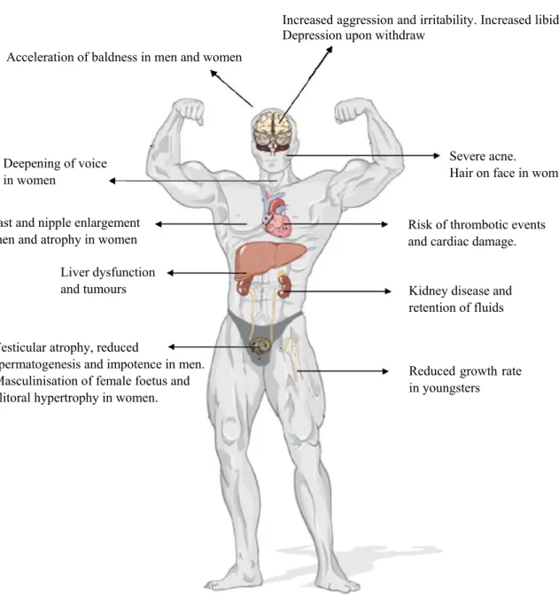
I.3.7 Adverse effects of AAS abuse 30
I.4 Analysis of AAS in doping control 32
I.4.1 Historical background 32
I.4.2 Detection of AAS in doping control 32
I.4.2.1 Analytical techniques 33
I.4.2.1.1 GC-MS 33
I.4.2.1.2 GC-C-IRMS 34
I.4.2.1.3 LC-MS (/MS) 35
I.4.2.2 Sample treatment 36
I.4.2.2.1 Hydrolysis 37
I.4.2.2.2 Extraction and clean-up of AAS from urine 37
I.4.2.2.3 Derivatisation 38
I.5 Other strategies for AAS Doping control 40
I.5.1 Mass spectrometry: MALDI-MS(/MS) strategies 40
I.5.1.1 Basics of mass spectrometry 40
I.5.1.1.1 Modes of ionisation 41
I.5.1.1.2 Mass analysers 43
I.5.1.1.3 Mass detectors 45
I.5.1.1.4 Mass spectrum 45
I.5.1.1.5 Tandem mass spectrometry 46
I.5.1.2 MALDI-TOF-MS 47
I.5.1.2.1 Principle of MALDI 47
I.5.1.2.2 MALDI matrix and sample preparation 48
I.5.1.2.3 Time-of-flight mass analyser 50
I.5.1.2.4 Tandem mass spectrometry 52
I.5.2 Sample preparation: the role of electromagnetic and acoustic irradiation 53
I.5.2.1 Ultrasonic Irradiation 53
I.5.2.1.1 Ultrasound 53
I.5.2.1.2 Sonochemistry 55
I.5.2.1.3 Factors affecting cavitation 55
I.5.2.1.3.1 Frequency 56
I.5.2.1.3.2 Intensity 56
I.5.2.1.3.3 Temperature and pressure 56
xv
I.5.2.1.3.5 Nature of the dissolved gas 57
I.5.2.1.4 Ultrasound source 57
I.5.2.1.5 Applications 58
I.5.2.1.6 Common instrumentation 59
I.5.2.1.6.1 Ultrasonic bath 59
I.5.2.1.6.2 Ultrasonic probe 60
I.5.2.6.1.3 Cup horn 60
I.5.2.2 Microwave Irradiation 61
I.5.2.2.1 Microwave 61
I.5.2.2.2 Thermal effects 61
I.5.2.2.3 Non-thermal effects 63
I.5.2.2.4 Microwave source 63
I.5.2.2.5 Applications 64
I.6 References 65
PART
TWO:
MALDI-MS(/MS) SCREENING METHODOLOGY
75Chapter II – Comparative study of matrices for their use in the rapid
screening of anabolic steroids by MALDI-TOF-MS
77
II.1 Abstract 77
II.2 Introduction 78
II.3 Experimental 79
II.3.1 Apparatus 79
II.3.2 Standards and Reagents 80
II.3.3 Sample preparation 80
II.3.3.1 Standard solutions 80
II.3.3.2 Urine hydrolysis procedure 80
II.3.3.3 SPE clean-up of hydrolysed samples 81
II.3.3.4 Derivatisation step procedure 81
II.3.3.5 SPE clean-up 81
II.3.3.6 MALDI-TOF-MS analysis 81
II.4 Results and Discussion 82
II.4.1 Direct analysis of AAS 82
II.4.2 Analysis of derivatised AAS 83
II.4.3 Influence of the organic matrix 84
II.4.4 Analytical sensitivity 85
II.4.5 Influence of the inorganic matrix 89
xvi
II.5 Conclusion 92
II.6 References 93
II.7 Supplementary material 95
Chapter III – MLibrary: a free software tool for doping screening by
MALDI based strategies
102
III.1 Abstract 102
III.2 Introduction 102
III.3 Experimental 104
III.3.1 Apparatus 104
III.3.2 Standards and Reagents 104
III.3.3 Sample preparation 105
III.3.3.1 Standard solutions 105
III.3.3.2 Urine hydrolysis procedure 105
III.3.3.3 Liquid-liquid extraction of target analytes 105
III.3.3.4 Derivatisation step procedure 106
III.3.3.5 SPE clean-up 106
III.3.3.6 MALDI-TOF-MS and MALDI-TOF-TOF-MS analysis 106
III.3.3.7 MLibrary software 107
III.4 Results and Discussion 108
III.4.1 MS Mode 108
III.4.1.1 Construction of MS database 108
III.4.1.2 Detecting the presence of possible AAS 108
III.4.2 MS/MS mode 111
III.4.2.1 Construction of MS/MS mode 111
III.4.2.2 Confirming the presence of AASs 112
III.4.3 Case study 115
III.5 Conclusion 117
III.6 References 119
PART
THREE:
IMPROVING SAMPLE TREATMENT
121Chapter IV – Improved ultrasonic based sample treatment for the
screening of anabolic steroids by GC-MS
123
IV.1 Abstract 123
IV.2 Introduction 124
xvii
IV.3.1 Apparatus 125
IV.3.2 Standards and Reagents 125
IV.3.3 GC-MS instrumentation and operating conditions 127
IV.3.3.1 GC-MS system and parameters used for the optimisation study 127 IV.3.3.2 GC-MS system and parameters used for the validation study 127
IV.3.4 Urine Samples 128
IV.3.5 Sample preparation 128
IV.3.5.1 Urine hydrolysis procedure 128
IV.3.5.2 SPE clean-up of urine samples 128
IV.3.5.3 Derivatisation procedure 130
IV.3.6 Experimental design 130
IV.3.6.1 Optimisation procedure 130
IV.3.6.2 Assessment of the ultrasonic procedure 131
IV.3.6.3 Application to real samples 131
IV.3.7 Statistical analysis 131
IV.4 Results and Discussion 131
IV.4.1 Effect of ultrasonication on the enhancement of the enzymatic hydrolysis 132
IV.4.1.1 Ultrasonic probe 133
IV.4.1.2 Sonoreactor 134
IV.4.1.3 Application to further steroids 134
IV.4.2 Effect of ultrasonication in the enhancement of derivatisation procedure 136
IV.4.2.1 Ultrasonic bath 137
IV.4.2.1.1 Effect of ultrasonication combined with temperature 137
IV.4.2.1.2 Effect of frequency 137
IV.4.2.2 Sonoreactor 138
IV.4.2.3 Application to further steroids 138
IV.4.3 Analytical application 139
IV.5 Conclusion 143
IV.6 References 144
Chapter V – Accelerated sample treatment for the screening of banned
doping substances by GC-MS: ultrasonication vs microwave energy
146
V.1 Abstract 146
V.2 Introduction 147
V.3 Experimental 149
V.3.1 Apparatus 149
xviii
V.3.3 Sample preparation 150
V.3.3.1 Urine hydrolysis procedure 150
V.3.3.2 Liquid-liquid extraction of target analytes 151
V.3.3.3 Derivatisation procedure 151
V.3.4 GC-MS instrumentation and operating conditions 151
V.3.5 Urine Samples 153
V.3.6 Experimental design 154
V.3.6.1 Optimisation of hydrolysis procedure 154
V.3.6.2 Optimisation of derivatisation procedure 155
V.3.6.3 Application to real samples 155
V.3.7 Statistical analysis 156
V.4 Results and Discussion 156
V.4.1 Effect of ultrasonic and microwave energy in the enhancement of enzymatic hydrolysis reaction
156
V.4.1.1 Effect of microwave energy 156
V.4.1.2 Effect of ultrasonic energy 159
V.4.1.3 Comparison between microwave and ultrasonic energy 159 V.4.2 Effect of ultrasonic and microwave energy in the enhancement of derivatisation
reaction
161
V.4.2.1 Effect of microwave energy 161
V.4.2.2 Effect of ultrasonic energy 164
V.4.2.3 Comparison between microwave and ultrasonic energy 164
V.4.3 Analytical application 167
V.5 Conclusion 167
V.6 References 169
Chapter VI – Unravelling the role of ultrasonic energy in the enhancement
of enzymatic kinetic
171
VI.1 Abstract 171
VI.2 Introduction 172
VI.3 Experimental 173
VI.3.1 Apparatus 173
VI.3.2 Standards and Reagents 174
VI.3.3 Chromatographic system and operating conditions 174
VI.3.4 Sample treatment 176
VI.4 Results and Discussion 176
xix
VI.4.2 β-glucuronidase activity/ stability under ultrasonic irradiation 178VI.4.3 β-glucuronidase kinetic parameters 180
VI.5 Conclusion 184
VI.6 References 185
PART FOUR: GENERAL DISCUSSION AND CONCLUSIONS
187Chapter VII 189
VII.1 General discussion and conclusions 189
xx
INDEX OF TABLES
Table I.1 Major androgen target tissues. 22
Table I.2 Common mass analysers. 44
Table II.1 Matrix concentration and matrix solution used to prepare the organic and inorganic matrices tested in this work.
82
Table II.2 Molecular weights of the free steroids and MALDI-TOF-MS ions after steroids derivatisation with Girard T hydrazine.
84
Table II.3 Concentration of the matrix HABA after dilution. 88
Table IV.1 Diagnostic ions (m/z) used in SIM mode for AAS detection. 129
Table IV.2 Hydrolysis yields (US procedure/ T conductivity×100) of AAS glucuronides from urine. Comparison of the thermal conductivity procedure (55ºC; 1 hour) and the ultrasonic procedure (sonoreactor at 50% of amplitude, 10 min).
136
Table IV.3 Derivatisation yield (US procedure/ T conductivity×100) of AAS with MSTFA. Comparison of the thermal conductivity procedure (78ºC; 30 min) and the ultrasonic procedure (sonoreactor at 50% of amplitude, 3min).
140
Table IV.4 Comparison of the classic and the ultrasonic procedures. The recoveries are calculated as the ratio yield between the ultrasonic method and the classic method and are expressed as percentages.
141
Table V.1 Diagnostic ions (m/z) used in SIM mode for detection of banned substances. 152 Table V.2 Hydrolysis yield of AAS glucuronides from urine. Comparison of the thermal
conductivity procedure (1hour at 55ºC) with the ultrasonic procedure (60% of amplitude, 10min) and the microwave procedure (240 Watts of power, 10min).
165
Table V.3 Derivatisation yield of TMS derivatives formation with MSTFA. Comparison of the thermal conductivity procedure (30 min at 78 °C) with the ultrasonic procedure (50% of amplitude, 3 min) and the microwave procedure (1,200 W of power, 3 min).
166
Table V.3 Comparison of the microwave and ultrasonic procedures with the classic procedure. The recoveries are calculated as the ratio yield between the microwave and ultrasonic method and the classic method and are expressed as percentages.
168
xxi
INDEX OF FIGURES
Figure I.1 Painting of the athletes in the Ancient Greek Olympic Games (Reproduced from Kristine Toohey and A. J. Veal, The Olympic Games: a social science perspective, CAB international, 2007).
5
Figure I.2 Ben Johnson at the Seoul Olympic Games (Reproduced from Kristine Toohey and A. J. Veal, The Olympic Games: a social science perspective, CAB international, 2007).
9
Figure I.3 The WADA Code and current components of the WADA International Standards (Adapted from www.wada-ama.org).
10
Figure I.4 Steroid`s carbon atoms numbering and ring labelling system. 13
Figure I.5 Biosynthetic pathway to form IPP and DMAPP. 14
Figure I.6 Biosynthetic pathway to form the C15 compound farnesyl pyrophosphate. The
condensation of IPP and DMAPP starts with the enzymatic formation of an allylic cation from DMAPP.
15
Figure I.7 Biosynthetic pathway to form Cholesterol. 16
Figure I.8 Biosynthetic pathway to form pregnenolone from cholesterol. 17
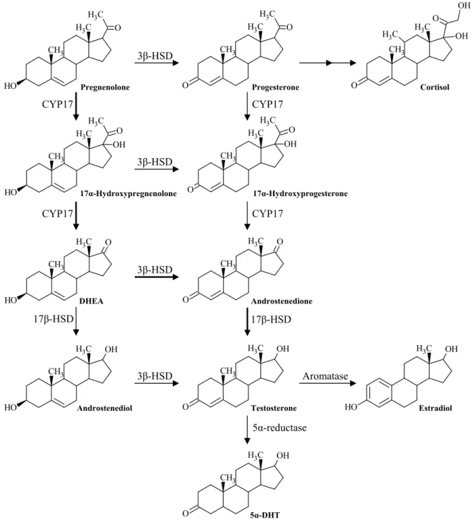
Figure I.9 Biosynthetic pathway to form testosterone and other steroid metabolites. The larger arrows represent the preferred path to form testosterone.
18
Figure I.10 Hypothalamic-pituitary-testicular and hypothalamic-pituitary-adrenal axis (adapted from J.B. Aragon-Ching et al., Frontiers in Bioscience, 12 (2007) 4957).
20
Figure I.11 Regulation of androgen receptor action. In the presence of androgens, particularly testosterone and 5α-DHT, the androgen receptor (AR) releases the heat shock protein (HSP) complex, which is responsible for its inactivation form, and binds to the androgen moiety. The activated receptor translocates from the cytoplasm to the nucleus and binds to the DNA. The activated AR recruits essential coactivators and promotes the action of the RNA polymerase, which responsible for the target gene expression.
23
Figure I.12 Phase I metabolism of testosterone. Androsterone and Etiocholanolone represent the major metabolites of testosterone.
24
Figure I.13 Phase II glucuronidation reaction of testosterone. Testosterone glucuronide is a minor metabolite of testosterone metabolic inactivation.
xxii
Figure I.14 (a) Common structural alterations introduced to testosterone molecule. (b) Chemical structures of some examples of pharmaceutical and designer AAS (1).
28
Figure I.15 Schematic representation of the primary adverse effects of AAS abuse (adapted from Lynette Rushton, The Endocrine System, Chelsea House Publications, 2009)
30
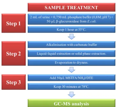
Figure I.16 Schematic sample treatment methodology for AAS analysis by GC-MS. 36
Figure I.17 Common EI ion source (Chhabil Dass, Fundamentals of Contemporary Mass Spectrometry, John Wiley & Sons, 2007).
41
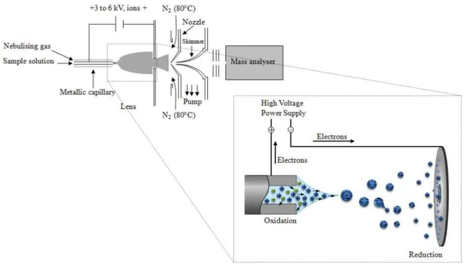
Figure I.18 Schematic representation of the processes that occur in electrospray ionisation source.
43
Figure I.19 Mass spectrum of testosterone TMS derivative. 45
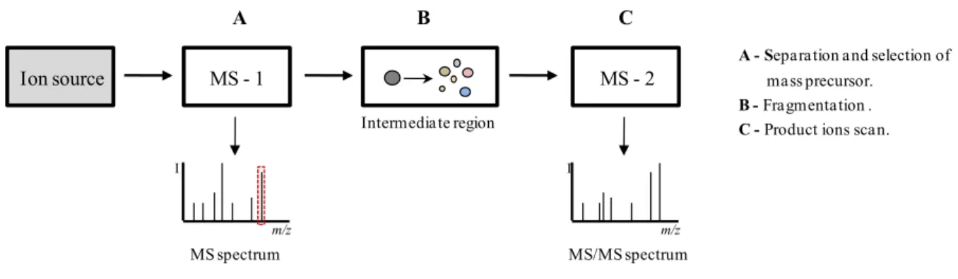
Figure I.20 Schematic representation of an in space tandem mass spectrometry system. 46
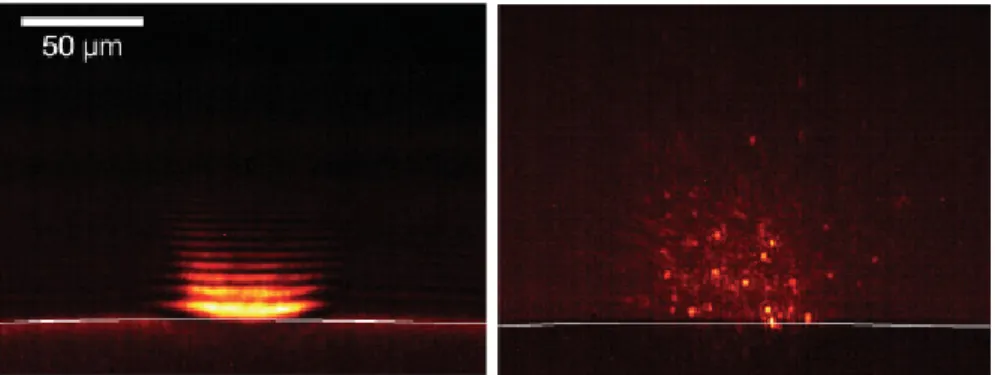
Figure I.21 High-speed photographs of UV-MALDI plumes generated with a Nd:YAG laser operating at 266nm (Reproduced from Franz Hillenkamp and Jasna Peter-Katalinic, MALDI-MS: A Practical Guide to Instrumentation, Methods and Applications, WILEY-VCH, 2007).
48
Figure I.22 Structures of most common MALDI matrices. 49
Figure I.23 Schematic representation of the TOF analyser. 50
Figure I.24 Schematic representation of the reflectron time-of-flight mass analyser. 52
Figure I.25 Schematic representation of the tandem time-of-flight mass analyser. 53
Figure I.26 Electromagnetic spectrum with the sound frequency ranges highlighted (Adapted from T.J. Mason and J.P Lorimer, Applied Sonochemistry: Uses of power ultrasound in chemistry and processing, Wiley-VCH, 2002).
54
Figure I.27 (a) Direct piezoelectric effect in which the application of mechanical stress generates a charge separation. (b) Reverse piezoelectric effect in which the application of a differential potential induces mechanical stress (Adapted from K. Kirk Shung, Diagnostic Ultrasound: Imaging and Blood Flow Measurements, Taylor & Francis, 2006).
58
Figure I.28 Relative intensities of the ultrasonic bath, cup horn and probe (Adapted from www.hielscher.com).
61
Figure I.29 Schematic representation of a magnetron (Adapted from
http://hyperphysics.phy-astr.gsu.edu/hbase/waves/magnetron.html).
xxiii
Figure II.1 MALDI mass spectra of 17α-methyltestosterone and 17β-hydroxy-17-methylandrosta-1,4-dien-3-one GT hydrazones, at concentrations of 1 µg/mL on the MALDI sample plate, obtained with different organic matrices. Ion identification: 1: 17β-hydroxy-17-methylandrosta-1,4-dien-3-one; and 2: 17α -methyltestosterone.
86
Figure II.2 MALDI mass spectra of the target steroid GT hydrazones, at concentrations of 0.025 µg/mL on the MALDI sample plate, obtained with the HABA matrix. Ion identification:1: 17β-hydroxy-17-methylandrosta-1,4-dien-3-one; 2: 17α -methyltestosterone; 3 (and 4): sum of 4-androsten-3,17-dione and boldenone; and 5: nandrolone.
87
Figure II.3 MALDI mass spectra of the target steroid GT hydrazones, at concentrations of 0.010 µg/mL on the MALDI sample plate, obtained with the HABA matrix diluted 2:5. Ion identification: 1: 17β -hydroxy-17-methylandrosta-1,4-dien-3-one; 2 (and 9): sum of 17α-methyltestosterone and mibolerone; 3 (and 4): sum of 4-androsten-3,17-dione and boldenone; 5: nandrolone; 6: 17α-trenbolone; 7: noretiocholanolone; 8: etiocholan-3α-ol- 17-one; 10 (and 11): sum of 2α -methyl-5β-androstane-3α-ol-17-one and mesterolone; 12: ethisterone; 13 (and 14): sum of bolasterone and calusterone; 15: fluoxymesterolone.
89
Figure II.4 MALDI mass spectra obtained after the analysis of human male urine with (b) and without spike (a) of five exogenous steroids. Steroid GT hydrazones ion identification: 2: 17α-methyltestosterone; 5: nandrolone; 6: 17α-trenbolone; 13: bolasterone; and 15: fluoxymesterolone.
91
Figure II.SM1 Chemical structures of the fifteen anabolic steroids analysed. 95
Figure II.SM2 Chemical structures of the nine organic matrices applied. 96
Figure II.SM3 MALDI mass spectra of 17-β-hydroxy-17-methylandrosta-1,4-dien-3-one, 17-α-methyltestosterone and 4-androsten-3,17-dione at a concentration of 5 μg/mL in the MALDI sample plate, obtained with different organic matrices. Peak identification: 1: 17-β-hydroxy-17-methylandrosta-1,4-dien-3-one; 2: 17-α-methyltestosterone; and 3: 4-androsten-3,17-dione.
97
Figure II.SM4 MALDI mass spectra of the target steroid GT hydrazones, at concentrations of 0.125 and 0.05 μg/mL in the MALDI sample plate, obtained with HABA, IAA, FA and 2,5-DHB matrices. Peak identification: 1: nandrolone; 2: sum of 4-androsten-3,17-dione and boldenone; 3: 17-β-hydroxy-17-methylandrosta-1,4-dien-3-one; and 4: 17-α-methyltestosterone.
xxiv
Figure II.SM5 MALDI mass spectra of: (a) the inorganic TiO2 matrix; (b) the inorganic Al2O3
matrix; (c) the target steroid GT hydrazones (0.250 μg/ml) obtained with the TiO2 matrix; (d) the target steroid GT hydrazones (0.250 μg/ml) obtained with
the Al2O3 matrix. Peak identification: 1: nandrolone; 2:
4-Androsten-3,17-dione and boldenone; 3: 17-β-hydroxy-17-methylandrosta-1,4-dien-3-one; 4: 17-α-methyltestosterone.
100
Figure II.SM6 MALDI mass spectra of: (a) the inorganic TiO2 matrix with the dispersing
solution; (b) the target steroid GT hydrazones (0.250 μg/ml) obtained with the TiO2 matrix with the dispersing solution. Peak identification: 1: nandrolone; 2:
4-Androsten-3,17-dione and boldenone; 3: 17-β -hydroxy-17-methylandrosta-1,4-dien-3-one; 4: 17-α-methyltestosterone.
101
Figure III.1 Schematic diagram of the MALDI-MS(/MS) strategy to analyse AAS using the MLibrary software.
107
Figure III.2 CSV file showing centroid mass and relative intensity of each peak 109
Figure III.3 MLibrary interface for MS spectrum data load. 109
Figure III.4 MLibrary MS spectrum analysis search window. Input information: Database, compound state, modification and mass tolerance.
110
Figure III.5 MLibrary MS spectrum analysis display window. Data identification: Blue line: experimental spectrum mass peaks; Green line: database reference masses; Red line: experimental mass that matched with the reference mass
111
Figure III.6 MLibrary interface for MS/MS spectrum data load 112
Figure III.7 MLibrary MS/MS spectrum analysis search window. Input information: Database, compound mass or compound name, compound state, modification, mass tolerance concerning the experimental and reference mass values, mass tolerance within the database spectra and discriminate power.
113
Figure III.8 MLibrary MS/MS spectrum analysis display window. 114
Figure III.9 MLibrary MS/MS spectra comparison analysis window. 114
Figure III.10 MLibrary urine sample MS spectrum analysis display window. Data identification: Blue line: experimental spectrum mass peaks; Green line: database reference masses; Red line: experimental mass that matched with the reference mass.
115
Figure III.11 (a) MLibrary MS/MS spectrum analysis display window for trenbolone. (b) MLibrary MS/MS spectrum analysis display window for 17α-methyltestosterone.
xxv
Figure IV.1 Comprehensive scheme of the classic and the ultrasonic protocols used in theoptimization study for the determination of androgenic anabolic steroids.
132
Figure IV.2 Effect of the ultrasonication on the hydrolysis yield of dehydrolisoandrosterone 3-glucuronide (DHEA) and etiocholanolone glucuronide using two devices, an ultrasonic probe (UP) at 60 % of ultrasonic amplitude and a sonoreactor (SR) at 50% of ultrasonic amplitude (n=5). The results are given in percentages (100% = result via the classic method).
133
Figure IV.3 Effect of the ultrasonication in the derivatisation yield of nandrolone (Nan), 4-androstes-3,17-dione (AD), boldenone (Bol) and 17β-hydroxy-17-methylandrosta-1,4-dien-3-one (MA), by means of two devices, a sonoreactor (SR) at 50% of ultrasonic amplitude and an ultrasonic bath (US) at 100 % of ultrasonic amplitude with two frequencies, 35kHz and 130kHz (n=5). The classic treatment consists of heating at 60°C for 1h. The results are given in
percentages (100% = result via the classic method).
138
Figure V.1 Comprehensive scheme of the classic, ultrasonic and microwave protocols for the determination of the banned compounds presented in table V.1.
154
Figure V.2 Effect of the microwave power on the hydrolysis yield of the natural endogenous steroids androsterone, etiocholanolone, keto-androsterone, 11-hydroxy-etiocholanolone and 11-hydroxy-androsterone (10 minutes of treatment; n=3). The results are given in percentages (100% = result via the classic method).
157
Figure V.3 Effect of the microwave time of treatment on the hydrolysis yield of the natural endogenous steroids androsterone, etiocholanolone, 11-keto-androsterone, 11-hydroxy-etiocholanolone and 11-hydroxy-androsterone (Microwave power: A- 180W, B- 240W; n=3). The results are given in percentages (100% = result via the classic method).
158
Figure V.4 Effect of microwave power in the derivatisation yield of 25 banned substances (3 min of treatment; n=3). The results are given in percentages (100% = result via the classic method).
162
Figure V.5 Effect of microwave time in the derivatisation yield of 25 banned substances (microwave power 1200 W; n=3). The results are given in percentages (100% = result via the classic method).
163
Figure VI.1 Calibration curve obtained following HPLC-UV analysis by plotting the area of the chromatographic peak corresponding to (a) PNP and (b) PNP-G as a function of concentration. The calibration equations obtained were y = (0.445 ± 0.002)x + (0.1 ± 0.3), (r2=0.9999), and y= (0.469 ± 0.006)x + (0.2 ± 0.9), (r2=0.9998), for PNP and PNP-G, respectively.
xxvi
Figure VI.2 Hydrolytic conversion of substrate PNP-G to the products PNP and D-glucuronic acid through the action of β-glucuronidase.
176
Figure VI.3 Representative chromatogram obtained after 4 minutes of incubation of the substrate PNP-G with the enzyme β-glucuronidase. The first peak elutes at 3.51 min and corresponds to PNP-G, whereas the second peak elutes at 6.67 min and corresponds to PNP.
177
Figure VI.4 (a) Appearance of PNP as a function of time for both incubation at 55ºC and ultrasonic irradiation essays. The experimental results were obtained using 450 µM of PNP-G. (c) Correspondent enzyme activity for both experiments. The unit of enzymatic activity was considered as the enzyme activity that increases the rate of release of 1µmol of PNP per minute. Data identification: : 55ºC; |: Ultrasonic irradiation.
178
Figure VI.5 Relative enzyme activity for the release of PNP from PNP-G. The concentration of PNP-G was fixed at 450 µM and the amount of β -glucuronidase varied from 0.22, 0.33 and 0.44 µg. Data identification: black line: 0.22 µg; blue line: 0.33 µg; red line: 0.44 µg.
180
Figure VI.6 Observed time courses for the hydrolytic conversion of PNP-G into PNP (a) at 55ºC and (b) under continuous ultrasonic irradiation, with increased PNP-G concentration. Data identification: ‡: 7.7 µM; ¢: 15.4 µM; Ì: 32.1 µM; ×: 62.9 µM; : 127.3 µM; Ç: 190.1 µM; : 257.0 µM; : 352.4 µM; : 448.3 µM; ¡: 539.3 µM. (c) Plot of the initial velocity values obtained as a function of the correspondent PNP-G concentration values. Data identification: line: 55ºC; | line: Ultrasonic irradiation.
181
Figure VI.7 Lineweaver-Burk plot for β-glucuronidase. Enzymatic activity was assayed with 0.22µg of enzyme at pH 6.5. The x and y axes indicate the reciprocals of the initial concentration of PNP-G and initial velocity, respectively. The value of Vmax is given from the intercept and the value of KM/Vmax from the slope.
Data identification: line: incubation at 55ºC essay; y= (6.3 ± 0.2)x + (0.067
± 0.003), (r2=0.9992); | line: ultrasonic irradiation essay; y=(5.5 ± 0.1)x + (0.059 ± 0.003), (r2=0.9979).
PART ONE
3
CHAPTER I
I.1 DEFINITION OF DOPING
4
I.2 HISTORICAL BACKGROUND
I.2.1 DOPING: ORIGIN OF THE WORD
Despite the ancient usage of physical enhancing substances, the word “doping” appeared for the first time in an English dictionary in 1889 [1]. At that time the word was used to describe a remedy containing opium commonly used to increase racehorse’s performance [1]. The origin of the word is still unclear. Some authors attribute its origin to the African Zulu warrior’s beverage called “Dope” or “doop”, which was used as a stimulant [1, 4]. Others claim that it comes from the slang word “dope” for opium, used by North Americans [4].
I.2.2 EARLY HISTORY OF DOPING
The use of doping substances to enhance the physical performance is as old as the beginning of civilization [4, 5]. Several reports point out the use by ancient people of substances extracted from plants and animals to increase their physical capacity. Those substances were used mainly to increase strength and to surpass fatigue and possible injuries, which were valuable attributes in war, society and athletic competition [1, 4, 6].
Extracts from Ephedra, one of the oldest medicinal plants know by man, has been used by the Chinese Traditional Medicine for more than 5000 years. The use of the Ma Huang, as it was known, was recommended for its stimulant and antiasthmatic effect by Chinese physicians [7].
South America tribes used to chew coca leaves as well as other plants containing powerful stimulants. Among those tribes, the Incas are an excellent example regarding the effect of those stimulants; their famous ability to run around 1750 Km between Cuzco and Quito in few days was only possible under the effects of cocaine [1, 6]. With the same purpose West Africans tribes made use of Cola acuminita and Cola nitida to enhance their running ability [6].
Early uses of “testosterone” are dated to the ancient Greeks [8]. The alteration of the animal’s behavior after castration provided them the evidence that the testicles were involved in the secondary male sex characteristics [4, 8]. Following a primitive medicine practice known as similia similibus that consisted of treating an organ with itself, the consumption of testicular tissue was frequently used to enforce virility [8]. For the same reason, the Huns were known to eat testicles before battles to increase their strength and performance [1]. During the time of the Roman Empire, gladiators as well as soldiers used stimulants to overcome fatigue and injury [4]. One of those stimulants was the extract from the plant Ephedra [7].
5
Figure I.1 – Painting of the athletes in the Ancient Greek Olympic Games (Reproduced from Kristine Toohey and A. J. Veal, The Olympic Games: a social science perspective, CAB international, 2007).Through ancient history, the use of enhancing substances is almost omnipresent in all civilizations. It was not a specific behaviour linked with sports, but to society in general as a way to achieve an advantage over others.
I.2.3 DOPING AND ITS EMINENT PROHIBITION
Despite the use of doping substances for centuries, their prohibition dates back to the beginning of the 20th century [1, 6]. Before that, the use of such substances was not considered cheating at all. For instance, it is remarkable that at the Ancient Olympic Games the widespread of doping among the athletes was not considered cheating, whereas other dishonest behaviours, such as bribing an opponent, was punished severely [1, 4, 6]. Those athletes were banished for life and their name alongside with their family’s were inscribed in stones so that everyone could know their offence. It has been also reported that even death penalties were given at that time to those who violate the Game’s rules [1]. From the old Olympic Games until the 19th century there is a gap of records concerning the use of enhancing substances or its prohibition, possibly because it was already an intrinsic behaviour in society.
6
improve the physical capacity was usual amongst athletes and without any kind of prohibition there was no need to cover its use. It was common for trainers to develop their own secret recipes with several enhancing substances [4, 6].
Around the end of the 19th and beginning of the 20th century, the professionalization of amateur sports gave an additional contribution to increase doping attempts. The beginning of the “Six Day” cycling races together with boxing and animal races demanded more physical strength and endurance from the competitors [4]. Caffeine, alcohol, ether, nitro-glycerine, and later on strychnine, cocaine and heroin were usual components of the enhancing mixtures taken by athletes [1, 4]. These levels of doping use and experimentation led to the death of the English cyclist, Arthur Linton, which was the first known death caused by extreme doping [1, 4].
At the beginning of the 20th century, doping abuse was an inevitable phenomenon within all sports [6]. At this point, for the first time, the use of doping substances was considered inappropriate and restrictions to their use were introduced [4]. Few years later, in 1928, the International Amateur Athletic Federation (IAAF) prohibited the use of doping in sports, specifically stimulants, which were the main doping agents at that time [1, 4]. Following the IAAF decision, other sport organizations joined the anti-doping cause, however official testing was not yet performed and the restrictions remained ineffective due to the absence of valuable tests [1, 4].
In 1935, testosterone, the main male sex hormone, was isolated from the testes as a crystallised pure substance by Ernst Lacqueur [4]. In that same year, the chemical synthesis of testosterone was achieved by two distinct studies carried out by Adolf Butenandt and Leopold Ruzicka [11, 12]. This discovery earned to both scientists the joint Nobel Prize for Chemistry for 1939 [1, 13]. The elucidation of the chemical synthesis of testosterone enabled its production worldwide and, given that the effect of these compounds had already been associated with the secondary male sex characteristics, it is unsurprising that their use was quickly introduced in sports, first as agents supporting recovery after massive stress and exhaustion and then as agents enhancing the physical performance of athletes [1].
Russian athletes were putatively the first to use anabolic steroids, in particular, testosterone, to improve athletic performance in international competitions [6, 8]. Nevertheless, its use quickly spread within sports and, soon after, research funded by pharmaceutical companies started in an attempt to find a compound with the anabolic effects of testosterone, but without its androgenic effects [6, 8]. Consequently, few years later, methandrostenolone (Dianabol) was synthesised by the pharmaceutical company Ciba and it became one of the first anabolic steroids marketed and introduced in sports [8]. In parallel, other pharmaceutical companies started to synthesise several testosterone derivatives [11]. The use of these substances has spread in such a way, that at the end of the 1950s this class of drugs became the principal group in the statistics of doping cases [1].
7
central nervous system stimulant [6]. The first systematic use of these compounds occurred within the military services with the aim to improve concentration and vigilance during the Second World War. Its use as strong-acting stimulants quickly spread to sports [1, 4]. Within these substances, amphetamine and methamphetamine were the most used amongst athletes. Their use was particularly established in cycling, reaching its peak between the 1960s and the 1970s [4, 6]. During this period of time, the death of the Danish cyclist Knut Jensen, in 1960, at the Olympic Games in Rome, and the first televised doping death of the English cyclist, Tom Simpson, during the Tour de France, in 1967, highlighted the urgency of an anti-doping policy [4, 6]. Soon after the death of the English cyclist, in that same year the International Olympic Committee (IOC) founded its own Medical Commission with the aim to ban the misuse of doping substances within sports [1, 14, 15]. Following its establishment, the IOC Medical Commission created its first list of prohibited substances [1, 2, 14]. Stimulants, analeptics, some alkaloids and narcotics, such as amphetamines, ephedrine, or cocaine became prohibited as the first classes of doping agents [1, 16]. Although the use of anabolic steroids was thought to be extremely extensive among sports, the first list of prohibited substances did not included that class of compounds because there was no viable analytical test available for them [3, 14]. IOC doping tests were first introduced during the Winter Games in Grenoble and at the Summer Olympic Games in Mexico in 1968 [1, 3, 6, 15, 17]. The doping control carried out in Mexico may be considered as a pilot project to the official and systematic antidoping tests performed in all sports at the Olympic Games in Munich in 1972 [15, 17]. It was at these Olympic Games that mass spectrometry (MS) was introduced coupled with gas chromatography (GC) as a confirmatory method to identify doping substances [17]. Professor Manfred Donike, from the Institute of Biochemistry at the German Sport University in Cologne, was the main initiator for the introduction of mass spectrometry in doping analysis [4, 16].In 1974, the anabolic steroids were finally banned, first by the IAAF and then by the IOC Medical Commission that included these compounds in the list of banned substances [2]. The screening method to detect the presence of anabolic steroids in the 1976 Montreal Olympics was based in radioimmunoassay (RIA) while GC/MS was used for confirmation purposes [18]. The introduction of reliable tests for the anabolic steroids determination resulted in numerous disqualifications in the late 1970`s, especially among strength related sports [2, 16].
8
I.2.4 HARMONISATION OF ANTIDOPING POLICIES
In the beginning of the 1980s a new challenge concerning the use of anabolic steroids arose within doping control agencies. Despite its capacity to detect anabolic steroids, anti-doping laboratories were not prepared to positively detect the misuse of exogenous testosterone, such as testosterone esters, which have the same excretion metabolites than its endogenous counterpart [17, 18]. It is perhaps unsurprising that until 1983, when Professor Donike, using GC-MS based methodologies, introduced a new screening method for testosterone, numerous testosterone abuses have gone unpunished [1, 17, 18]. The method purposed by Donike, established a testosterone to epitestosterone ratio threshold, above which it was considered a doping offence [1, 17, 18].
Along with the use of endogenous anabolic steroids administered exogenously, the anti-doping laboratories were also confronted with the emergence of new drugs and new methods, such as the human growth hormone (hGH) and blood transfusion [16]. At the time blood doping was already considered unethical but it was not officially prohibited for inexistence of a valuable test [2]. However, right before the opening of the 1984 Los Angeles Games, the authorities uncovered a clandestine operation carried out by some cyclists that were transfused with whole blood [2, 6]. The impact of this behaviour on the media and consequently on society, led the medical commission of the IOC to ban these drugs and methods, although no tests were available for their detection [1, 2, 6]. Along with the introduction of blood doping, beta-blockers in 1985, diuretics in 1987 and the growth hormone in 1989 were also included in the doping list [16].
In addition to the challenges faced by doping laboratories, a new setback arose to anti-doping organizations with the suspicion of several state sponsored anti-doping activities. Most of these suspicions were centred on the Eastern Bloc countries, such as the Soviet Union, Poland, Czechoslovakia and the German Democratic Republic (GDR), but allegations that other countries like England, Australia and Canada were covering up some of its athletes doping abuse cases were also emerging [16, 19]. Regarding the GDR, these suspicions have been substantiated with the finding of several studies concerning the development of ideal drug regimens to improve performance, after its collapse in 1990 [19].
In 1988 at the Seoul Olympic Games, the increasing public awareness that new measures should be employed was catapulted by another scandal; the 100-metres champion, Ben Johnson (see Figure I.2), tested positive for stanozolol, an anabolic steroid [3].
9
Figure I.2 – Ben Johnson at the Seoul Olympic Games (Reproduced from Kristine Toohey and A. J. Veal, The Olympic Games: a social science perspective, CAB international, 2007).Before the Doping Convention, the Council of Europe had already adopted the Anti-Doping Resolution in 1967, stating that doping was the opposite of sport values and that the sport organizations should be responsible for doping control, and draw the European Anti-Doping Charter for Sport, which had a recommendation status and contained guidelines for governments concerning anti-doping policies. The Anti-Doping Convention arose as a sign of governmental motivation to reduce and, if possible, eliminate doping from sports [16, 20].
I.2.5 THE WORLD ANTI-DOPING AGENCY (WADA)
In the 1990´s, the IOC medical commission established, as a condition to assign the IOC accreditation to anti-doping laboratories, the laboratory’s previous accreditation by the International Organization for Standardization (ISO) [2]. This demand, by the IOC, was an important development within anti-doping laboratories; not only was it useful to standardize methods and procedures as it markedly strengthened the idea of high quality anti-doping laboratories.
10
In 1998, one of the biggest scandals in the history of sports drove the anti-doping community toward the creation of the World Anti-Doping Agency (WADA).
Right before the beginning of the 1998 Tour de France, a large number of prohibited medical substances, in which were included several dosages of EPO and anabolic steroids, were found by French Customs police inside the car of the longtime masseur for the former Festina road-racing team, Willy Voet [4, 16, 22-24]. The Festina team was expelled from the race and other six teams quit during the Tour [4, 23]. Moreover, the arrestment of Voet uncovered one of the major doping schemes involving athletes, coaches, team physicians and other team staff [6, 16]. In the subsequent investigations, carried out by the French and Italian authorities, several other top teams and athletes were also implicated in that highly organized scheme [6].
Following these events and with a growing pressure on the IOC demanding new measures, a World Conference on Doping, proposed by the IOC, was held in February 1999 in Lausanne [1, 9, 16]. The need for an independent international agency for doping became clear, which led to the establishment of the World Anti-Doping Agency on November 10 of that same year [1, 3, 16]. WADA was established as a Swiss foundation [1].
The involved parts agreed that the WADA would be equally financed by sports organizations and governments [25]. Its main mission was to coordinate and promote the development of international standardized anti-doping rules, facilitate the coordination between sport organizations and governmental authorities, ongoing with doping control research, and promote doping prevention activities [1, 3, 16].
In 2002, WADA moved its headquarters to Montreal in order to distance itself from the IOC and mark its position as an independent body [1, 9]. The first major project of the WADA was the elaboration and implementation of the World Anti-Doping Code (WADC) that was adopted in the second World Conference on Doping in Copenhagen in 2003 [1, 9]. The Code was developed after intensive collaboration with several stakeholders and it became the first worldwide accepted document providing a set of harmonized anti-doping policies, rules and regulations within sport organisations and public authorities [1].
11
The WADC was part of the World Anti-Doping Program (WADP) presented by WADA on that conference that included two other proceedings (see Figure I.3), the International Standards for the Prohibited List, for the Laboratories, for Testing and for Therapeutic Use Exemptions (TUE), and models of good practice and guidelines [1-3]. From the three levels of the WADP, only the models of the best practice and guidelines was not mandatory for all Code signatories [3]. The Code and the International standards entered into force on January 2004 in time for the 2004 Athens Olympic Games. At those Games, a significant change within the Prohibited List was the inclusion of gene doping as a prohibited method [1].Despite the developments in the fight against doping, the beginning of the twenty-first century was also marked by another scandal involving the USA Corporation BALCO (Bay Area Laboratory Co-operative) [9]. BALCO was providing athletes with a new enhancing drug undetectable by the GC/MS screening methods used at that time [9]. That unknown substance was later isolated by the United States Anti-Doping Agency (USADA) from a used syringe and it was found to be a designed anabolic steroid which was called tetrahydrogestrinone (THG) [9]. Alongside with the development of new pharmaceutical enhancing drugs, BALCO also produced combined drugs containing the enhancing substance and its masking agent to prevent an adverse analytical finding [6].
Regarding the WADP, a legal problem still persisted; many governments could not formally take part in legal agreements with nongovernmental entities [1, 16, 26]. This situation was overcome in 2005, with the UNESCO Anti-Doping Convention that led to the first global treaty against doping in sport [16, 26]. The Convention guaranteed the effectiveness of the World Anti-Doping Code and supported the WADA activities, providing the legal framework for governments to act. The Convention came into effect in 2007 [1, 3, 16, 26].
In 2006 WADA started the revision process of the World Anti-Doping Code that was presented and adopted in the third Anti-Doping World Conference in November 2007, held in Madrid [3]. The revised World Anti-Doping Code and its International Standards were implemented in 2009 [2, 3].
12
I.3 THE ROLE OF ENDOGENOUS AND SYNTHETIC AAS IN THE
HUMAN BODY
The human body is an extremely complex structure that contains a variety of systems, each one playing a specific role. The perfect functioning of the human body implies that all these systems work together. This means that it has to have the ability to self regulate and that the different organs and tissues that constitute those systems also communicate between themselves. The main control mechanisms, which regulate all aspects of physical life, are the nervous and the endocrine system [27]. With regard to the endocrine system, this consists of glands that release chemical messengers, called hormones, through the blood to act in distant sites [27-29]. Compared with the time of action of the nervous system, the time of action of these compounds in the body is long and, therefore, they are responsible for long-lasting generalized physiological effects, such as growth, development, reproduction and metabolic rate [27, 28].
The hormones will generate different responses depending on the organs in which they operate and although they can be transported through the bloodstream to any cell in the body, only certain cells, called target cells, express highly specific receptors for hormone’s recognition [27, 28]. After the signal recognition, several chemical reactions are triggered inside the target cells, leading to a modification in the output of those cells. A common modification is, for example, the synthesis of specific proteins [27].
Knowing that hormones play a key role in the regulation of the human body, it becomes clear that any alteration of these compounds in the organism will trigger a set of mechanisms that will change the physiological processes of the body.
Hormones can be divided in two classes according to their chemical composition, the steroids and the non steroids hormones that comprises, mainly, proteins and amines [28]. Their effects have long been recognized by athletes that, for several decades, use these compounds to enhance their athletic performance. Within steroids, the male sex hormones, or androgens, and their synthetic derivatives are responsible for the muscular development and strength.
This chapter pretends to illustrate the major aspects concerning the biochemistry of the androgenic anabolic steroids, its mechanism of action and its effects in the human body.
I.3.1 STRUCTURE
13
ring junction [30]. Other common substitutions are the hydroxyl or carbonyl group at C-3 and C-17 carbon atoms. In addition, an alkyl side chain may be found at the C-17 carbon atom [30].Figure I.4 – Steroid`s carbon atoms numbering and ring labelling system.
I.3.2 BIOSYNTHESIS AND SECRETION OF ANDROGENS
In the human body, the biosynthesis of most steroid hormones occurs mainly in a specific set of tissues, which are the adrenal cortex gland and the gonads that comprises the testes and ovaries. Within these specific tissues the biological pathways to produce the steroid hormones are common. Nevertheless, the secretion of steroid hormones is different in the different tissues, mainly due to the different distribution, level of expression and activities of the involved enzymes [28].
In normal men, the production rate of testosterone is approximately 3-10 mg/ day of which more than 95% is produced in the testes, whereas in women this value is about 10 times lower and it’s mainly secreted in the adrenal cortex [1, 28].
Regarding other androgens, the steroid 5α-dihydrotestosterone (DHT), a more potent androgen than testosterone, is secreted in the testes but most of the circulating DHT is formed in the peripheral tissues by conversion of testosterone, catalysed by the enzyme 5α-reductase [1, 31]. The aromatisation of the A ring of testosterone, catalysed by the enzyme aromatase, to produce 17β-estradiol in the brain and in the male reproductive organs has important consequences in the sexual dimorphism [32, 33]. Epitestosterone, which is the epimer of testosterone, is also thought to be primarily formed in the testes [1].
Dehydroepiandrosterone (DHEA) and androstenedione that can be precursors of other androgens are mostly synthesised in the adrenal cortex as well as the 11β-hydroxyandrostenedione and its metabolites [1, 34, 35].
It is important to stress that steroidogenesis of some of these androgens has also been reported to take place in peripheral tissues, either by interconversion or by complete biosynthesis [34, 35].
14
I.3.2.1 BIOSYNTHESIS OF CHOLESTEROL
Cholesterol is the biochemical precursor of all steroid hormones [36]. In this chapter it is presented the
de novo synthesis of cholesterol, although cholesterol may be obtained from other sources, such as low-density lipoprotein (LDL) and high-density lipoprotein (HDL) as well as from the hydrolysis of esterified cholesterol stored as lipid droplets [1, 37].
The elucidation of the biosynthetic pathway to cholesterol was achieved in the 1960s and it starts with the biosynthesis of mevalonic acid [30, 36]. The first step in the mevalonate pathway involves a Claisen-type condensation between two molecules of acetyl-CoA, catalysed by the enzyme acetoacetyl-CoA thiolase, to form acetoacetyl-CoA [36, 38]. The second step is catalysed by the enzyme hydroxymethylglutaryl-CoA synthase, and it involves an aldol-type reaction between acetoacetyl-CoA and another molecule of acetyl-CoA to produce the hydroxymethylglutaryl-CoA [36, 38]. Mevalonic acid is formed after hydroxymethylglutaryl-CoA reduction with two molecules of NADPH. This last step is catalised by the enzyme hydroxymethylglutaryl-CoA reductase and is the rate-limiting step in cholesterol biosynthesis [30, 36].
The next step in the Mevalonate pathway is the conversion of mevalonic acid, a six carbon compound, into the five carbon isopentenyl pyrophosphate (IPP), after successive phosphorylation and decarboxylation [30, 36, 38]. Following its formation, isopentenyl pyrophosphate suffers an enzymatic isomerisation that gives rise to dimethylallyl pyrophosphate (DMAPP), establishing an equilibrium that makes both compounds available to the cell (see Figure I.5).
![Figure I.12 – Phase I metabolism of testosterone [1]. Androsterone and Etiocholanolone represent the major metabolites of testosterone](https://thumb-eu.123doks.com/thumbv2/123dok_br/16476232.732040/54.892.90.758.621.1096/figure-metabolism-testosterone-androsterone-etiocholanolone-represent-metabolites-testosterone.webp)
![Figure I.13 – Phase II glucuronidation reaction of testosterone. Testosterone glucuronide is a minor metabolite of testosterone metabolic inactivation [1]](https://thumb-eu.123doks.com/thumbv2/123dok_br/16476232.732040/55.892.137.805.585.757/glucuronidation-testosterone-testosterone-glucuronide-metabolite-testosterone-metabolic-inactivation.webp)