Bárbara Martins Paiva da Cunha
Degree in Biochemistry
Expression of the human carboxylesterase 2 enzyme
(CES2) in mammalian cells
Dissertation to obtain a Master Degree in Biotechnology
Supervisor: Ana Luísa Simplício, Ph.D, IBET/ITQB-UNL
Co-Supervisor: Ana Sofia Coroadinha, Ph.D, IBET/ITQB-UNL
Júri:
Presidente: Prof. Doutor Rui Manuel Freitas de Oliveira
Arguente: Profª. Doutora Alexandra Fernandes
Vogal: Doutora Ana Luísa Ferreira Simplício
Bárbara Martins Paiva da Cunha
Degree in Biochemistry
Expression of the human carboxylesterase 2 enzyme
(CES2) in mammalian cells
Dissertation to obtain a Master Degree in Biotechnology
Supervisor: Ana Luísa Simplício, Ph.D, IBET/ITQB-UNL
Co-Supervisor: Ana Sofia Coroadinha, Ph.D, IBET/ITQB-UNL
Júri:
Presidente: Prof. Doutor Rui Manuel Freitas de Oliveira
Arguente: Profª. Doutora Alexandra Fernandes
Vogal: Ana Luísa Ferreira Simplício
Copyright
"Expression of the human carboxylesterase 2 enzyme (CES2) in mammalian cells".
Copyright Bárbara Martins Paiva da Cunha, FCT/UNL, UNL
Acknowledgments
Acknowledgments
Gostaria de agradecer a todas as pessoas que directa ou indirectamente contribuíram para a realização deste trabalho e sem as quais este não teria sido possível.
Gostaria de agradecer à Doutora Ana Luísa Simplício pela oportunidade que me deu para fazer este trabalho, pelas palavras certas nos momentos oportunos, pelo apoio incondicional, pela troca de ideias, pelo sempre à-vontade e disponibilidade, bem como por me ter protegido quando mais precisei.
À Doutora Ana Sofia Coroadinha por tudo o que me ensinou, pela disponibilidade e apoio, por também me ter recebido neste projecto, por todas as ideias e procura de soluções no decorrer deste trabalho.
Um especial obrigado à Doutora Cristina Peixoto pelas horas intermináveis, pela sempre palavra amiga, pela boa disposição, por tudo o que ensinou e está sempre disposta a ensinar, obrigado! É um prazer trabalhar contigo e claramente que este ano sem ti tinha sido muito mais difícil!
Gostaria de agradecer também ao Eng. Marcos Sousa, por tudo o que me ensinou ao longo deste ano, bem como pelos seus conselhos e sempre boa disposição.
Indiscutivelmente, o meu maior e sincero obrigado é para a Joana Lamego. Ao longo deste ano foste minha mentora, mini-chefe, colega e amiga. És um estímulo constante e um exemplo a seguir. Ensinaste-me e continuas a ensinar-me, incansavelmente, todos os dias, e soubeste sempre dizer a palavra certa no momento certo. Obrigado não fica nem perto da gratidão que sinto para contigo. Obrigado por tudo o que partilhámos, por tudo o que aprendi, por todos os puxões de orelhas, por saberes quando parar, por acreditares em mim e por me protegeres. Se todos tivessem uma Joana na vida, o Mundo era melhor. É um orgulho e prazer trabalhar ao teu lado.
Gostaria também de agradecer a todos os membros do TCA, em particular à Patrícia (für alles!), Vanessa (pelas cantorias!), Hélio, Miguel, Tiago, Rute, Joana, e, claro, à Carina Brilha, que sempre me ouviu, ajudou e que tem um valor inestimável!
A todos os meus amigos, os old school, os que estão longe (mas sempre perto!) e ao meu núcleo duro, por serem um dos meus pilares, pela galhofa, mas também pelo apoio e confiança incondicionais. Todos vocês são uma parte central de quem eu sou. Obrigada!
Ao Luís, que partilhou esta aventura comigo, pelo ânimo, incentivo, apoio e carinho e por sempre ter acreditado no dia de amanhã, mesmo quando eu não o fiz. Sem ti não era o mesmo, obrigada!
Por fim, gostaria de agradecer à minha família: avós e os fantásticos 10! Sou tão sortuda por ter uma família como vocês. A presença e apoio incondicional fazem de mim uma pessoa melhor todos os dias. Em especial à minha mãe e irmão por me acompanharem na minha jornada, pela paciência, apoio, carinho e por fazerem de mim quem eu sou hoje. Obrigada por sempre terem acreditado em mim, me terem dado tudo, mesmo nas situações mais difíceis, por me mostrarem que é sempre possível dar a volta e que por muito más que as situações sejam, vão sempre melhorar.
Abstract
Abstract
The carboxylesterase 2 enzyme (CES2), the main carboxylesterase (CES) expressed in human intestine, has a key role in the metabolism of ester containing xenobiotics including the activation of several prodrugs and therefore, it can influence significantly the bioavailability of these compounds. CES2 is receiving an increasing attention due to its potential application in anti-cancer combined therapies, for the treatment of different pathologies like colon adenocarcinoma and malignant glioma.
Unlike carboxylesterase 1 (CES1), the structure of this enzyme is not yet known. Since the knowledge of the structure of this protein will be crucial for the understanding of its properties, it becomes relevant the establishment of a process for the production and purification at the milligram level.
The development and optimization of human recombinant CES2 production process, using human embryonic kidney cells (HEK-293T cells), is described in this thesis. First, the C-terminal 10xHistidine tag CES2 (CES2-10xHis) was produced using adherent cultures, and then the process was transferred to suspension cultures with serum-free media and finally scaled up to a five-litre stirred tank bioreactor. An affinity chromatography purification process was applied to obtain the CES2-10xHis protein, with a high purity grade.
Despite being an Endoplasmic Reticulum (ER) anchored protein, secretion of CES2 to serum free media was achieved due to the presence of the in frame C-terminal 10xHistidine tag. With this elegant procedure we avoided the addition of extra N-terminal signaling sequences or the mutation or deletion of the C-terminal Histidine - Threonine - Glutamic acid - Leucine (HTEL) motif responsible for anchoring the protein in the lumen of the ER.
Secretion contributed to a good production yield; however, the global process yield was quite low, due to difficulties in the purification step, which we attributed to protein aggregation.
Resumo
Resumo
O enzima carboxilesterase 2 (CES2), o carboxilesterase (CES) maioritário no intestino humano, tem um papel fundamental no metabolismo de xenobióticos que contenham grupos éster e também na activação de diversos pró-fármacos, podendo influenciar significativamente a sua biodisponibilidade. CES2 tem vindo a receber uma atenção constante e crescente devido ao seu potencial papel em terapias combinadas anti-cancerígenas para o tratamento de diferentes patologias, tal como adenocarcinoma do cólon ou ainda glioma maligno.
A estrutura deste enzima ainda não é conhecida, ao contrário da do carboxilesterase 1 (CES1). O conhecimento da sua estrutura poderá ser crucial para compreender as suas propriedades, pelo que se torna relevante o estabelecimento de um processo para a sua produção e purificação (ao nível de miligrama).
Nesta tese de mestrado, é descrito o desenvolvimento e optimização do processo de produção de CES2 recombinante humano, utilizando células derivadas de rim humano (Human Embryonic Kidney cells, HEK-293T cells). Numa primeira fase, a proteína com um tag de dez histidinas a C-terminal (CES2-10xHis) foi produzida em cultura aderente, tendo posteriormente o processo sido transferido para culturas em suspensão com meio sem soro, finalizando com o aumento de escala para 5 L em bioreactor de tanque agitado. Foi aplicado um processo de purificação com cromatografia de afinidade para obter a proteína CES2-10xHis, com um elevado grau de pureza.
Embora esta proteína esteja ancorada ao retículo endoplasmático (ER), a sua secrecção para meio sem soro foi alcançada através da presença do tag in frame de dez histidinas a C-terminal. Com este elegante procedimento, evitou-se a adição de sequências extra de sinalização a N-terminal ou mutar ou delectar o motivo HTEL a C-terminal, responsável pela ancoragem da proteína ao lúmen do ER.
A secreção contribuiu para um bom rendimento na produção, contudo, o rendimento do processo global foi baixo, devido a dificuldades no passo de purificação, que foi atribuído a agregação da proteína.
Table of Contents
Table of Contents
1. Introduction ... 1
1.1 - Animal Cell culture ... 1
1.1.1 - Bioprocessing ... 3
1.1.1.1 - Upstream: relevant culturing systems and process modes ... 3
1.1.1.2 - Downstream: recombinant protein purification ... 4
1.1.2 - Cell line ... 5
1.1.3 - Protein processing ... 5
1.1.4 - Expressing heterologous proteins in mammalian cells ... 6
1.2 - Carboxylesterases ... 8
1.2.1 - CES characterization ... 9
1.2.2 - CES2 manufacturing in different systems ... 11
2. Goals ... 13
3. Materials and Methods ... 15
3.1 Biological Material and Culture Media ... 15
3.1.1 Bacterial Strain ... 15
3.1.2 Mammalian cell line ... 16
3.1.3 Mammalian Expression vectors ... 17
3.2 Transfection assays ... 18
3.2.1 Static culture ... 18
3.2.2 Suspension culture ... 18
3.3 Brefeldin A assay ... 19
3.4 Protein Purification ... 19
3.4.1 CES2 purification from cell extracts of transfected HEK-293T adherent cells ... 19
3.4.2 CES2 purification from cell extracts of transfected HEK-293T suspension cells ... 20
3.5 Analytical tests ... 20
3.5.1 Quality of the purified plasmids ... 20
3.5.2 Determination of cell concentration and viability ... 21
3.5.3 Metabolite quantification ... 21
3.5.4 Protein Quantification ... 21
3.5.5 Enzymatic Activity Assay ... 21
3.5.6 Electrophoresis ... 22
3.5.7 Western Blot ... 22
4. Results and Discussion ... 25
4.1 Development of a mammalian expression vector plasmid ... 25
4.2 CES2 production in transfected HEK-293T adherent cells ... 33
4.3 CES2 purification from transfected HEK-293T adherent cell extracts ... 36
Table of Contents
4.5 CES2 secretion from transfected HEK-293T cells in suspension ... 46
4.6 CES2 purification from supernatant samples of transfected HEK-293T cells in suspension . 50
4.7 CES2 production in bioreactor with transfected HEK-293T cells in suspension ... 53
5. Final Remarks ... 57
6. Future Work ... 59
Figure Index
Contents
Figure index
Figure 1.1: Illustrative scheme of the DNA cloning process.. ... 7
Figure 1.2: Illustrative representation of the internal localization of CES2 protein.. ... 9
Figure 1.3: Illustrative representation of the tridimensional structure of the human carboxylesterase 1 trimer (A, B and C monomers) complexed with tacrine (potent inhibitor of human acetylcholinesterase). ... 10
Figure 2.1: Flow diagram for the expression and purification of CES2. ... 13
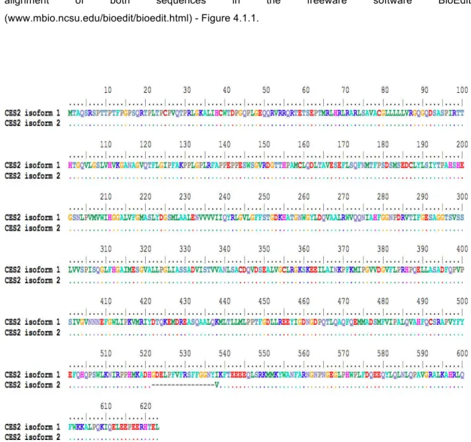
Figure 4.1.1: Sequence alignment of CES2 protein isoforms 1 and 2 (NCBI database). ... 26
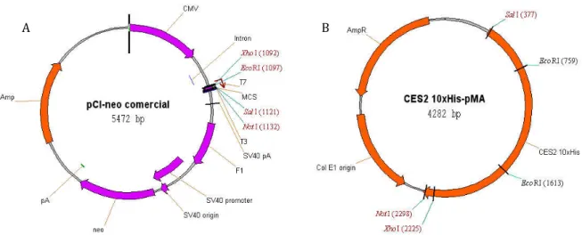
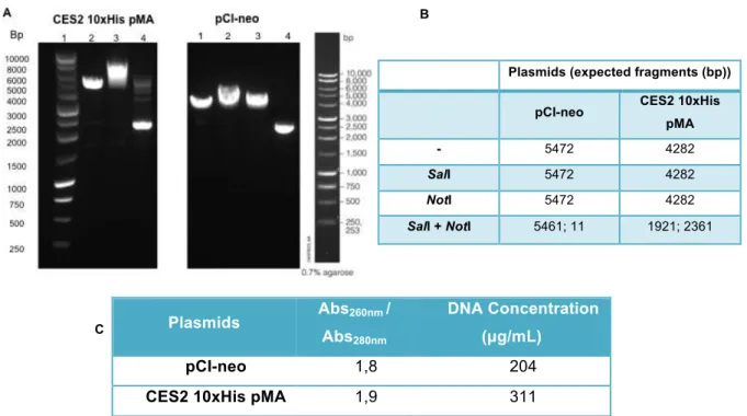
Figure 4.1.2: Schematic representation of commercial pCI-neo and CES2-10xHis-pMA plasmids. .... 28
Figure 4.1.3: pCI-neo and CES2-10xHis-pMA characterization. ... 29
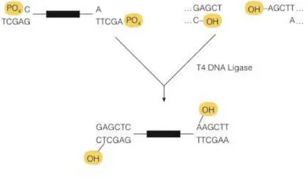
Figure 4.1.4: Illustrative representation of T4 DNA Ligase reaction mechanism.. ... 30
Figure 4.1.5: Illustrative representation of pCI-neo-CES2-10xHis and pCI-neo-CES2 plasmids. ... 31
Figure 4.1.6: Confirmation of the ligation product (pCI-neo-CES2-10xHis).. ... 32
Figure 4.2.1: Adherent HEK-293T cell growth kinetics. ... 33
Figure 4.2.2: Carboxylesterase kinetic profiles in different cell extracts - transfected HEK-293T cells with pCI-neo-CES2-10xHis and non-transfected cells ... 34
Figure 4.2.3: Recombinant CES2 expression in cell extracts. ... 35
Figure 4.3.1: Purification chromatograms of recombinant CES2 purification. ... 37
Figure 4.3.2: Analysis of purified fractions. ... 39
Figure 4.4.1: Growth of transfected HEK-293T cells in suspension culture with different DNA concentrations. ... 40
Figure 4.4.2: Total enzymatic activity of transfected HEK-293T suspension cells with different DNA concentrations (0, 2, 5, 10 and 20 µg/mL) in different time points (24, 48, 72 and 96 h) for cellular extracts. ... 41
Figure 4.4.3: Recombinant CES2 production in cell extracts from HEK-293T cells in suspension. ... 42
Figure 4.4.4: Total enzymatic activity of transfected HEK-293T cells in suspension with different DNA concentrations (0, 2, 5, 10 and 20 µg/mL) in different time points (24, 48, 72 and 96 h) for supernatant samples.. ... 43
Figure 4.4.5: Recombinant CES2 production in the supernatant and cellular extracts from HEK-293T cells in suspension. ... 44
Figure 4.4.6: CES activity towards 4-MUBA ... 45
Figure 4.4.7: Matrix effect of the recombinant CES2 production in supernatants and cellular extracts from HEK-293T cells in suspension.. ... 46
Figure Index
Figure 4.6.1: Purification chromatogram of recombinant CES2 purification from supernatant samples
of transfected HEK-293T cells in suspension with pCI-neo-CES2-10xHis. ... 51
Figure 4.6.2: Purified Recombinant CES2 protein. ... 52
Figure 4.6.3: Purified Recombinant CES2 protein. ... 53
Figure 4.7.1: Characterization of CES2 production in bioreactor. ... 54
Figure 4.7.2: Characterization of CES2 production in the 5L-bioreactor.. ... 55
Figure Index
Contents
Table index
Table 1.1: Chromatographic methods applied according to differences in protein properties. ... 4
Table 4.1: CES2 codifying gene sequence (Entrez Gene database) ... 25
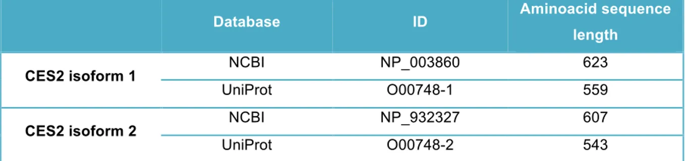
Table 4.2: Available data present in two different databases regarding the two CES2 isoforms ... 27
Table 4.3: Total protein concentration of the collected samples during the protein purification process.
... 38
Table 4.4: CES2 activity in transiently HEK-293T transfected cells, 24 h post-transfection, represented as the relative percentage of the total activity (intra and extracellular) of pCI-neo-CES2-10xHis sample. ... 48
Table 4.5: Total protein concentration of the collected samples during protein the purification process and their quantification with the Micro BCA™ Kit ... 51
Table 4.6: Characterization of the purification samples (SA and SB) ... 52
Abbreviations
Abbreviations
10xHis - Ten Histidine Tag
4-MUB - 4-methylumbelliferone
4-MUBA - 4-methylumbelliferyl acetate
Abs 260nm - UV absorbance read at 260 nm wavelength
Abs 280nm - UVabsorbance read at 280 nm wavelength
AMP - Adenosine monophosphate
Amp - Ampicillin
Anti-CES2 - Antibody raised to recognize a CES2 epitope
Anti-His - Antibody raised to recognize Histidine tagged proteins
Anti-mouse - Antibody raised to recognize a primary antibody produced in mice
Anti-rabbit - Antibody raised to recognize a primary antibody produced in rabbit
ATCC - American Type Culture Collection
ATP - Adenosine Triphosphate
BCA - Bicinchoninic acid
BHK - Baby hamster kidney cells
bp - Base Pairs
BSA - Bovine Serum Albumin
BuChE - Butyrylcholinesterase
CaPi - Calcium Phosphate
CES - Carboxylesterases
CES1 - Carboxylesterase 1
CES2 - Carboxylesterase 2
CES2-10xHis - Carboxylesterase 2 with a C-terminal ten-histidine tag
CES3 - Carboxylesterase 3
CES5 - Carboxylesterase 5 (cauxin)
CES6 - Carboxylesterase 6
CHO - Cells fromChinese hamster ovary
CMV - Cytomegalovirus
COS-7 - Kidney fibroblast cell line from African Green Monkey
CPT-11 - Irinotecan or Camptothecin-11
DMEM - Dulbecco’s Modified Eagle’s Medium
DMSO - Dimethyl sulfoxide
DNA - Deoxyribonucleic acid
DSMZ - Deutsche Sammlung von Mikroorganismen und Zellkulturen GmbH
ECACC - European Collection of Animal Cell Cultures
ECL - Enhanced chemiluminescence
Abbreviations
E. coli - Escherichia coli
EDTA - Ethylenediaminetetraacetic acid
ER - Endoplasmic reticulum
Erl - Erlenmeyer
FBS - Fetal Bovine Serum
FW - Forward
Glu - Glutamic acid
HEK-293 (T) - Human embryonic kidney cells
HeLa - Human cervical carcinoma cells
HIEL - Histidine - Isoleucine - Glutamic acid - Leucine aminoacid sequence
His - Histidine
HTEL - Histidine - Threonine - Glutamic acid - Leucine aminoacid sequence
HTIEL - Histidine - Threonine - Isoleucine - Glutamic acid - Leucine aminoacid sequence
IMAC - Immobilized metal-affinity chromatography
KDEL - Lysine - Aspartic acid - Glutamic acid - Leucine aminoacid sequence
LB - Luria Bertani
MRC-5 - Human fetal lung fibroblast cells
mRNA - Messenger Ribonucleic acid
NAG - N-acetylglucosamines
NBT/BCIP - The combination of NBT (nitro-blue tetrazolium chloride) and BCIP (5-bromo-4-chloro-3'-indolyphosphate p-toluidine salt)
NSO - Murine myeloma cells
OD - Optic density
PBS - Phosphate Buffer Saline
pCI-neo-CES2 - pCI-neo plasmid with the CES2 gene
pCI-neo-CES2-10xHis - pCI-neo plasmid with the CES2-10xHis gene
PEI - polyethylenimine
PER.C61 - Cells derived from a single human cell ρ-NPA - ρ-nitrophenyl acetate
PVDF - Polyvinylidene Fluoride
QEDL - Glutamine - Glutamic acid - Aspartic acid - Leucine aminoacid sequence
RT - Room temperature
RV - Reverse
SDS-PAGE - Sodium Dodecyl Sulfate Polyacrylamide Gel Electrophoresis
Ser - Serine
Sf21 - Insect cell line from Spodoptera frugiperda
Sf9 - Insect cell line from Spodoptera frugiperda
SIA - Sialic Acids
SN-38 - 7-Ethyl-10-Hydroxycamptothecin
Abbreviations
SV40 - Simian virus 40
T-25 - T-flask with 25 cm2
TAE - Tris-acetate-EDTA
TB - Terrific Broth
TCA - tricarboxylic acid cycle
tPA - Human tissue plasminogen activator
TTBS - Tris buffered saline with 0.05% Tween20
UV - Ultraviolet radiation
vvm - Gas volume flow per media volume per minute
Introduction
1. Introduction
1.1 - Animal Cell culture
Animal cell culture appeared more than a hundred years ago, but it was the initial need for human viral vaccines in the 1950s that accelerated the design of large-scale bioprocesses with mammalian cells (1). The enhanced interest in mammalian cell culture bioprocesses has also been associated with recombinant protein development in the 1970s and 1980s. In 1986, human tissue plasminogen activator (tPA, Activase; Genentech) (2) became the first therapeutic protein produced from mammalian cells (CHO cells) to obtain market approval. Today, 60-70% of all therapeutical recombinant proteins are produced in mammalian cells (3).
Animal cell cultures require a high level of laboratory material and continuous maintenance, which may lead to morphological and functional changes in cellular growth and in their karyotype (4). Authenticated stocks of continuous cell lines (more homogenous, more stable and more reproducible) can be acquired from a recognized animal cell culture repositories, such as the American Type Culture Collection (ATCC), Deutsche Sammlung von Mikroorganismen und Zellkulturen GmbH (DSMZ) in Germany, the Riken Gene Bank in Japan, or the European Collection of Animal Cell Cultures (ECACC) in the United Kingdom (5). The cells or tissues may be freshly isolated from animal or human donors (primary cells) or may comprise a laboratory-adapted strain or line that has been serially propagated and maintained in continuous culture (cell line) (5).
Depending on their applications, animal cells can also be grouped as (6):
(i) Cell producing proteins employed in the production of complex therapeutics, subunit vaccines, and diagnostic products, such as CHO, BHK, HEK-293, WI-38, MRC-5, SP2/0, NS0, and insect cells (7);
(ii) Cells producing viruses used in gene therapy and viral gene vaccines (for instance, Vero, HEK-293, and PER.C61 cells);
(iii) Normal cells, tumor cells, and stem cells used in research and development, specifically in the discovery of new products and for in vitro study and toxicology models (e.g. nerve cells, fibroblasts, Caco-2, MRC-5, and endothelial cells);
(iv) Human cells for subsequent use in cell therapy and regenerative medicine (e.g. embryonic and adult stem cells);
Introduction
Animal cell culture techniques are similar to those employed for bacteria, fungi, and yeast, although there are some differences. In general, animal cells are more vulnerable to mechanical damage and present lower growth rates, in some cases lower productivities and require more complex culture media and special substrates (8). Also, cell culture has to be performed under rigorous aseptic conditions, since animal cells grow more slowly than most usual microorganisms, such as bacteria and fungi. In vitro, animal cell growth is dependent on several factors, such as pH, temperature, osmolality and gas concentration, mainly oxygen (O2) and carbon dioxide (CO2) (9, 10). Culture medium needs to be buffered to compensate for CO2 and lactic acid derived from glucose metabolism.
In animal cells, glucose, a monosaccharide sugar that functions as an important energy source for metabolism, may be stored (as a polysaccharide such as glycogen or starch), oxidized to pyruvate via glycolysis to provide ATP and metabolic intermediates, or oxidized via the pentose phosphate pathway with the oxidation of NADH and FADH2 from tricarboxylic acid cycle (TCA), being the last the major metabolic pathway involved in energy production (11).
Cells in culture have a modified metabolism when compared with the same cell in the organism of origin, showing a high glycolytic flux and being unable to completely oxidize glucose, generating high amounts of lactate as an end product, leading to a rise of osmolality and decay of the pH, which combined lead to cellular growth inhibition, even under fully aerobic conditions (11). These high consumption levelsof glucose and also in glutamine (major source of energy, carbon, and nitrogen for mammalian cells), due to their inefficient utilization and consequent production of toxic metabolites such as lactate and ammonia, are a clear evidence of the unregulated metabolism. These toxic metabolites interfere in the cellular growth and in the expression of recombinant proteins (11-13). As the productivity of mammalian cell lines is some times lower compared with that of other production systems (bacteria, insect cells, etc.), it is desirable to improve the productivity per cell or the total cell yield per unit volume of culture. The knowledge of mammalian cell metabolism can help in the development of strategies for productivity enhancement, as changes in the culture conditions can affect both metabolism andproductivity (12).
To help monitor pH variation (cell culture usually from pH values between 7.0 and 7.4) some commercial media formulations have incorporated a pH indicator, such as phenol red, that enables immediate visual inspection of media pH – phenol red is rose colored at more basic pH values (7.8) becoming red, orange and yellow with the increase of the acidic conditions (7.4, 7.0 and 6.5 respectively) (14). For the control of the pH in in vitro culture systems, sodium bicarbonate is usually used in media formulation, to maintain physiological pH.
Regardless the scale or culture method (T-flask, Schott bottle, Erlenmeyer, spinner flask, or bioreactor), the temperature of the culture medium is always a fundamental variable, since it interferes not only with cellular growth and the production process, but it also affects the solubility of various
medium components, especially gases such as CO2 and O2, which have low solubilities (15). Most mammalian cells have optimal growth rates within the range of 35–37 ºC.
Introduction
also increase due to evaporation, since culture flasks are generally not sealed (to allow equilibrium between culture medium and the CO2–air gas mixture) (4).
The most important components of the gaseous phase are CO2 and O2. Monitoring and controlling these gases in the culture medium are essential procedures for the success of in vitro animal cell culturing. O2 is frequently the first component to limit achieving high cell densities due to its low solubility in aqueous medium; therefore, ideally it needs to be supplied continuously to the culture medium (15).
1.1.1 - Bioprocessing
To bring to the market a product of pharmaceutical interest successfully produced in the laboratory, further process development is needed (1), as well as the development and optimization of methods and techniques for the separation and purification of these biological macromolecules. A successful bioprocess is the one where upstream (production) and downstream (purification) find and complete each other leading to the production of the desired biopharmaceutical product with high purity and quality at a cost-effective price.
1.1.1.1 - Upstream: relevant culturing systems and process modes
Laboratory scale processes are great platforms for process development and optimization. Normally the process starts with small flasks (with adherent cells) and then it is transferred to spinner flasks or erlenmeyers (attempting to adapt the cells to grow in suspension) (1), but ultimately the process needs to be scaled-up to bioreactor, to assure it is transferable to industrial scale.
The production processes must be cost-effective and need to be well controlled in order to produce a consistent product. To obtain a high quality product, cell proliferation and product biosynthesis have to be efficient. As a practical consequence of these requirements, several types of equipment designed to control environmental culture conditions have been developed, giving rise to controlled bioreactor systems, currently well established and used in the biopharmaceutical industry (1, 3, 16), such as stirred tank (most employed), Roller Bottles, Cell Factories, Wave™, among others.
An ideal culture system has to meet requirements such as (a) control of the temperature and of the acid-base equilibrium of the culture medium; (b) to provide gas exchange by supplying O2 to the cells and promote CO2 stripping (removal of the excess of CO2), which alters the pH of the media; (c) allow an adequate supply of nutrients through the use of specifically designed formulations of culture media (9, 17); and (d) should maintain aseptic conditions, therefore avoiding contamination by microorganisms, viruses, or other cells (1, 6).
Although many different culture modes can be adopted for bioreactors, the most general is the one that considers the following operation modes: batch (discontinuous mode), fed-batch (semi-continuous), continuous, and perfusion (which is a continuous mode with cell retention) (14). The batch operation mode is the simplest to carry out, and therefore it is widely employed (1).
Introduction
and cell growth occurs without any additional supplementation of nutrients after inoculation of cells. While substrates are metabolized, the cell population grows, forming the product and other metabolites. The volume is maintained constant throughout the whole process. Due to the low solubility of oxygen, this gas must be supplied continuously, instead of just at the start of the culture (1). Control of pH is carried out through addition of base and by varying the CO2 concentration in the gas phase, since most animal cell culture media contain sodium bicarbonate, in order to allow this type of buffering control of the medium pH (6).
1.1.1.2 - Downstream: recombinant protein purification
For certain applications, biological products can be used as crude extracts with little or no purification at all (11). However, biopharmaceuticals typically require exceptional purity, making downstream processing a critical step of the overall bioprocess. Currently, proteins are the most important biopharmaceuticals (18).
Purity requirements are different for each therapeutic agent, since it depends on the intended use of the biopharmaceutical - the dose, the risk–benefit ratio, etc. The most common methods for preparative purification of proteins involve chromatography, since (a) it provides very high separation efficiencies, which allow the resolution of complex mixtures with very similar molecular properties; (b) chromatography columns packed with high capacity adsorbents are ideal for capturing molecules from the dilute solutions encountered in bioprocessing; (c) can be performed in an almost closed system and the stationary phase can be easily regenerated; and (d) chromatographic methods are well established in many practical biopharmaceutical manufacturing processes and suitable equipment and packing materials are readily available and are suitable for scale-up (18, 19).
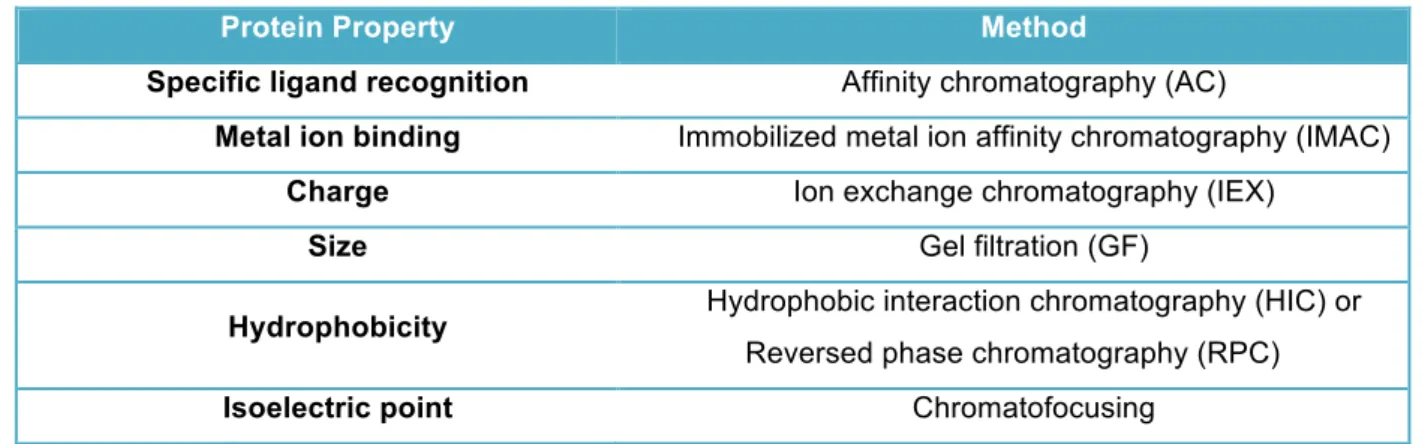
Different methods can be applied in chromatography process depending on different protein properties (Table 1.1), and allow to separate according to differences between the properties of the protein to be purified and the properties of other substances in the sample.
Table 1.1: C hrom atographic m ethods applied according to differences in protein properties.
Protein Property Method
Specific ligand recognition Affinity chromatography (AC)
Metal ion binding Immobilized metal ion affinity chromatography (IMAC)
Charge Ion exchange chromatography (IEX)
Size Gel filtration (GF)
Hydrophobicity Hydrophobic interaction chromatography (HIC) or
Reversed phase chromatography (RPC)
Isoelectric point Chromatofocusing
Introduction
will not bind or will bind weakly. IMAC is a versatile method that can be utilized to rapidly purify polyhistidine affinity-tagged proteins with high yields and up to 95% of purity (20). This method is based on the interactions between a transition metal ion (Co2+, Ni2+, Cu2+, Zn2+) immobilized on a matrix and specific amino acid side chains (20, 21). These matrices securely coordinate metal ions through four coordination sites while leaving two of the transition metal coordination sites exposed to interact with histidine residues in the affinity tag (20, 22).
1.1.2 - Cell line
In order to obtain high amounts of a desirable recombinant protein, the choice of the host cell line is crucial - it should be susceptible to transfection, be able to transcribe, translate, fold, and process the protein and, if possible, to secrete it to the culture medium (7). On the other hand, it is advisable
that the selected cell line grows in a serum-free media, since recovery of the recombinant protein from culture media with a low protein content is simpler (23) and the risk of contamination with animal pathogens is smaller, reducing the burden of quality control analytics and the approval process simpler (24). Post-translational modifications, such as glycosylation, are fundamental to insure the full biological activity of proteins (11). Eukaryotic cells have the metabolic capability to perform these modifications and so, mammalian cells have become the host cells of choice to produce recombinant proteins with complex post-translational modifications (16, 25).
One of the most used cell lines in industry for recombinant protein is human embryonic kidney cell line (HEK-293). HEK-293 cell line exhibits epithelial morphology and is derived from human embryonic kidney cells, by the transformation of these cells with a fragment of adenovirus type 5 DNA.
This cell line is well suited for large-scale production and has desirable features such as its quick and easy reproduction and maintenance, amenability to transfection using a wide variety of methods, high efficiency of transfection and protein production, faithful translation and processing of proteins (26), growing easily in suspension and being adaptable to serum-free medium (7, 26). They also stably express the adenovirus 13-S-E1a protein that has been shown to significantly enhance transcription from the cytomegalovirus (CMV) promoter (further discussed in Section 1.1.4) (27).
The HEK-293T cell line has been explored for viral vector production for gene therapy and for obtaining human recombinant proteins with normal glycosylation profiles (4). In addition, this cell line is a highly transfectable derivative of the HEK-293 cell line into which the temperature sensitive gene for SV40 T-antigen was inserted (28), which allow episomal replication of transfected plasmids containing the SV40 origin of replication.
1.1.3 - Protein processing
Introduction
In eukaryotes, proteins are synthesized on ribosomes in the cytosol and then are directed to their cellular destinations. This process uses a short sequence of amino acids called a signal sequence, whose function is to direct a protein to its appropriate location in the cell and, for many proteins, is removed during transport or after the protein has reached its final destination. For instance, most lysosomal, membrane, or secreted proteins have an amino-terminal signal sequence that marks them for translocation into the lumen of the endoplasmic reticulum. The signal sequence has a cleavage site where proteases remove the sequence after the protein is imported into the ER (11).
In the ER lumen, newly synthesized proteins are further modified in several ways, following the removal of signal sequences, polypeptides folding, disulfide bonds formation, and many proteins glycosylated to form glycoproteins. Proteins travel from the ER to the Golgi complex in transport vesicles and once there, the Golgi complex also sorts proteins and sends them to their final destinations (29).
1.1.4 - Expressing heterologous proteins in mammalian cells
Cell lines can be generated following delivery of the gene of interest and the selection gene into host cells by transfection (30). In fact, to develop a successful production process, several aspects need to be optimized, namely, correct selection of the expression vector, host cell, and culture medium and also the deoxyribonucleic (DNA) acid quality, the transfection vehicle and the culture medium (4, 7, 28). Moreover, the genetic manipulation of the first two and the choice of the appropriate method for transfection and genetic amplification may influence positively the specific cellular productivity. An increase in the specific productivity could involve enhancement at the genetic level by gene amplification or by the addition of an inducer to enhance the transcription of a gene.
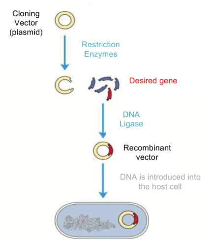
Cloning (Figure 1.1 (11, 31)) is the basic procedure in molecular biology required to move inserts from one vector to another to gain the desired functionality and thus creating a functional vector with the desired gene in it, which later will be transferred to the cells (31).
For the expression of heterologous proteins, it is not enough just to have the sequence of DNA encoding for the protein of interest. The presence of sequences that regulate gene expression are essential. The promoter is the sequence located upstream from the beginning of the transcription, whose function is also to regulate the initiation of the transcription of the adjacent gene (32). The CMV promoter allows for strong, constitutive expression in a variety of cell types (7). Also, the presence of introns (non-coding sequences further excised from the precursor mRNA by specific cleavage in a process known as splicing) (32) significantly improves the efficiency of transcription and thus the expression of recombinant proteins, by increasing mRNA stability and export from the nucleus (7).
Introduction
Figure 1.1: Illustrative schem e of the D N A cloning process. A dapted from (11).
Nowadays, physical, biological and chemical methods are applied to introduce genetic material into the cells and this process may comprise temporary introduction of DNA into the host cell (transient transfection) or permanent integration into the genome (stable transfection) with clone selection through the use of antibiotics. Transfection may be performed by physical methods such as electroporation or microinjection, which require special instruments and their application is often restricted to special cell lines or tissues (4); by biological systems for nucleic acid delivery, which often employ viruses. Viral transfection methods use genetically modified viruses that are no longer pathogenic, however their application is limited by viral-related immunogenicity and the size limitation of the transgene. This system uses complex methods that are not easy to use for general applications (4). The most widely applied transfection method (transient) is the chemical one using cationic lipids, cationic polymers or Calcium Phosphate (CaPi) precipitates. These vehicles should allow the DNA condensation, promote its binding to the cell membrane and facilitate its entry into the cell and nucleus (7). Available cationic lipids may be applied, which result in high expression levels, although CaPi and the polycation polyethylenimine (PEI) are currently the two most cost-effective and efficient transfection vehicles used. These methods are easy to use and do not require any additional laboratory instruments. PEI offers additional advantages over CaPi, since it is simpler to use, has compatibility with serum-free medium (7), and is also efficient in suspension cultures (34).
Cloning
Vector
(plasmid)
Desired gene
Restriction
Enzymes
DNA
Ligase
Recombinant
vector
Introduction
1.2 - Carboxylesterases
Carboxylesterases (CES) comprise a multigene superfamily (35) and are categorized as phase-I drug-metabolizing enzymes, since is their responsibility the detoxification of a wide a range of ester containing xenobiotics (biotransformation of these compounds to polar products to facilitate their elimination) (36), such as heroin and cocaine (37, 38), but also drugs and prodrugs (39) as well as insecticides, such as pyrethroids (40, 41). They are also involved in several lipid metabolic reactions (42, 43) and may also be connected to the assembly of low-density lipoprotein particles in the liver (44). An increased interest in the CES field arise due to their application to design herbicides with selective toxicity (45, 46), to their potential use for treatment of drug overdose, addiction, and chemical warfare, as well as cancer prodrug combined therapy (47).
CES are a subset of esterases (48) (EC 3.1.1.1) (38), which are hydrolytic enzymes that catalyze the conversion of carboxylic esters to their corresponding alcohols and carboxylic acids. Thus, drugs such as heroin, cocaine, irinotecan, capecitabine, oseltamivir (Tamiflu), lidocaine, and meperidine (Demerol) are all hydrolyzed by CES (49). Besides these, several other clinically used compounds are esterified, since this is an approach commonly used by the pharmaceutical industry to improve the water solubility of molecules, and thus are also hydrolyzed by CES.
Mammalian CES genes usually contain 12-14 exons of DNA encoding CES enzyme sequences, which may be shuffled during mRNA synthesis, generating several CES transcripts and enzymes encoded by each of the CES genes (36). CES were initially classified by their substrate specificity and isoelectric point (pI) (50). However, this classification is ambiguous in overlapping substrate specificities, for instance, a single esterolytic reaction can be mediated by several kinds of enzymes. Since CES comprises a superfamily of genes, identification of homology and similarity of characteristics are acceptable factors to classify these enzymes. Thus, mammalian CES nomenclature has been recently reviewed (51) and gives (a) a name for each human (CES) or mouse and rat (Ces) gene; (b) names and identifies the gene family of origin for identified CES pseudogenes; and (c) provides a system for naming transcript isoforms derived from each of the CES genes. This new classification for mammalian CES comprises at least five gene families including carboxylesterase 1 (CES1), the major liver enzyme (42, 52), CES2, the major intestinal enzyme (53, 54), CES3, expressed in the brain, liver, and colon (55), CES5 (also called CES7 or cauxin), a major urinary protein of the domestic cat also present in human tissues (56), and CES6, a predicted CES-like enzyme in the brain (57).
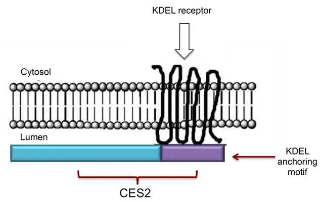
Introduction
Figure 1.2: Illustrative representation of the internal localization of C E S 2 protein. A dapted from (36).
1.2.1 - CES characterization
The two major expressed human CES are CES1 and CES2. CES1 is highly expressed in the liver and also observed in macrophages, human lung epithelia, heart, testis and other tissues (59). CES2 is present in the small intestine, colon, kidney, liver, heart, brain and testis (36). CES1 and CES2 are two different gene products and the two genes have only 48% amino acid sequence homology (41, 59).
Mammalian liver is predominantly responsible for drug detoxification (where CES1 is present in higher amounts than CES2), with CES1 and CES2 playing major roles, following absorption of drugs into the circulation (37, 59), while mammalian intestine (where CES2 is present in higher amounts than CES1), has a key role in first-pass clearance of several drugs, predominantly via CES2 in the ileum and jejunum (51).
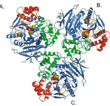
To date, from human carboxylesterases, only the CES1 crystallographic structure is available (60) (Figure 1.3), confirming that this enzyme is found in trimers composed of approximately 60 kDa monomers. This enzyme has also been described to occur as hexamers (60), existing in a trimer-hexamer equilibrium that could be shifted towards trimer or trimer-hexamer, depending on the substrate present (60).
The three-dimensional structural analysis of CES1 (Figure 1.3), revealed that this enzyme exhibits the α/β-hydrolase-fold, typical of serineesterases, but also contains a large substrate binding area with both rigid and flexible pockets (60). In fact, it pointed to three major ligand binding sites, including the broad-specificity active site (which is lined largely by hydrophobic residues which surround a serine esterase catalytic triad composed of Serine-221, Histidine-468, and Glutamic acid-354), the
KDEL receptor
Cytosol
Lumen
CES2
KDEL
anchoring
Introduction
"side door" and the "Z-site", where substrates, fatty acids and cholesterol analogs, respectively, are bound (60).
Figure 1.3: Illustrative representation of the tridim ensional structure of the hum an carboxylesterase 1 trim er (A , B and C m onom ers) com plexed w ith tacrine (potent inhibitor of hum an acetylcholinesterase). The catalytic dom ains, α/β dom ains, and regulatory dom ains of each m onom er are in blue, green, and red, respectively, the novel Ω loops are in orange, the N-acetylglucosam ines (N AG ) are in cyan, and the sialic acids (SIA) are in dark green (60).
A Ν-glycosylation site was described for CES1, contributing to its stability and maintenance of catalytic efficiency (61), also potential Ν-glycosylation sites were observed although in different positions for other CES enzymes and may perform similar functions as to the one on CES1 (51).
Mammalian CES families show promiscuity toward a wide range of substrates, exhibiting a broad substrate specificity, which leads to difficulties in establishing specific roles for these enzymes (36, 51). CES1 preferentially catalyzes the hydrolysis of compounds esterified with a small alcohol and a large acyl group such as clopidogrel, while CES2 hydrolyzes compounds with a large alcohol group and a small acyl group (59), such as cocaine, 4-methyumbelliferyl acetate, heroin and 6-monoacetylmorphine (37).
Introduction
1.2.2 - CES2 manufacturing in different systems
Recombinant active CES expression has already been reported in the literature, for several mammalian (rat, rabbit, porcine, human) CES, in different cell types, such as yeasts (67, 68), insects (68-71) and mammalian cells (65, 68, 72). Purification using bacterial strains has proved to be unsuccessful since despite significant amounts of recombinant CES were expressed, however very low level of enzyme activity was observed (68). For enzymes that are secreted from or sequestered within the endoplasmic reticulum of mammalian cells, expression in bacteria may yield inappropriately folded and/or glycosylated proteins (68).
The expression of recombinant CES is mainly reported intracellularly (68-70, 72). However, the secretion of CES has been previously reported in yeast (73), insect (68) and mammalian cells (39), always comprising the modification/deletion of the retention signal and/or the addition of signalling N-terminus sequences. An alternative pig liver esterase containing the α-factor signal sequence of Saccharomyces cerevisiae and missing the five last C-terminus amino acid residues was produced in liquid cell culturing of Pichia pastoris (73). A rabbit liver carboxylesterase without the six last C-terminus amino acid residues was produced in serum-free Sf21 insect cells (68) and human CES2 was reported to be produced, with 60% purity grade, also in a secreted form in Sf9 insect cells (71), and in Sf21 cells, with 98% purity grade (38). A secreted form of human CES2 was reported in COS-7 cells, without the C-terminal ER anchoring motif and with an Ig κ-chain leader sequence (directs proteins to the secretory pathway) (39) and in human cervical carcinoma (HeLa) cells, without the KDEL C-terminal amino acid residues (65).
Previous reports of CES purification produced in insect cells show a wide range on protein yield and purity. Human CES2 produced intracellularly in Sf9 insect cells was purified with a 0.8% yield (70). Rabbit liver carboxylesterase, produced extracellularly using Sf21 insect cells originated an active protein with, according to the authors, 98% purity grade and 61.6% yield (68).
Goals
Development of a mammalian expression
vector plasmid
Mammalian cells transfection
Adherent cell culture
(T-Flask)
Suspension cell culture (Erlenmeyer
Bioreactor
Process optimization
Activity Assays
Western Blot
Purified CES2
CES2 purification
Activity Assays
Western Blot
2. Goals
The main goal of this project was the development and optimization of a process for the production and purification of the human carboxylesterase enzyme (CES2).
This protein has a key role in the metabolism of ester containing xenobiotics (41) and also in the activation of several prodrugs (49). Therefore, it can influence significantly their bioavailability or contribute to the reduction of their secondary effects (65).
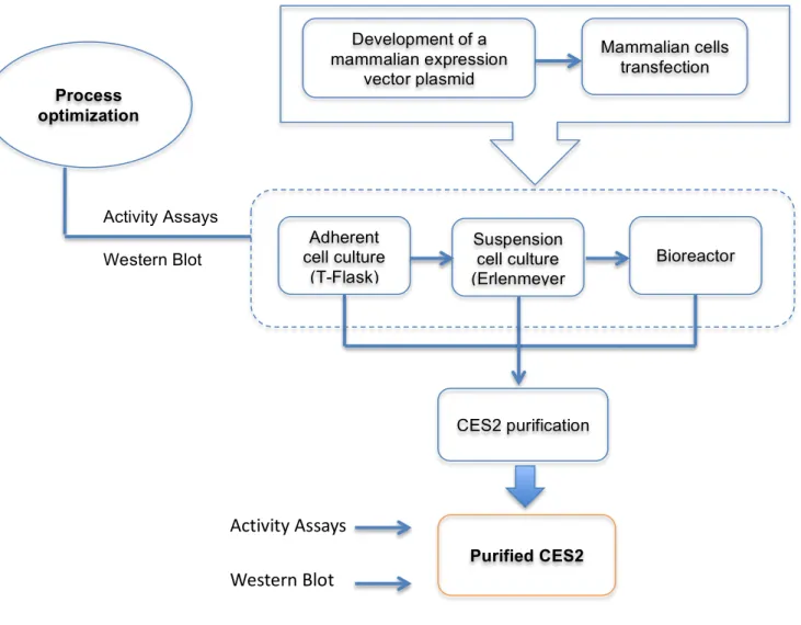
The knowledge of this protein structure will be crucial for the understanding of its properties, as well as to further understand the well described similarities and differences between CES1 and CES2 in their behaviour towards different substrates and inhibitors (60, 78). However, one of the main bottlenecks for these studies is the unavailability of purified CES2, whose structure is not yet known, unlike CES1. Therefore, it becomes relevant the establishment of a robust platform for the production of this protein (the workflow is represented in Figure 2.1), in order to have the enough protein amount (1 - 10 mg of highly pure protein) to proceed for the crystallographic studies.
Goals
Materials and Methods
3. Materials and Methods
3.1 Biological Material and Culture Media
3.1.1 Bacterial Strain
Escherichia coli (E. coli) DH5‐α Library Efficiency (Invitrogen, Carlsbad, USA) competent cells were used for the production of the DNA plasmids. The bacteria transformation was performed following the manufacturer's instructions. Working banks of transformed bacteria for each used plasmid were created - the bacteria were grown until optical density approximately of 2 was reached at 600 nm (OD 600 nm). The bacteria suspension was mixed with 15% of glycerol, aliquoted and frozen at -80 ºC.
The bacterial culture was performed with Luria Bertani (LB) (NZYTech, Lisboa, Portugal) medium, supplemented with 100 µg/mL of Ampicillin (Sigma, St. Louis, U.S.A.) and with Terrific Broth (TB) (Fast-Media® Amp TB from Invivogen, California, U.S.A.). LB medium was sterilized for 30 minutes at 121 ºC and 1.2 bar, and afterwards supplemented with ampicillin. This medium was used for low cell density culture of transformed bacteria. Sterile TB medium was used for high cell density culture of transformed bacteria. Both media were prepared accordingly the manufacturer's instructions and used for extraction of high quantity and quality of DNA plasmids. For the preparation of both media, ultrapure water (Millipore, Billerica, U.S.A.) was used.
Materials and Methods
3.1.2 Mammalian cell line
HEK-293T cell line is derived from HEK-293 cell line in which the SV40 T-antigen gene was inserted (7). This cell line was purchased from ATCC (CRL‐11268) and used in this work.
In a first stage of this work, HEK-293T cell line was cultured in adherent monolayers in T-flasks with 25, 75 and 175 cm2 (BD, New Jersey, U.S.A.) using high glucose DMEM media (Dulbecco’s Modified Eagle’s Medium, Gibco®, Grand Island, U.S.A.), containing 0.11 g/L of pyruvate, 4.5 g/L of glucose, 0.58 g/L of L-glutamine and 3.7 g/L of sodium bicarbonate (NaHCO3), supplemented with 10% Fetal Bovine Serum (Gibco) (FBS) at 37 ºC with 5 % CO2. Cells were detached from T-flask surface with Trypsin 0.05%- EDTA (Gibco), and were split twice a week after reaching 70 - 80% confluence.
Afterwards, in-house suspension adapted HEK-293T were cultured in Freestyle™ 293 (Gibco) serum-free, animal origin-free, chemically defined culture media formulated with Glutamax™-I (contains a dipeptide, L-alanyl-L-glutamine, a stabilized form of L-glutamine, Invitrogen). This medium was specifically developed for the high-density, suspension culture and transfection of 293 cells grown in suspension conditions and was stored, protected from light, at 4 ºC.
The cells were sub-cultured when their density was approximately 2 - 3 x 10⁶ viable cell/mL, typically every 3 - 4 days. Briefly, cellular suspension was centrifuged at 200 g for 10 minutes (min) at room temperature (RT) and the sediment was carefully ressuspended with fresh Freestyle™ 293 medium. The obtained cellular suspension was used to inoculate new Erlenmeyers with a cellular concentration of 0.3 - 0.5 x10⁶ cell/mL. The centrifuging step was performed in every 2 - 3 cell passages. Cells were maintained either in 125 or 500 mL polycarbonate, disposable, sterile Erlenmeyer flask (Corning) or glass, sterile Erlenmeyer flasks (Shott), containing 20 to 25 mL or 80 to 90 mL total working volume of cell suspension, respectively. The erlenmeyer flasks were incubated at 37 ºC in a humidified atmosphere of 8 % CO2, using orbital agitation (130 rpm). The assays for optimization of the transfection conditions were performed in Erlenmeyers with 90 mL (working volume).
Materials and Methods
Mammalian cells, when continuously cultured in vitro, may suffer morphological and functional changes in their growth pattern or even in their karyotype (4). When properly frozen, the cells may be preserved for a long-term period of time without suffering these changes. Thus, a cell bank with cells properly frozen and preserved allows to repeat the cell culture, from a certain cell lot, with identical characteristics.
Adherent HEK-293T cells were frozen with a concentration of 2 x 10⁶ cell/mL per cryovial. A specific freezing solution was used, constituted by 95% of FBS and 5% of dimethyl sulfoxide (DMSO, Sigma). DMSO is a conventional cryoprotector that is toxic at RT (79), reason why the cell freezing process needs to be done at low temperatures (achieved by using cooled down cryovials and freezing solutions). Cells were frozen at a -1 ºC/minute cooling rate using 5100 Cryo 1 ºC Freezing Container, “Mr. Frosty” (Nalgene Nunc, New York, U.S.A.). Briefly, the cells grew until the exponential phase was reached and then they were sedimented by centrifugation at 200 g for 10 min at 4 ºC. The cells were carefully ressuspended in the freezing solution. The cellular suspension was aliquot in cryovials (Nalgene Nunc) and remained for 20 minutes at 4 ºC. The cryovials were then transferred to the "Mr. Frosty" at -80 ºC. After 24 h, one cryovial was thawed to confirm the freezing efficiency. The thawed cryovial was incubated at 37 ºC and immediately after thawing the cellular suspension was carefully added to fresh medium and to a T-Flask. After 3 h, the medium from the T-Flask was removed (containing DMSO) and replaced by fresh medium.
The HEK-293T suspension cells were frozen with an average concentration of 10 x 106 cell/mL or 4.5 x 10⁷ cell/mL to thaw directly into a 125 mL or to a 500 mL Erlenmeyers, respectively. The cells grew until the exponential phase was reached and then they were sedimented by centrifugation at 200 g for 10 min at 4ºC. The cell pellet was carefully ressuspended in the freezing solution CryoStor™CS10 (STEMCELL Technologies, Grenoble, France). The cellular suspension was aliquoted in cryovials (Nalgene Nunc) and remained for 20 minutes at 4 ºC. The cryovials were then
transferred to the "Mr. Frosty" at -80 ºC. After 24 h, one cryovial was thawed to confirm the freezing efficiency. The thawed cryovial was incubated at 37 ºC and immediately after thawing the cellular suspension was carefully added to fresh medium and centrifuged at 200 g for 10 min at 4 ºC. Due to DMSO's toxicity to the cells at RT, its quick removal from the cell suspension is critical. The cell sediment is then ressuspended in fresh medium and then added to the Erlenmeyer.
3.1.3 Mammalian Expression vectors
Materials and Methods
During this project, working DNA banks for each of these plasmids were generated and stored at -20 ºC.
Plasmid purification was performed in different scales. Small-scale purification (yields around 20 µg of DNA) was performed with QIAprep® Miniprep (Qiagen, Hilden, Germany). A larger scale purification (yields around 500 µg of DNA) was performed with Genopure Plasmid Maxi Kit (Roche, Basel, Switzerland) following manufacturer’s instructions. Large-scale purification was performed with QIAprep® Gigaprep (Qiagen) (yield of 25 mg of DNA) following the manufacturer's instructions. The isolation procedures are based on a modified alkaline lysis protocol followed by absorption of DNA onto silica in the presence of high salt. The bacteria are partially lysed, allowing the plasmid DNA to escape the cell wall into the supernatant, while the larger E. coli chromossomal DNA is trapped in the cell wall. Then the lysate is cleared of cellular debris by centrifugation and the plasmid DNA containing fraction is added to a column. The bound plasmid DNA is washed to remove contaminating bacterial components and then is eluted in a low-salt buffer. The DNA is eluted and precipitated to remove salt and to concentrate the eluate.
3.2 Transfection assays
The transfection assays were carried out with the polycation polymer polyethylenimine (PEI, Linear 25 kDa from Polysciences, Eppelheim, Germany) at 1:3 ratio of DNA:PEI, based on previous studies (80). For the preparation of the transfection solution, which contains fresh serum-free-media, DNA and PEI, it was used a PEI stock solution with 1 mg/mL. This solution was prepared by the addition of the medium, DNA and PEI. This solution was vigorously stirred and incubated for 10 min at RT before being added drop wise to the cell culture. HEK-293T cells were transiently transfected with pCI-neo-CES2 or pCI-neo-pCI-neo-CES2-10xHis plasmids.
3.2.1 Static culture
HEK-293T cells were seeded in 100 mm sterile Petri dishes, with a seeding inoculum of 5 x 10⁴ cell/cm2 and transfected 24 h after cell seeding. Transfection was performed using 30 µg (16.6 µg/mL) of plasmid DNA (pCI-neo-CES2-10xHis) and 90 µg of PEI in a final volume of 1.8 mL per plate. Cells were harvested (by centrifugation at 200 g, 10 min, 4 ºC) 48 h after transfection and chemically lysed with 750 µL of M-PER® Mammalian Extraction Reagent (Pierce Biotechnology, Rockford, U.S.A.). After lysis the lysate was centrifuged at 12000 g at 4ºC for 15 min and the supernatant was stored at -20 ºC.
3.2.2 Suspension culture
Materials and Methods
suspension volume. Different plasmid DNA concentrations were tested: 2, 5, 10 and 20 µg/mL to optimize the transfection assay. Cell growth and viability was followed throughout a culture time period of 96 h. In each time point (24, 48, 72 and 96 h post-transfection) a sample of 10 mL was taken from the Erlenmeyer, centrifuged at 200 g, at 4 ºC for 10 min. The supernatant was stored at -20 ºC and the sedimented cell pellet was lysed with 200 µL of M-PER® Mammalian Extraction Reagent (Pierce Biotechnology) and further clarified at 12000 g for 10 min at 4 ºC. The cells extracts were stored at -20 ºC. No protease inhibitors were added for both samples.
After optimizing the conditions, all the transfections assays were performed only with 5 µg/mL DNA concentration. Cells and supernatant were harvested after 96 h.
3.3 Brefeldin A assay
Inhibition of protein secretion was performed with brefeldin A antibiotic (99.9%, Aldrich, Steinheim, Germany), as described before (81), with some modifications. HEK-293T suspension cells were transiently transfected with 5 µg/mL of pCI-neo-CES2-10xHis or pCI-neo-CES2 expression vectors, as described in section 3.2.2. At 24, 48, 72 and 96 h post-transfection, two samples of 10 mL from each transfection type were centrifuged at 200 g, for 10 min. The cellular pellet was ressuspended in FreeStyle™ Expression medium or in the same medium, but containing 10 µg/mL of brefeldin A and were incubated, for 4 h. Cells extracts and supernatants were processed as described above.
3.4 Protein Purification
For the purification of CES2-10xHistidine, platform ÄKTAexplorer™ 10S System (GE Healthcare, Little Chalfont, U.K.), a purification platform based on chromatographic analysis and separation, was used, at 4 ºC.
3.4.1 CES2 purification from cell extracts of transfected HEK-293T adherent cells
Materials and Methods
Purified samples were loaded into a desalting column (Sephadex™ G-25 desalting column HiPrep™ 26/10, GE Healthcare) to remove imidazole (since it damages the enzyme activity). Samples were eluted in Storage Buffer (20 mM CH3COONa (≥ 99.0%, Fluka, Seelze, Germany), 600 mM NaCl pH 5.0), and then filtered under sterile conditions with 0.2 µm filters (low protein binding, PALL Corporation, New York, U.S.A.). For proper storage it was added 20% of sterile glycerol (Sigma) to each sample and the samples were stored at -80 ºC.
3.4.2 CES2 purification from cell extracts of transfected HEK-293T suspension cells
Culture supernatant containing the soluble human recombinant CES2-10xHis protein was analysed and stored at -20 ºC until the beginning of the purification process.
Purification of human recombinant CES2-10xHis was performed using a 5 mL HiTrap Chelating High Performance column (GE Healthcare), charged with 100 mM of nickel sulphate and equilibrated with 20 mM sodium phosphate buffer, pH 7.4, containing 10 mM imidazole and 500 mM sodium chloride. 210 mL of cultures supernatants, obtained as described above and further clarified by centrifugation at 10000 g, for 10 min at 4 ºC, were diluted in an equal volume of 20 mM sodium phosphate buffer, pH 7.4, containing 20 mM imidazole and 500 mM NaCl, and loaded onto the column. A washing step was performed with the same buffer until stabilization of the baseline. Elution of the His-tagged bound proteins was performed with a two-step imidazole gradient, from 10 to 500 mM. The first slower step was performed until 250 mM of imidazole with 10 column volumes. The following step, until 500 mM of imidazole was performed with 5 column volumes. A flow rate of 5 mL/min was used. Buffer exchange and concentration with 20 mM sodium acetate buffer, pH 5.0, containing 600 mM sodium chloride (Storage Buffer), was performed to all eluted fractions using Vivaflow cassettes (Sartorius Stedim Biotech) with a membrane cut off of 30 kDa. After imidazole removal, all samples were sterilized by filtration (0.2 µm filters (low protein binding, PALL)). After the addition of 20 % glycerol, all samples were aliquoted and stored at -80 ºC. All fractions were analysed by SDS-PAGE and western blot.
3.5 Analytical tests
3.5.1 Quality of the purified plasmids
The DNA concentration was determined by the Lambert-Beer law (A = ε x l x C, where ε is the extinction coefficient, l is the path length and C the sample concentration) with absorbance
Materials and Methods
Ethidium Bromide, and visualized under UV light (in a transilluminator from Uvitec, Cambridge, England).
Restriction analysis was performed with restriction enzymes from New England Biolabs - SalI, NotI and XhoI. In short, all the necessary components - nuclease-free water (Promega), 10X restriction buffer, Bovine Serum Albumin (BSA), DNA and enzyme(s) - were mixed by pipetting. These reactions were performed at 37 ºC for 3 h.
3.5.2 Determination of cell concentration and viability
Cell concentration and viability were determined by the trypan blue exclusion method using a 0.1% (v/v) solution prepared in Phosphate Buffer Saline (PBS, Gibco) and counting cells in a Fuchs-Rosenthal haemacytometer (Brand, Wertheim, Germany). Each counting was performed at least twice and a 15% error percentage was assumed.
3.5.3 Metabolite quantification
Glucose and lactate were quantified in an automated analyzer (YSI 7100 Multiparameter Bioanalytical System (Dayton, Ohio, USA). These measurements have a 5% error associated.
3.5.4 Protein Quantification
Total protein quantification in cell extracts was performed by the bicinchoninic acid (BCA) assay with BCA™ Protein Assay Kit (Pierce Biotechnology), using BSA as standard, according to the manufacturer’s instructions. The BCA Protein Assay is a detergent-compatible formulation based on bicinchoninic acid for the colorimetric detection and quantitation of total protein. This method combines the well-known reduction of Cu2+ to Cu+ by protein in an alkaline medium (the biuret reaction) with the highly sensitive and selective colorimetric detection of the cuprous cation (Cu+) using a reagent containing bicinchoninic acid. The purple-colored reaction product of this assay is formed by the chelation of two molecules of BCA with one cuprous ion. This water-soluble complex exhibits a strong absorbance at 562 nm that is nearly linear with increasing protein concentrations over a broad working range (20 - 2000 µg/mL).
In some cases, it was used the Micro BCA™ Kit (Pierce Biotechnology) to quantify more dilute protein samples (0.5 - 20 µg/mL), following the manufacturer's instructions.