ABSTRACT
Introduction: The diagnosis of Primary Sjögren's Syndrome (pSS) is sometimes challenging,
taking into account the clinical heterogeneity and the variability of diagnostic criteria fulfillment. This study aims to evaluate the tear meniscus (TM) and corneal sub-basal nerve plexus (SBNP) in a population with pSS.
Material and Methods: Cross-sectional study of 50 patients with pSS and 20 healthy controls.
Conventional dry-eye disease evaluation was performed, using Schirmer test I without anaesthesia, tear break-up time and corneal fluorescein staining. TM height and area were analyzed using anterior segment optical coherence tomography (AS-OCT) and corneal SBNP was evaluated using in vivo Confocal Microscopy (IVCM), with study of nerve density, length and tortuosity. Differences in pSS ophthalmic features were analyzed according to phenotype profile. Data analysis was conducted by IBM-SPSS Statistics 25.0.
Results: TM height was significantly lower in pSS in contrast to the controls (p < 0.001).
Corneal SBNP density and length were significantly lower and tortuosity significantly higher in pSS patients comparing to controls (p < 0.001, p = 0.004 and p = 0.001, respectively).
Conclusion: AS-OCT and IVCM may be useful in the evaluation of lachrymal unit
dysfunction in pSS, complementing the conventional methods used in the evaluation of dry-eye disease.
RESUMO
Intrudução: O diagnóstico de Síndrome de Sjögren’s Primário (SSp) é, por vezes, desafiador,
atendendo à variabilidade da sintomatologia e do cumprimento dos critérios diagnósticos. Este estudo tem como objectivo a avaliação do menisco do lacrimal (ML) e plexo do nervoso sub-basal da córnea (PNSB) na SSp.
Materais e Métodos: Estudo transversal de 50 doentes com SSp e 20 controlos saudáveis.
Foram realizados o teste de Schirmer I, BUT e coloração corneana com fluoresceína como avaliação convencional de síndrome de olho seco. O ML foi analisado quanto à altura e área, através da tomografia de coerência óptica de segmento anterior (OCT-SA); e o PNSB da córnea foi avaliado por Microscopia Confocal in vivo (MCIV), com estudo da densidade, comprimento e tortuosidade nervosa. A análise estatísticas foi conduzida IBM-SPSS Statistics 25.0.
Resultados: A altura do ML revelou-se significativamente menor nos doentes com SSp
comparativamente ao grupo controle. O PNSB da córnea revelou uma densidade e comprimento significativamente menores e tortuosidade significativamente maior nos doente com SSp comparativamente ao grupo controle (p <0,001).
Conclusão: O OCT-SA e a MCIV podem ser úteis na avaliação da disfunção da unidade
funcional lacrimal na SSp, complementando a metodologia convencional utilizada na avaliação da síndrome de olho seco.
INTRODUCTION
Primary Sjögren’s syndrome (pSS) is a chronic
progressive autoimmune disease characterized by
lymphocytic infiltration, damage and consequent loss of function of the exocrine glands.1 The combination of xerostomia, xerophthalmia, fatigue and joint pain is the characteristic presentation form of this exocrinopathy, which affects mainly caucasian women, beginning around the fourth to fifth decade of life.2
Ocular symptoms and signs are preponderant points of the American–European Consensus Group (AECG) criteria for pSS classification,3 the most widely used since 2002. However, the two ocular objective signs considered in these criteria, namely Schirmer’s test I and ocular staining score, are not perfect for diagnosis, since they have low to moderate reproducibility in assessing dry-eye disease and might be insufficient in differentiating pSS from other dry-eye conditions, as well as to assess disease severity and activity.4–10
In this sense, the interest in developing novel objective and reproducible tools for pSS assessment is growing.9,11,12 Regarding the aqueous tear deficiency and the resulting ocular surface damage associated with pSS dry-eye, the objective evaluation of tear meniscus (TM), using anterior segment optical coherence tomography (AS-OCT) and corneal sub-basal nerve plexus (SBNP) by in vivo Confocal
Microscopy (IVCM), may reveal important information about the status of the ocular surface of these patients.11,13–16
Therefore, in this study, we aimed to analyse the usefulness of TM AS-OCT and SBNP-IVCM in detecting pSS, as well as in distinguishing pSS patients according to disease activity, immunological and histological profiles.
METHODS
Study design and Population
Data were collected from a cross-sectional study of fifty patients with pSS and twenty healthy volunteer women.
pSS was confirmed following the AECG criteria,3 and patients were considered to have clinical active disease if they scored in any domain of the clinical European League
Against Rheumatism-(EULAR) Sjögren’s Syndrome
Disease Activity Index (ClinESSDAI). The ClinESSDAI was developed by removing the Biological domain from ESSDAI (C3 and C4 complement factors, gammaglobulin and cryoglobulins) and intends to be independent from B-cell derived biomarkers.17 However, there seems to be a high concordance between both indexes.18
The exclusion criteria proposed by the AECG were applied to all subjects in study, as well as other systemic or organ-specific autoimmune disease; history of IgG4-related
disease; neurodegenerative diseases; treatment with drugs of known corneal toxicity or xerogenic effect; eyelid malposition or movement disorders; lacrimal drainage pathway obstruction; ocular surface diseases other than DED; history of previous corneal LASER procedures, refractive, glaucoma or retinal surgeries; contact lens wear within 2 weeks from the ophthalmic evaluation. Additionally, patients were instructed not to use artificial tears within eight hours before their ophthalmic evaluation.
Healthy volunteers, without any sicca symptoms or signs, fulfilling the eligibility criteria, constituted the control group.
A complete ophthalmologic (described in the next section) was performed. Demographic, clinical and laboratorial data were collected from the clinical files, upon which the ClinESSDAI was assessed. Written informed consent has been obtained from all subjects prior to their participation in the study. Approval by the Institutional Ethics' committees was obtained and the tenets of the Declaration of Helsinki were accomplished.
Dry Eye Assessment
All the subjects were evaluated by the same examiner, who was unaware of subject condition, in the same examination room, under standardized conditions of low illumination and temperature (20 - 25°C).19 Both eyes were examined, however, only the eye recording the highest corneal staining score (CSS) or, in case of equal CSS, the lower score on Schirmer’s test I, was selected for statistical analysis.
Firstly, TM was assessed using Visante® OCT (Carl Zeiss Meditec, Dublin, CA), as described by Ibrahim et al.15 TM scans were captured with a vertical beam, set at a 90º angle, in the protocol “AC Biometry PreOp” and posteriorly lower tear meniscus images were analysed concerning to height and area. Lower TM height (TMH) was manually measured using the software callipers of the device, corresponding to the vertical line formed by the union of the touch points between TM and the corneal surface and the lid margin. TM area (TMA) was measured using Image J software (Java software program developed by the National Institutes of Health, Bethesda, MD). Three images were analysed and the mean value was obtained.
Then, Schirmer’s test I without anaesthesia and tear break up time (BUT) were evaluated has previously
defined8 and CSS was assessed using only fluorescein and graded in six groups according to severity, based on corneal surface staining of the Oxford grading scale: 0 - absent, I - minimal, II - mild, III - moderate, IV - marked and V – severe.20
Finally, corneal SBNP - IVCM (Heidelberg® Retina Tomograph III, Rostock Cornea Module) was performed, according to the protocol established by Tavakoli et al.21 SBNP was characterized in terms of nerve density, length and tortuosity. Nerve fibre density was defined as the total number of major nerves per square millimetre of corneal tissue; nerve fibre length, was defined as the total length of all nerve fibres and branches, in mm per square millimetre of corneal tissue;22 and nerve fibre tortuosity was classified as grade 0 –almost straight; grade 1 - slightly tortuous; grade 2 –moderately tortuous; grade 3- quite tortuous; Grade 4 - very tortuous.23
Data analysis
A descriptive analysis of patients’ demographic and immunological features was performed. Frequency analyses were also conducted to assess the distribution of the variables and the non-responses. The comparison between the group of patients and the control group was tested by the Independent Samples t-Test t and the Median test. The Independent t-test was also used to examine differences on ophthalmic features according to the disease activity, histological and immunological dichotomous profiles. A categorical multiple regression (CATREG) was done because it was necessary to accommodate categorical and quantitative independent and dependent variables in small samples.24,25 Data analysis was conducted by IBM-SPSS Statistics 25.0.
RESULTS
Fifty patients with a mean age of 58.2 years (SD = 11.9 years) were included in the pSS group. Twenty subjects with a mean age of 50.9 years (SD = 6.5 years) were included in the control group. All the subjects were female. The characteristics of pSS patients are reported in Table 1.
Table 1 - Patients’ Demographic and Immunological Features
pSS (N=50)
Age (years) a 58.2 (11.9)
Gender (Female) (%) 100.0%
Symptoms Onset (years) a 46.0 (11.4)
Diagnosis (years) a 51.7 (11.8)
Xerostomia, n (%) 48 (96.0%)
Xerophthalmia, n (%) 47 (94.0%)
Minor salivary gland biopsy (FS≥ 1), n (%) c 37 (77.1%) Unstimulated salivary flow (<0.1 ml/min) c 20 (71.4%)
Abnormal Submaxillary gland scintigraphy b e 2.0
Abnormal Parotid scintigraphy b e 2.0
Extraglandular disease ever 21 (42.0%)
Clinical active disease 23 (46.0%)
Anti-Ro/SSA, n (%) 33 (66.0%)
Anti-La/SSB, n (%) f 16 (35.6%)
ANA, n (%) 46 (92.0%)
Rheumatoid factor, n (%) g 22 (51.2%)
Gammaglobulin ≥ 1.6 g/dl, n (%) 11 (22.0%)
pSS –primary Sjögren’s syndrome. a Mean and standard deviation were reported. b Median was reported (ordered variable, ranged from 0 to 4). c N valid = 48. d N valid = 20. e N valid = 25. f N valid = 45. g N valid = 43
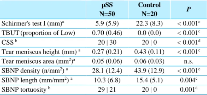
The results obtain from dry-eye assessment are reported in are reported in Table 2. Schirmer’s test I reported means were significantly lower in pSS group comparing with controls (p < 0.001). The proportion of low BUT was significantly higher in pSS patients in contrast to normal controls (p < 0.001). CSS was significantly higher in pSS group in contrast to the control group (p < 0.001).
Concerning to TM evaluation, TMH reported means were significantly lower in pSS group in contrast to the controls (p < 0.001). No significant differences were found between the two groups regarding TMA.
In turn, corneal sub-basal nerve plexus density and length were significantly lower and tortuosity was significantly higher in pSS group in contrast with the controls (p < 0.001, p = 0.004 and p = 0.001, respectively).
Table 2 - Patients’ Ophthalmic Characteristics - Dry-eye assessment profile pSS
N=50
Control
N=20 P
Schirmer's test I (mm)a 5.9 (5.9) 22.3 (8.3) < 0.001c TBUT(proportion of Low) 0.70 (0.46) 0.0 (0.0) < 0.001c
CSS b 20 | 30 20 | 0 < 0.001d
Tear meniscus height (mm) a 0.27 (0.21) 0.43 (0.11) < 0.001c Tear meniscus area (mm2)a 0.05 (0.06) 0.06 (0.03) n.s. SBNP density (n/mm2) a 28.1 (12.4) 43.9 (12.9) < 0.001c SBNP length (mm/mm2) a 10.3 (6.8) 15.4 (5.1) 0.004c
SBNP tortuosity b 29 | 21 20 | 0 0.001d
pSS – Primary Sjögren’s syndrome TBUT – Tear break up time; CSS – Corneal staining score; SBNP – Sub-basal nerve plexus; n.s. – non-significant. a Mean and standard deviation were reported. b Number of cases <= Median | > Median were reported. CSS is an ordered variable ranged from 0 to 3. c t-Test (two independent samples). d Median test with Monte Carlo estimation
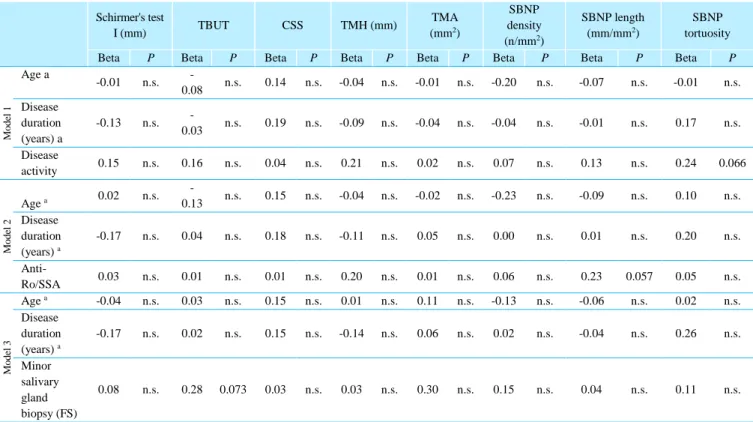
Demographic data of pSS patients according to clinical activity disease, immunological and histological profiles are reported in Table 3, whereas the results obtain from of the comparison between ophthalmic characteristics according to clinical activity disease, immunological and histological profiles are reported in Table 4.
pSS patients with positivity to anti-SSA antibodies presented earlier onset of Sicca complains and were diagnosed at an younger age than SSA-negative patients (p= 0.058 and p= 0.021, respectively). pSS patients with positive minor salivary gland biopsy, defined as focus score ≥1, were older (p= 0.029), presented disease complains later and were diagnosed at an older age, with significant differences compared to pSS patients with negative minor salivary gland biopsy (p= 0.029, p=0.023 and p= 0.016, respectively).
The effect of disease activity on SBNP tortuosity was marginally significant (Beta = 0.24, p = 0.066). Patients with clinical active disease showed higher SBNP tortuosity scores (Mrank for disease activity += 26.2 and Mrank for disease activity – = 24.9).
The effect of SSA on SBNP length was marginally significant (Beta = 0.23, p = 0.073). Patients with positive for SSA antibodies showed higher results of SBNP length (MSSA+ = 9.25 and MSSA- = 12.3). Finally, the effect of minor salivary gland biopsy on BUT was marginally significant (Beta = 0.28, p = 0.057). Patients with negative minor salivary gland biopsy had higher frequency of low BUT (90.9% of patients with FS < 1 had low BUT and that percentage decreased to 62.2% for patients with FS ≥ 1).
Table 3 – Ophthalmic characteristics according to disease activity, immunological and histological profiles
Disease activity + Disease activity -
P SSA+ SSA- P FS ≥ 1 FS < 1 P
M M M M M M
Age (years) 56.7 59.5 n.s. 57.1 60.5 n.s. 61.6 49.4 0.029
Symptomsonset (years) 42.5 49.0 0.040 43.8 50.2 0.058 48.5 39.8 0.023
Diagnosis (years) 48.5 54.3 n.s. 48.9 56.9 0.021 54.4 44.8 0.016
Legend: M – Mean; n.s. – non-significant. Disease activity + - clinical active disease. FS – Focus score. The independent samples t test was performed to compare means.
Table 4 – Regression results on ophthalmic features according to disease activity, histological and immunological profiles
Schirmer's test I (mm) TBUT CSS TMH (mm) TMA (mm2) SBNP density (n/mm2) SBNP length (mm/mm2) SBNP tortuosity
Beta P Beta P Beta P Beta P Beta P Beta P Beta P Beta P
M o d el 1 Age a -0.01 n.s. -0.08 n.s. 0.14 n.s. -0.04 n.s. -0.01 n.s. -0.20 n.s. -0.07 n.s. -0.01 n.s. Disease duration (years) a -0.13 n.s. -0.03 n.s. 0.19 n.s. -0.09 n.s. -0.04 n.s. -0.04 n.s. -0.01 n.s. 0.17 n.s. Disease activity 0.15 n.s. 0.16 n.s. 0.04 n.s. 0.21 n.s. 0.02 n.s. 0.07 n.s. 0.13 n.s. 0.24 0.066 M o d el 2 Age a 0.02 n.s. -0.13 n.s. 0.15 n.s. -0.04 n.s. -0.02 n.s. -0.23 n.s. -0.09 n.s. 0.10 n.s. Disease duration (years) a -0.17 n.s. 0.04 n.s. 0.18 n.s. -0.11 n.s. 0.05 n.s. 0.00 n.s. 0.01 n.s. 0.20 n.s. Anti-Ro/SSA 0.03 n.s. 0.01 n.s. 0.01 n.s. 0.20 n.s. 0.01 n.s. 0.06 n.s. 0.23 0.057 0.05 n.s. M o d el 3 Age a -0.04 n.s. 0.03 n.s. 0.15 n.s. 0.01 n.s. 0.11 n.s. -0.13 n.s. -0.06 n.s. 0.02 n.s. Disease duration (years) a -0.17 n.s. 0.02 n.s. 0.15 n.s. -0.14 n.s. 0.06 n.s. 0.02 n.s. -0.04 n.s. 0.26 n.s. Minor salivary gland biopsy (FS) 0.08 n.s. 0.28 0.073 0.03 n.s. 0.03 n.s. 0.30 n.s. 0.15 n.s. 0.04 n.s. 0.11 n.s.
TBUT – Tear break up time; CSS – Corneal staining score; SBNP – Sub-basal nerve plexus; n.s. – non-significant; TMH – Tear Meniscus Height; TMA – Tear Meniscus Area a Age and disease duration (years)– control variables
DISCUSSION
Primary Sjögren’s syndrome is a challenging disorder, in terms of diagnosis, severity and activity assessment.10 The objective ocular signs considered for pSS diagnosis3
are highly variable with ambient and patient
characteristics, which has led to widespread dissatisfaction among the medical community.4,5,26 Thus, novel objective and reproducible tools to respond to the unmet needs in pSS ocular assessment are needed.
In this study, 50 pSS patients and 20 healthy controls were evaluated in order to demonstrate the value of TMH
and corneal SBNP in detecting pSS. We also aimed to demonstrate if ophthalmic features were affected by clinical active disease, immunological and histological profiles of pSS patients. To our best knowledge, and to this date, this is the first study evaluating the differences of conventional dry-eye tests, TM and corneal SBNP in pSS patients, according to disease phenotype.
The results confirmed the value of TMH and corneal SBNP density, length and tortuosity in detecting pSS comparing to healthy individuals. In fact, the diagnostic value of TM for dry-eye disease and pSS has been proven. AS-OCT enables non-invasive, cross sectional
high-resolution images of the TM, thus considered to be a valid tool for the evaluation and diagnosis of aqueous deficient dry-eye disease.12,14,15,27 Concerning the corneal nerve plexus evaluation, high-resolution and reproducible images of the ocular surface, obtained by IVCM16 enable the objective assessment of corneal SBNP and the detection of nerve variations associated to dry-eye disease, associated or not with pSS11,28,29
However, the value of the referred tools in assessing pSS should not be taken as absolute, but integrated with the clinical picture and with other items in the AECG classification criteria. Indeed, rheumatic diseases are usually identified by the presence of a combination of clinical and laboratory manifestations.3
In turn, the effect of disease activity, immunological and histological profiles, corrected for disease duration, had only a marginal effect on patients’ ophthalmological features. Patients with clinical active disease showed higher SBNP tortuosity, whereas patients with positive SSA antibodies showed higher results of SBNP length and patients with negative minor salivary gland biopsy had higher frequency of low BUT. This could mean that TM and SBNP are independent parameters, not influenced by phenotype profile, but also that they may have limited value in assessing disease activity, immunological and histological profile, which have been previously demonstrated to be associated with important clinical consequences.30–32
TM may change with different blink intervals, with lid position and ambient conditions,15,33 whereas SBNP variations are indicators of nerve fibre damage, thus, not specific of dry-eye disease.22,34,35 We also acknowledge that, being the sample limited to pSS, the interpretation of the results in terms of TM and SBNP diagnostic value may be limited. Also, the availability of TM AS-OCT and SBNP IVCM assessment equipment, as well as examination duration, still limit the applicability of these techniques in routine practice. However, their use in a scientific setting has brought valuable information about in
differential diagnosis, follow-up and therapeutic
approaches of pSS.11,12,36
CONCLUSION
Novel objective and reproducible tools for the assessment of the ocular component of pSS are needed. AS-OCT and IVCM may be useful in the evaluation of lachrymal unit dysfunction in pSS, complementing the conventional methodology used in the evaluation of dry-eye disease. More studies are needed to clarify the value of these ophthalmic features in the assessment of disease impact and severity, as well as their possible use as a biomarker for the diagnosis and classification of pSS.
REFERENCES
1. Psianou K, Panagoulias I, Papanastasiou AD, et al. Clinical and immunological parameters of Sjögren’s syndrome. Autoimmun Rev. 2018 Oct;17(10):1053-1064.
2. Mariette X, Criswell LA. Primary Sjögren’s Syndrome. N Engl J Med. 2018;378:931-939.
3. Vitali C, Bombardieri S, Jonsson R, Moutsopoulos HM A, Al. E et. Classification criteria for Sjögren’s syndrome: a revised version of the Erupean criteria proposed by the American- European Consensus Group. Ann Rheum Dis. 2002;61:554-558.
4. Sullivan BD, Crews LA, Sönmez B, et al. Clinical Utility of Objective Tests for Dry Eye Disease. Cornea. 2012;31:1000-1008.
5. Nichols KK, Nichols JJ, MPH M, et al. The Lack of Association Between Signs and Symptoms in Patients With Dry Eye Disease. Cornea. 2004;23:762-770.
6. Hirsch JD, Reis BL, Services C, et al. Reliability and validity of the Ocular Surface Disease Index. Arch Ophthalmol. 2000;118:615-621.
7. Sullivan BD, Whitmer D, Nichols KK, et al. An Objective Approach to Dry Eye Disease Severity. Investig Opthalmology Vis Sci. 2010;51:6125.
8. Lemp MA. Report of the National Eye Institute/Industry Workshop on clinical trials in dry eye. CLAO J. 1995. 9. Versura P, Frigato M, Cellini M, et al. Diagnostic
performance of tear function tests in Sjogren’s syndrome patients. Eye. 2007;21:229-237.
10. Gallo A, Baldini C, Teos L, et al. Emerging trends in Sjögren’s syndrome: Basic and translational research. Clin Exp Rheumatol. 2012;30:779-784.
11. Gabbriellini G, Baldini C, Varanini V, et al. In vivo confocal scanning laser microscopy in patients with
primary sjögren’s syndrome: A monocentric experience. Mod Rheumatol. 2015;25:585-589.
12. Chen Q, Zhang X, Cui L, et al. Upper and lower tear menisci in Sjögren’s syndrome dry eye. Investig Ophthalmol Vis Sci. 2011;52:9373-9378.
13. Cui L, Shen M, Wang J, et al. Age-related changes in tear menisci imaged by optical coherence tomography. Optom Vis Sci. 2011;88:1214-1219.
14. Mainstone JC, Bruce AS, Golding TR. Tear meniscus measurement in the diagnosis of dry eye. Curr Eye Res. 1996;15:653-661.
15. Ibrahim OMA, Dogru M, Takano Y, et al. Application of visante optical coherence tomography tear meniscus height measurement in the diagnosis of dry eye disease. Ophthalmology. 2010;117:1923-1929.
16. Kalteniece A, Ferdousi M, Adam S, et al. Corneal confocal microscopy is a rapid reproducible ophthalmic technique for quantifying corneal nerve abnormalities. PLoS One. 2017;12:1-10.
17. Brito-Zerón P, Theander E, Baldini C, et al. Early diagnosis of primary Sjögrens syndrome: EULAR-SS task force clinical recommendations. Expert Rev Clin Immunol. 2016;12:137-156.
18. Dumusc A, Ng WF, James K, et al. Comparison of ESSDAI and ClinESSDAI in potential optimisation of trial outcomes in primary Sjögren’s syndrome: Examination of data from the UK Primary Sjögren’s Syndrome Registry. Swiss Med Wkly. 2018;148:1-8.
19. Whitcher JP, Shiboski CH, Shiboski SC, et al. NIH Public Access. 2012;149:405-415.
20. Sook Chun Y, Park IK. Reliability of 4 clinical grading systems for corneal staining. Am J Ophthalmol. 2014;157:1097-1102.
21. Tavakoli M, Malik RA. Corneal Confocal Microscopy: A Novel Non-invasive Technique to Quantify Small Fibre Pathology in Peripheral Neuropathies. J Vis Exp. 2011:1-7. 22. Malik RA, Kallinikos P, Abbott CA, et al. Corneal confocal microscopy: A non-invasive surrogate of nerve fibre damage and repair in diabetic patients. Diabetologia. 2003;46:683-688.
23. Oliveira-Soto L, Efron N. Morphology of corneal nerves using confocal microscopy. Cornea. 2001;20:374-384. 24. Models ML. Department of Statistics , UCLA Department
of Statistics Papers. 2004.
25. van der Kooij AJ. 2. Local Minima in Regression with Optimal Scaling Transformations. Predict Accuracy Stab Regres with Optim Scaling Transform. 2007:15-36.
26. Serruya LG, Nogueira DC, Hida RY. Schirmer test performed with open and closed eyes: variations in normal individuals. Arq Bras Oftalmol. 2009;72:65-67.
27. Penner V, Rocha G. Use of the Visante for anterior segment ocular coherence tomography. Tech Ophthalmol. 2007;5:67-77.
28. Benítez Del Castillo JM, Wasfy MAS, Fernandez C, et al. An in vivo confocal masked study on corneal epithelium and subbasal nerves in patients with dry eye. Investig Ophthalmol Vis Sci. 2004.
29. Labbé A, Liang Q, Wang Z, et al. Corneal nerve structure and function in patients with non-sjögren dry eye: Clinical correlations. Investig Ophthalmol Vis Sci. 2013;54:5144-5150.
30. Quartuccio L, Baldini C, Bartoloni E, et al. Anti-SSA/SSB-negative Sjögren’s syndrome shows a lower prevalence of lymphoproliferative manifestations, and a lower risk of lymphoma evolution. Autoimmun Rev. 2015;14:1019-1022.
31. Baldini C, Pepe P, Quartuccio L, et al. Primary sjögren’s syndrome as a multi-organ disease: Impact of the serological profile on the clinical presentation of the disease in a large cohort of Italian patients. Rheumatol (United Kingdom). 2014;53:839-844.
32. Theander E, Vasaitis L, Baecklund E, et al. Lymphoid organisation in labial salivary gland biopsies is a possible predictor for the development of malignant lymphoma in primary Sjögren’s syndrome. Ann Rheum Dis. 2011;70:1363-1368. doi:10.1136/ard.2010.144782. 33. Palakuru JR, Wang J, Aquavella J V. Effect of blinking on
tear dynamics. Investig Ophthalmol Vis Sci. 2007;48:3032-3037.
34. El-Fayoumi D, Youssef MM, Khafagy MM, et al. Assessment of Corneal and Tear Film Parameters in Rheumatoid Arthritis Patients Using Anterior Segment Spectral Domain Optical Coherence Tomography. Ocul Immunol Inflamm. 2016;3948:1-7.
35. Dabbah MA, Graham J, Petropoulos IN, et al. Automatic analysis of diabetic peripheral neuropathy using multi-scale quantitative morphology of nerve fibres in corneal confocal microscopy imaging. Med Image Anal. 2011;15:738-747. 36. Villani E, Magnani F, Viola F, et al. In Vivo Confocal
CONTACT
Joana CardigosHospital de Santo António dos Capuchos Alameda de Santo António dos Capuchos 1169-050 Lisboa
E-mail: [email protected]
No funding was received for this research.
All authors certify that they have no affiliations with or involvement in any organization or entity with any financial interest or non-financial interest in the subject matter or materials discussed in this manuscript.