Ciência Rural, v.48, n.3, 2018.
Canine visceral leishmaniosis in an area of fishing tourism,
Bonito Municipality, Mato Grosso do Sul, Central-West Brazil
Leishmaniose visceral canina em área de turismo pesqueiro, município de Bonito, Mato Grosso do Sul, Centro-Oeste do Brasil. Andreia Fernandes Brilhante1* Maria Elizabeth Cavalheiros Dorval2
Eunice Aparecida Bianchi Galati1 Vivianne de Oliveira Landgraf de Castro3 Maria Elizabeth Ghizzi Rocca4 Gilberto Gonçalves Facco5
Celeste da Silva Freitas de Souza6 Vânia Lúcia Brandão Nunes5
ISSNe 1678-4596
Received 05.18.17 Approved 02.02.18 Returned by the author 03.06.18
INTRODUCTION
In Brazil, visceral leishmaniasis (VL) is a zoonosis of public health importance, and its etiological agent is Leishmania (Leishmania) infantum chagasi. As
main vectors of this parasite, two phlebotomine sandfly
species, Lutzomyia longipalpis, widespread in the
country, and Lutzomyia cruzi registered in Mato Grosso do Sul and Mato Grosso States, (SANTOS et al., 1998; LAINSON & SHAW, 2005; MISSAWA et al., 2011; BRASIL, 2014) have been implicated, and the domestic dog has been considered the common reservoir, with 1Faculdade de Saúde Pública, Universidade de São Paulo (USP), Av. Dr. Arnaldo, 715, 01246-904, São Paulo, SP, Brasil. E-mail:
brilhanteaf@usp.br. *Corresponding author.
2Laboratório de Análises Clínicas (LAC), Universidade Federal de Mato Grosso do Sul (UFMS), Campo Grande, MS, Brasil. 3Hospital Universitário Maria Aparecida Pedrossian (HUMAP/EBSERH), Campo Grande, MS, Brasil.
4Núcleo de Controle de Zoonoses, Bonito, MS, Brasil.
5 Laboratório de Parasitologia Humana, Universidade Anhanguera, Campo Grande, MS, Brasil. 6Laboratório de imunomodulação e protozoologia, Instituto Oswaldo Cruz, Rio de Janeiro, RJ, Brasil.
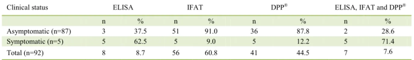
ABSTRACT: The study aimed to investigate Leishmania infection in a population of dogs (92 animals) in a fishing area of Bonito Municipality,
and evaluate comparatively the serological methods used, immunoenzymatic assay (ELISA), indirect immunofluorescence antibody test (IFAT), and rapid Dual Path-Platform test (DPP®). Blood and aspirate of bone marrow samples were used and parasitological investigation was also performed, such as parasite isolation in Neal, Novy, Nicolle (NNN) medium culture, Woo technique, Giemsa stained smears and specific identification by polymerase chain reaction (PCR). IFAT revealed 56/92 seropositive, the ELISA 8/92 and the DPP® 41/92. Regarding the
agreement between the serological tests by the Kappa index, there was a slight agreement between ELISA/IFAT and ELISA/DPP®, and fair
agreement in IFAT/DPP®. The NNN culture was positive in three out of 21 dogs, and identified by PCR as Leishmania infantum chagasi. No samples were positive by the Woo technique. Our results showed low agreements between the serological tests recommended by the Brazilian Ministry of Heath, and it is necessary to associate other diagnostic techniques, such as parasitological tests and PCR, to increase the sensitivity for canine visceral leishmaniosis diagnosis, mainly regarding asymptomatic dogs in endemic areas.
Key words: domestic dogs, serodiagnosis, ecotourism.
RESUMO: O estudo teve como objetivo verificar a infecção por Leishmania em uma população de cães (n=92 animais) de uma área de turismo
de pesca no município de Bonito e avaliar comparativamente os métodos sorológicos utilizados, ensaio imunoenzimático (ELISA), reação de
imunofluorescência indireta (RIFI) e teste rápido de plataforma dupla (DPP®). Testes parasitológicos também foram realizados, como o
isolamento do parasita em meio de cultura Neal, Novy, Nicolle, técnica de Woo, esfregaços em lâminas coradas com Giemsa e identificação
específica pela reação em cadeia da polimerase (PCR). A RIFI revelou sorologia positiva de 56/92, ELISA 8/92 e DPP® 41/92. Quanto à
concordância entre os testes sorológicos pelo índice Kappa, houve um ligeiro acordo entre ELISA/RIFI e ELISA/DPP®, e uma concordância
razoável entre RIFI/DPP®. A cultura NNN foi positiva em três cães, e identificada por PCR como Leishmania infantum chagasi. Nenhuma
amostra foi positiva pela técnica de Woo. Nossos resultados mostraram baixas concordâncias entre os testes sorológicos recomendados pelo Ministério da Saúde, sendo necessário associar a outras técnicas de diagnóstico, como testes parasitológicos e PCR, para aumentar a sensibilidade ao diagnóstico de leishmaniose visceral canina, principalmente, com relação aos cães assintomáticos em áreas endêmicas. Palavras-chave: cães domésticos, sorodiagnóstico, ecoturismo.
2 Brilhante et al.
canine cases normally preceding the occurrence of
human cases (GONTIJO & MELO, 2004).
The first cases of canine and human
visceral leishmaniasis in Mato Grosso do Sul date
back to the 80s at the Corumbá municipality(NUNES
et al., 1988; YAMAMOTO et al., 1988). By the 90s, the visceral leishmaniasis spread widely across the
state being diagnosed in 56 of its 78 municipalities, affecting rural and urban populations of different age groups and the vectors have being reported in almost all the municipalities (CORTADA et al., 2004; OLIVEIRA et al., 2006; NUNES et al., 2008; LIMA-JÚNIOR et al., 2009; ANDRADE et al., 2009; MATO GROSSO DO SUL, 2014).
Researches undertaken in Bonito District
have reported natural infection by Leishmania in
sandflies and dogs in both rural and urban areas (NUNES
et al., 2001; 2008; GALATI et al., 2006; SAVANI et al., 2009, ANDRADE et al., 2009; BRILHANTE et al., 2015b), and a moderate incidence of the disease in both human and canine populations (ANDRADE et al., 2009; MATO GROSSO DO SUL, 2014).
According to the Brazilian Ministry of
Health, serological methods are used to diagnose and control programs of canine visceral leishmaniasis
(CVL) in the country (BRASIL, 2014). However, the complexity of the diagnosis of CVL should be considered due to the low sensitivity of the tests, variance of clinical signs, presence of asymptomatic dogs and social factors caused by the culling of dogs
(DE SANTIS et al., 2013).
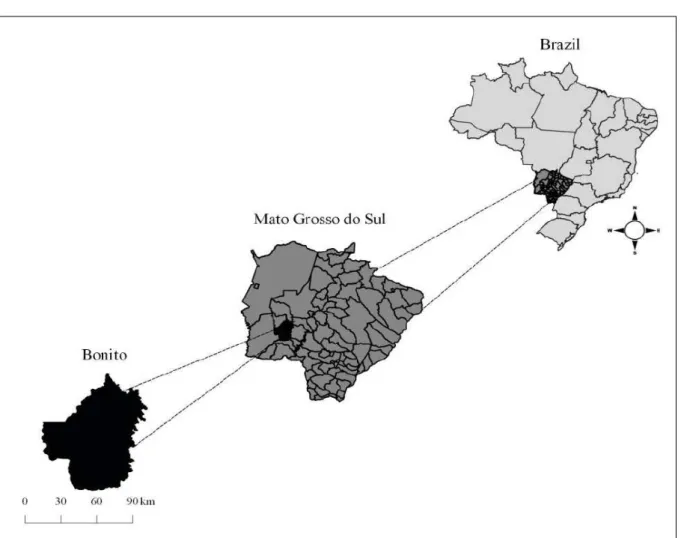
In Águas do Miranda District, one of the major tourist resorts for ecotourism and sporting
fishing in midwestern Brazil, the notification of CVL led us to investigate the infection by Leishmania in the canine population, and compare the performance of serological methods used, whose results are
reported in this study.
MATERIALS AND METHODS
Study area
Bonito Municipality, one of the Mato Grosso
do Sul municipalities, is situated in the southwest of Mato Grosso do Sul in the geomorphological unit known as Bodoquena Plateau, and consists of two
districts: Bonito, containing the municipal offices,
and Águas do Miranda, both considered important regional, national and international tourist resorts (IBGE 2016; BONITO, 2016).
The District of Águas do Miranda (20º45’44.4” S, 56º05’42.8” W) is 75 km from the Municipality of Bonito District containing
the municipal offices (21º07’16” S, 56º28’55” W), and
180 km from Campo Grande, the capital of the Mato Grosso do Sul State (IBGE, 2016) (Figure 1). The human population consists of 450 inhabitants, which
may rise to as many as 10,000 in the fishing season, from March to October. The local economy is based mainly on fishing and ecotourism (BONITO, 2009).
Sampling
With information about the occurrence of CVL cases and the help of the Zoonosis Control
Center of Bonito Municipality, domiciliary visits
were made in the Águas do Miranda District from
June to December 2009. By consent of the owners,
the 92 adult dogs, which constituted the local canine population, were submitted to clinical evaluation
for the identification of manifestations suggestive
of leishmaniosis. About 5 ml of venous blood was
collected by jugular or cephalic vein puncture and
stored in plastic tubes without and with anticoagulant
(ethylenediaminetetraacetic acid - EDTA). Serum samples were stored at −20°C until the performance
of the serological techniques.
For the 41 positive dogs in the Dual Path-Platform test (DPP®), a second visit was made to
their owner’s domicile to collect bone marrow blood
samples. However, only for 21 of the dogs it was
possible to obtain samples.
Serological tests
Serum samples were used for serological tests which were carried out in the
laboratory of Imunodulação e Protozoologia of
the Oswaldo Cruz Institute/FIOCRUZ in Rio de Janeiro. The techniques used for the diagnosis
of CVL were indirect fluorescent antibody test
(IFAT) (kit IFI Canine Visceral Leishmaniosis
Bio-Manguinhos/FIOCRUZ); enzyme-linked
immunosorbent assay (kit ELISA Canine Visceral
Leishmaniosis Biomaguinhos/FIOCRUZ), and immunochromatographic rapid test (kit TR DPP®
Canine Visceral Leishmaniosis-Bio-Manguinhos/
FIOCRUZ). The latter is a lateral flow test for the detection of antibodies specific for Leishmania,
using recombinant proteins K26 and K39 specific
for Leishmania infantum chagasi. Serological
techniques and immunochromatographic assay
were performed according to the manufacturer’s
instructions. In order, to classify the positive results,
the following criteria were considered: IFAT titres equal to or higher than 1:40, ELISA readings over the cut-off, and for DPP®, the emergence of two
Parasitological diagnosis
The Woo technique was applied to identify trypanosomatid flagellates from total blood samples in
accordance with WOO (1969). The method consisted
of filling two capillary tubes with approximately 0.06 ml of peripheral blood, which were flamed sealed
at one end and centrifuged at 12, 000 rpm (Make of centrifuge) for 4 minutes. These tubes were then
placed in a capillary tube holder and examined under
a microscope using a 10 X objective to investigate
trypanosomes at the junction of the plasma and buffy layer in the centrifuged blood (WOO, 1969).
For observation of amastigote forms, part of the bone marrow blood sample was used to prepare Giemsa stained smears (n=21). The remainder
of each sample was inoculated intraperitoneally
into two hamsters (Mesocricetus auratus) (n=42). Nine months after inoculation, the hamsters were
necropsied and fragments of their liver and spleen were seeded in NNN culture medium supplemented with 20% fetal bovine serum. Samples were seeded in duplicate, with a total of 84 cultures.
DNA extraction and molecular test
Positive samples of the NNN culture had their DNA extracted using a DNAzol® kit in
accordance with the manufacturer’s instructions,
and then analyzed by polymerase chain reaction
(PCR) for the Leishmania infantum chagasi using the initiators RV1- CTTTTCTGGTCCCGCGGGTAGG e RV2- CACCTGGCCTATTTTACACCA. The PCR conditions were undertaken according to LIMA-JÚNIOR et al. (2009). The DNA reference strain
was L. infantum chagasi (MHOM/BR/74/PP/75),
supplied by the laboratory of Leishmaniasis of the René
4 Brilhante et al. Statistical analysis
To evaluate the agreement between serological techniques (ELISA, IFAT and DPP®), the Kappa coefficient (κ) was calculated with confidence interval of 95%. The test was performed by software Bioestat version
5.3 (AYRES et al., 2007). Values of κ were considered
according to SOLANO-GALLEGO et al. (2014) as: no
agreement (κ < 0), slight agreement (0 < κ < 0.2), fair agreement (0.2 < κ <0.4), moderate agreement (0.4 < κ < 0.6), substantial agreement (0.6 < κ <0.8) and almost perfect agreement (κ > 0.8).
RESULTS
The total number and percentages of positive animals in serological tests and parasitological
examination according to clinical symptomatic or asymptomatic status are shown in table 1. The IFAT
revealed 56/92 seropositive, the ELISA 8/92, and the DPP® 41/92. Regarding the agreement between the serological tests by the Kappa index, there was a
slight agreement between ELISA/IFAT and ELISA/ DPP, and fair agreement in IFAT/DPP (Table 2).
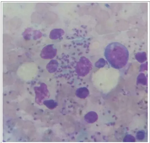
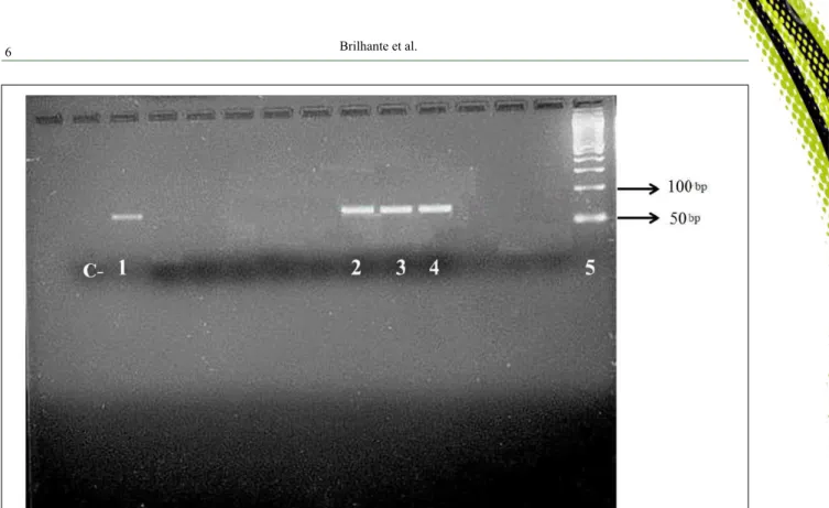
Amastigote forms were observed in the Giemsa stained smears of the bone marrow from three dogs (3/21). Promastigote forms were also observed in three cultures seeded with the spleen and/or liver fragments of the inoculated hamsters. Flagellates from
the positive cultures samples were identified by PCR
as Leishmania infantum chagasi. It is noteworthy that
three dogs (3/92) were positive in all tests performed. No
flagellates were observed in the peripheral blood by
Woo’s technique. The photograph of imprint smear from bone marrow of a positive dog demonstrating the amastigote forms and the electrophoresis in agarose gel
are available in this research as supplementary material
(Figure 2; Figure 3).
In the canine survey, five (5.4%) dogs
showed clinical signs suggestive of CVL, such as
weight loss, alopecia and onychogryphosis, and were
positive in all serological tests, while 87 (94.6%) were
asymptomatic. In other words, they did not present any of the symptoms described above. Of the 87 asymptomatic
dogs investigated, 36 (41.4%)were positive in at least one of the diagnostic tests performed.
DISCUSSION
In Mato Grosso do Sul, a large number of
seropositive asymptomatic dogs have been reported,
both in rural settlements and urban areas of Bonito District (NUNES et al., 2001; ANDRADE et al., 2009) in
Anastácio, a municipality adjacent to Bonito (CORTADA
et al., 2004) and Campo Grande (MATO GROSSO DO SUL, 2014). Similar results were reported in Mato Grosso State(MATO GROSSO DO SUL, 2014; DE SANTIS et al., 2013) and other areas in the Brazilian south-eastern and north-eastern regions (DANTAS-TORRES et al., 2006; COURA-VITAL et al., 2011). Some studies suggested that animals without apparent clinical signs of CVL could be the source of Leishmania for phlebotomine
sandflies (MORENO & ALVAR, 2002; LAURENTI
et al., 2013); although, VERÇOSA et al. (2008) have
demonstrated that asymptomatic dogs are not efficient
sources of Leishmania for vectors.
Low agreement between the serological
tests in our study can also be observed in researches
carried out in Monte Negro – Rondônia (RO) State (AGUIAR et al., 2010), between ELISA and IFAT test, in Panorama - São Paulo (SP) State (LOPES et al., 2017), between ELISA and DPP tests. However,
also high and moderate agreements have already
been observed between the ELISA and IFAT tests in
other localities of Brazil, as in Campo do Goycatazes
- Rio de Janeiro State (TÁVORA et al., 2007) and Ilha Solteira (SP) (ASSIS et al., 2010; QUEIROZ et al., 2010). These observations demonstrated the need to better assess the diagnostic techniques of CVL, aiming at standardization, considering the evolution of the disease in the dog and its immune response.
Table 1 - Number and percentage of positive dogs in serological techniques for Leishmaniaby clinical status, Águas do Miranda District,
Bonito, Mato Grosso do Sul, Brazil, 2009.
Clinical status ELISA IFAT DPP® ELISA, IFAT and DPP®
n % n % n % n %
Asymptomatic (n=87) 3 37.5 51 91.0 36 87.8 2 28.6
Symptomatic (n=5) 5 62.5 5 9.0 5 12.2 5 71.4
Total (n=92) 8 8.7 56 60.8 41 44.5 7 7.6
In this research, the use of molecular
tools was only possible in positive samples of NNN
cultures. Previous studies have shown that the combination of serological and molecular techniques increase the detection of the parasite in the canine
population, indicating failures in the official protocol
of diagnosis of CVL in Brazil (QUEIROZ et al., 2010; LOPES et al., 2017). However, molecular tests are
not available in all laboratories in the public system,
and there is still no gold standard for the diagnosis of CVL, since for this technique there is the application of different protocols.
Until the year 2011, the ELISA and IFAT are methods recommended by the Brazilian Health Ministry (BHM) for CVL diagnosis. The ELISA was employed for screening large populations, and
Table 2 - Agreement analysis by Kappa index between serological techniques (ELISA/IFAT, ELISA/DPP and IFAT/DPP) for diagnosis of canine visceral leishmaniosis (CVL) in domestic dogs, Águas do Miranda, Bonito, Mato Grosso do Sul, Brazil.
Comparison
between tests +/+ +/- -/+ -/- Total κ CI p Agreement
ELISA/IFAT 8 0 48 36 92 0.115* -0.214-0.445 0.008 Slight
ELISA/DPP 7 1 34 50 92 0.164* -0.135-0.464 0.005 Slight
IFAT/DPP® 34 22 7 29 92 0.384* 0.190-0.578 <0.001 Fair
+: Positive; -: Negative; * Statistically significant (P ≤ 0.05), confidence interval 95%. ELISA: enzyme-linked immunosorbent assay; IFAT:
indirect immunofluorescence antibody test; DPP®: Dual Path-Platform test.
6 Brilhante et al.
the IFAT for the confirmatory diagnosis. After some
studies, and through a BHM technical note (Nota
Técnica nº 01/2011 – CGDT/CGLAB/DEVIT/SVS/ MS), this context has been modified to DPP® for screening and ELISA for confirmatory, allowing
this screening to be performed in all Brazilian municipalities (BRASIL, 2014).
However, if in our study we had adopted the strategy recommended by the BMH, a low number of infected dogs, as confirmed by ELISA
(8/92), would have escaped detection. Regarding the
DPP®, it is considered easy to apply in field research. In the studies undertaken by GRIMALDI JÚNIOR et al. (2012), this test showed high sensitivity in symptomatic dogs. This result is corroborated in our study since all symptomatic animals were
positive in this diagnostic test. The differences
reported in the present study between the reactivity of the serum samples may result from false positive reactions due to cross-reactivity with agents other
than Leishmania and the sensitivity thresholds of
other tests (LIRA et al., 2006; SANTOS et al., 2010). However, some studies have reported no
cross-reactivity between Leishmania and other pathogens
(OLIVEIRA et al., 2008; GUIMARÃES et al.,
2009). Another possibility is that the levels of
anti-Leishmania antibodies were below the cut-off point
used as positive, as shown by SILVA et al. (2009), which IFAT assay using a 1:20 dilution showed a better contingency coefficient.
The use of the Woo technique is justified,
given the occurrence of Trypanosoma evansi in domestic animals at the Bodoquena Plateau, in the Pantanal of
the Mato Grosso do Sul and in Bonito Municipality
(STEVENS et al., 1989; NUNES et al., 1994; SAVANI et al., 2005), also present in a dog from the rural area of the São Paulo State (COELHO et al., 2013).
The isolation of the parasite by NNN
culture has its importance for obtaining parasite “in-mass” for use of techniques such as monoclonal antibodies and biochemical characterization, as
observed in studies by DE SANTIS et al. (2011).
In Águas do Miranda, the evidence of
dogs carrying L. infantum chagasi and the presence
of Lutzomyia longipalpis circulating in the peridomicile
environment (BRILHANTE et al., 2015a) together
with insufficient knowledge of the population about the disease, its severity and the forms of transmission, points to the possibility of introducing visceral leishmaniasis to
the residents of that district, as well as for tourists, like other areas of canine cases preceding human cases of parasitosis (GONTIJO & MELO, 2004).
Figure 3 - PCR amplification products with primers RV1/RV2. C-: Negative control, MilliQ water; 1 Positive control, reference strain
CONCLUSION
Our results showed low agreement between
the serological tests recommended by the BHM, and it is necessary to associate other diagnostic techniques,
such as parasitological tests and PCR, to increase the
sensitivity for CVL diagnosis, mainly regarding the asymptomatic dogs in endemic areas. Although, these
methods are expensive and require high professional
qualification, the proposal should be discussed among
Brazilian health authorities, since euthanasia of positive dogs is one of the main measures of control of VL in Brazil.
In addition, our findings may help the
surveillance agencies in Bonito, because the high prevalence of seropositive dogs in this endemic region
may contribute to an increase in the prevalence of infection in humans, since the municipality of Bonito is
considered a moderate transmission area of leishmaniasis.
ACKNOWLEDGEMENTS
The authors thank Universidade
Anhanguera-Uniderp for the financial support, and the Zoonosis Control Center of Bonito Municipality for the logistic support.
BIOETHICS AND BIOSECURITY COMMITTEE APPROVAL
This study was approved by the Ethics Committee of
Animal Use of the Universidade Anhanguera/Uniderp, Unidade Agrárias, Campo Grande, MS (authorization: 76-011/09-MS). Results of this research were sent to the Center of Zoonosis Control of Bonito
Municipality, so that control measures could be taken.
DECLARATION OF CONFLICTING INTERESTS
The authors declare no conflicts of interest.
REFERENCES
AGUIAR, D.M. et al. Seroprevalence of anti-Leishmania spp.
antibodies in rural dogs from the city of Monte Negro, State
of Rondônia, Brazil. Revista Brasileira de Parasitologia
Veterinária, v. 19, p. 73-74, 2010. Available from: <http://dx.doi. org/10.4322/rbpv.01901014>. Accessed Jul. 07, 2017.
ANDRADE, A.R.O et al. Epidemiological study on leishmaniasis in
an area of environmental tourism and ecotourism, state of Mato Grosso do Sul, 2006-2007. Revista da Sociedade Brasileira de Medicina
Tropical, v. 42, p. 488-493, 2009. Available from: <http://dx.doi. org/10.1590/S0037-86822009000500003>. Accessed Nov. 10, 2015.
ASSIS, J. et al. Comparative study of diagnostic methods for visceral
leishmaniasis in dogs from Ilha Solteira, SP. Revista Brasileira de
Parasitologia Veterinária, v. 19, p. 17-25, 2010. Available from:
<http://dx.doi.org/10.4322/rbpv.01901004>. Accessed Jul. 07, 2017.
AYRES, M. et al. BioEstat 5.0: aplicações estatísticas nas áreas das
ciências biológicas e médicas. Belém: MCT; IDSM; CNPq, 2007. 364 p. Available from: <http://www.mamiraua.org.br/pt-br/downloads/ programas/bioestat-versao-53/>. Accessed: 01 Aug. 2017.
BONITO. Prefeitura Municipal de Bonito. Relatório de
planejamento da Prefeitura Municipal de Bonito. Bonito; Mato Grosso do Sul, 2009.
BONITO. Prefeitura Municipal de Bonito. Secretaria de Indústria,
Turismo e Comércio. Available from: <http://www.bonito.ms.gov. br/>. Accessed: Jun. 29, 2016.
BRASIL. Ministério da Saúde. Manual de Vigilância e Controle da Leishmaniose Visceral, Brasilia-DF, 1ª ed. 5ª reimpressão,
2014. Available from: <http://bvsms.saude.gov.br/bvs/publicacoes/ manual_vigilancia_controle_leishmaniose_visceral_1edicao.pdf>
Accessed: Nov. 08, 2015.
BRILHANTE, A.F. et al. Natural infection of phlebotomines
(Diptera: Psychodidae) by Leishmania (Leishmania) amazonensis
in an area of ecotourism in Central-Western Brazil. Journal of
Venomous Animals Toxins Including Tropical Diseases, v. 31,
p. 1-3, 2015b. Available from < http://dx.doi.org/10.1186/s40409-015-0041-8>. Accessed: 12 Jul., 2016.
BRILHANTE, A.F. et al. Phlebotomine fauna (Diptera:
Psychodidae) in an area of fishing tourism in Central-Western Brazil.
Revista do Instituto de Medicina Tropical de São Paulo, v. 57, p. 233-238, 2015a. Available from:
<http://dx.doi.org/10.1590/S0036-46652015000300009>. Accessed: Jul. 12, 2016.
COELHO, W.M.D. et al. Detection of co-infections by Leishmania
(L.) chagasi, Trypanosoma evansi, Toxoplasma gondii and
Neospora caninum in dogs. Ars Veterinaria, v. 29, p.
169-174, 2013. Available from <http://dx.doi.org/10.15361/2175-0106.2013v29n3p169-174>. Accessed: Jul. 07, 2017.
CORTADA, V.M.C.L. et al. Canine visceral leishmaniasis in Anastácio, Mato Grosso do Sul State, Brazil. Veterinary Research
Communications, v. 24, p. 365-374, 2004. Available from: <http:// dx.doi.org/10.1023/B:VERC.0000035014.80785.b7>. Accessed:
Jul. 20, 2016.
COURA-VITAL, W. et al. Prevalence and factors associated with Leishmania infantum infection of dogs from an urban area
of Brazil as identified by molecular methods. PLoS Neglected Tropical Diseases, v. 8, e1291, 2011. Available from: <http://dx.doi. org/10.1371/journal.pntd.0001291>. Accessed: Sept. 24, 2016.
DANTAS-TORRES, F. et al. Seroepidemiological survey on canine leishmaniasis among dogs from an urban area of Brazil. Veterinary
Parasitology, v. 140, p. 54-60, 2006. Available from: <http://dx.doi. org/10.1016/j.vetpar.2006.03.008>. Accessed Sept. 24, 2016.
DE SANTIS, B. et al. Characterization of Leishmania infantum
species in dogs from the urban area of Cuiabá, State of Mato Grosso, Brazil. Revista da Sociedade Brasileira de Medicina Tropical, v. 44, n. 6, p. 771-773, 2011. Available from: <http://dx.doi.
org/10.1590/S0037-86822011000600022>. Accessed: Jul. 10, 2017.
DE SANTIS, B. et al. Performance of DPP™ immunochromathographic rapid test (IRT) for canine visceral leishmaniasis: comparison with other serological methods in suspected dogs from Cuiabá, Mato Grosso State, Brazil. Brazilian Journal of Veterinary Research and
8 Brilhante et al.
dx.doi.org/10.11606/issn.1678-4456.v50i3p198-205>. Accessed: Jul.
12, 2016.
GALATI, E.A.B. et al. Phlebotomines (Diptera: Psychodidae) in
forested areas of the Serra da Bodoquena, state of Mato Grosso do Sul, Brazil. Memórias do Instituto Oswaldo Cruz, v. 101, p.
175-193, 2006. Available from: <http://dx.doi.org/10.1590/S0074-02762006000200010>. Accessed: Aug. 14, 2016.
GONTIJO, C.M.F.; MELO, M.N. Visceral Leishmaniasis in Brazil: current status, challenges and prospects. Revista Brasileira de
Epidemiologia, v. 7, p. 338-349, 2004. Available from: < http://dx.doi. org/10.1590/S1415-790X2004000300011>. Accessed: Jul. 10, 2016.
GRIMALDI JÚNIOR, G. et al. Evaluation of a novel chromatographic
immunoassay based on Dual-Path Platform technology (DPP® CVL
rapid test) for the serodiagnosis of canine visceral leishmaniasis.
Transactions of Royal Society and Tropical Medicine and Hygiene,
v. 106, p. 54-59, 2012. Available from: <http://dx.doi.org/10.1016/j. trstmh.2011.10.001>. Accessed: Aug. 14, 2016.
GUIMARÃES, A.M. et al. Fatores associados à soropositividade para Babesia, Toxoplasma, Neospora e Leishmania em cães atendidos em nove clínicas veterinárias do município de Lavras, MG. Revista Brasileira de Parasitologia Veterinária, v. 18, p. 49-53, 2009. Available from: <http://dx.doi.org/10.4322/
rbpv.018e1009>. Accessed: Jul. 10, 2016.
IBGE – Instituto Brasileiro de Geografia e Estatística. Available from: <http://cidades.ibge.gov.br/painel/historico. php?codmun=500220>. Accessed: Jun. 21, 2016.
LAINSON, R.; SHAW, J.J. New World Leishmaniasis. In: FEG
Cox, JP Kreir, D Wakelin (eds). Microbiology and Microbial Infections, Parasitology. Topley & Wilson’s, Arnold, Sydney,
Auckland; p. 313-349, 2005.
LAURENTI, M.D. et al. Asymptomatic dogs are highly competent
to transmit Leishmania (Leishmania) infantum chagasi to the natural vector. Veterinary Parasitology, v. 196, p. 296–300, 2013.
Available from: <http://dx.doi.org/10.1016/j.vetpar.2013.03.017> .
Accessed: Jul. 12, 2017.
LIMA-JÚNIOR, M.S.C. et al. Identification of Leishmania species
isolated in human cases in Mato Grosso do Sul, by means of the polymerase chain reaction. Revista da Sociedade Brasileira de Medicina Tropical, v. 42, p. 303-308, 2009. Available from:
<http://dx.doi.org/10.1590/S0037-86822009000300012>.
Accessed: Sept. 02, 2016.
LIRA, R.A. et al. Canine visceral leishmaniosis: A comparative
analysis of the EIE-leishmaniose visceral canina BioManguinhos and
the IFI leishmaniose visceral canina BioManguinhos kits. Veterinary
Parasitology, v. 137, p. 11-16, 2006. Available from: <http://dx.doi. org/10.1016/j.vetpar.2005.12.020>. Accessed: Sept. 20, 2016.
LOPES, E.G. et al. Serological and molecular diagnostic tests for canine visceral leishmaniasis in Brazilian endemic area: one out of
five seronegative dogs are infected. Epidemiology and Infection,
v. 20, p. 1-9, 2017. Available from: <http://dx.doi.org/10.1017/ S0950268817001443>. Accessed: Aug. 10, 2017.
MATO GROSSO DO SUL (Estado). Secretaria de Estado de Saúde. Coordenadoria Estadual de Vigilância Epidemiológica. Informe epidemiológico das leishmanioses no 1/2014. Campo
Grande, MS, 2014.
MISSAWA, N.A. et al. Evidence of transmission of visceral
leishmaniasis by Lutzomyia cruzi in the municipality of Jaciara,
State of Mato Grosso, Brazil. Revista da Sociedade Brasileira
de Medicina Tropical, vol. 44, p. 76-78, 2011. Available from:
<http://dx.doi.org/10.1590/S0037-86822011000100017>.
Accessed 22 Aug., 2017.
MORENO, J.; ALVAR, J. Canine leishmaniasis: epidemiological risk and the experimental model. Trends in Parasitology, v. 18, p.
399–405, 2002. Available from: <http://dx.doi.org/10.1016/S1471-4922(02)02347-4>. Accessed: Sep. 15, 2016.
NUNES, V.L.B. et al. Study of phlebotomines (Diptera, Psychodidae) in the urban area of Bonito municipality, Mato
Grosso do Sul, Brazil. Revista Brasileira de Entomologia, v.
52, p. 446-451, 2008. Available from: <http://dx.doi.org/10.1590/ S0085-56262008000300019>. Accessed: Jul. 15, 2017.
NUNES, V.L.B. et al. Estudos epidemiológicos sobre leishmaniose tegumentar (LT) e Mal das Cadeiras no município de Corguinho, Mato Grosso do Sul - Estudo de reservatórios, 1992-1994. Revista
Brasileira Parasitologia Veterinária, v. 3, p. 29-35, 1994. Available
from: <http://cbpv.org.br/rbpv/documentos/311994/r3129_35.pdf>.
Accessed: Jul. 05, 2016.
NUNES, V.L.B. et al. Estudos epidemiológicos sobre leishmaniose visceral em cães de Corumbá, Mato Grosso do Sul. Pesquisa
Veterinária Brasileira, v. 8, p. 17-21, 1988.
NUNES, V.L.B. et al. Occurrence of canine visceral leishmaniasis in an agricultural settlement in the State of Mato Grosso do Sul, Brazil. Revista da Sociedade Brasileira de Medicina Tropical, v.
34, p. 301-302, 2001. Available from: <http://dx.doi.org/10.1590/ S0037-86822001000300014>. Accessed: Jul. 05, 2016.
OLIVEIRA, A.L.L. et al. Emergent outbreak of visceral leishmaniasis in Mato Grosso do Sul State. Revista da Sociedade Brasileira de Medicina
Tropical, v. 39, p. 446-450, 2006. Available from: <https://dx.doi. org/10.1590/S0037-86822006000500005>. Accessed: Jul. 30, 2017.
OLIVEIRA, T.M.F.S. et al. A study of cross-reactivity in serum samples from dogs positive for Leishmania sp., Babesia canis and
Ehrlichia canis in enzyme-linked immunosorbent assay and indirect
fluorescent antibody test. Revista Brasileira de Parasitologia Veterinária, v. 17, p. 7-11, 2008. Available from: <http://dx.doi.
org/10.1590/S1984-29612008000100002>. Accessed: Jul. 30, 2017.
QUEIROZ, N.M.G.P. et al. Canine Visceral Leishmaniasis diagnosis
by immunohistochemistry and PCR in skin tissues in association
with RIFI and ELISA-test. Revista Brasileira de Parasitologia
Veterinária, v. 19, p. 32-38, 2010. Available from <http://dx.doi. org/10.4322/rbpv.01901006>. Accessed: Jul. 30, 2017.
SANTOS, J.M.L. et al. Prevalence of anti-Leishmania spp antibodies in dogs from Garanhuns, in the middle scrub zone (Agreste) of Pernambuco. Revista da Sociedade Brasileira de Medicina
Tropical, v. 43, p. 41–45, 2010. Available from: <http://dx.doi.
org/10.1590/S0037-86822010000100010>. Accessed Aug. 12, 2016.
SANTOS, S.O. et al. Incrimination of Lutzomyia cruzi as a vector of American Visceral Leishmaniasis. Medical and Veterinary
Entomology, v. 12, p. 315-317, 1998. Available from: <http://dx.doi. org/10.1046/j.1365-2915.1998.00104.x>. Accessed: Jul. 30, 2017.
SAVANI, E.S.M.M. et al. Occurrence of co-infection by Leishmania
dog in the state of Mato Grosso do Sul, Brazil. Memórias do Instituto
Oswaldo Cruz, v. 100, p. 739-741, 2005. Available from: <http:// dx.doi.org/10.1590/S0074-02762005000700011>. Accessed: Aug.
14, 2016.
SAVANI, E.S.M.M. et al. The finding of Lutzomyia almerioi
and Lutzomyia longipalpis naturally infected by Leishmania
spp in a cutaneous and canine visceral leishmaniases focus in serra da Bodoquena, Brazil. Veterinary Parasitology, v. 160,
p. 18-24, 2009. Available from: <https://doi.org/10.1016/j. vetpar.2008.10.090>. Accessed: Aug. 14, 2016.
SILVA, R.M. et al. TG-ROC analysis of immunofluorescence assays in canine visceral leishmaniasis diagnosis. Revista de Saúde Pública,
v. 43, p. 1-9, 2009. Available from: <http://dx.doi.org/10.1590/S0034-89102009005000063>. Accessed: Oct. 02, 2016.
SOLANO-GALLEGO, L. et al. Serological diagnosis of canine leishmaniosis: comparison of three commercial ELISA tests (Leiscan®, ID Screen® and Leishmania 96®), a rapid test (Speed
Leish K®) and an in-house IFAT. Parasites & Vectors, v. 7, p.
1-10, 2014. Available from <http://dx.doi.org/10.1186/1756-3305-7-111>. Accessed: Aug. 10, 2017.
STEVENS, J.R. et al. Isoenzyme characterization of Trypanosoma evansi isolated from capybaras and dogs in Brazil. Acta Tropica,
v. 46, p. 213-222, 1989. Available from: <https://projects.exeter.
ac.uk/meeg/sites/default/files/publications/Stevens%20et%20
al%201989%20Acta%20Tropica.pdf>. Accessed: Jul. 05, 2016.
TÁVORA, M.P.F. et al. Comparative validation study between the
ELISA and RIFI techniques for diagnosing Leishmania sp in stray
dogs caught in the municipality of Campos de Goytacazes, State
of Rio de Janeiro. Revista da Sociedade Brasileira de Medicina
Tropical, v. 40, p. 482-483, 2007. Available from: <http://dx.doi. org/10.1590/S0037-86822007000400023>. Accessed: Jul. 15, 2017.
VERÇOSA, B.L, et al. Transmission potential, skin inflammatory
response, and parasitism of symptomatic and asymptomatic dogs
with visceral leishmaniasis. BMC Veterinary Research, v. 4, p.
45, 2008. Available from: <https://dx.doi.org/10.1186/1746-6148-4-45>. Accessed: Jul. 15, 2017.
WOO, P.T.K. The haematocrit centrifuge technique for the
detection of Trypanosomes in blood. Canadian Journal of Zoology, v. 47, p. 921-923, 1969. Available from <https://dx.doi. org/10.1139/z69-150>. Accessed: Jul. 10, 2017.
YAMAMOTO, Y.Y. et al. Estudo da eficiência das reações de
imunofluorescência e de hemaglutinação passiva no diagnóstico de
leishmaniose visceral em cães. Revista da Faculdade de Medicina
Veterinária e Zootecnica da Universidade de São Paulo, v. 25, p. 143-152, 1988. Available from <http://dx.doi.org/10.11606/