Leishmania major
or
L. infantum
Antigens for the
Diagnosis of Canine Visceral Leishmaniasis in Brazil
Mauro Maciel de Arruda1*, Fabiano Borges Figueiredo2, Fernanda Alvarenga Cardoso3, Roberto
Mitsuyoshi Hiamamoto4, Ju´lia Cristina Macksoud Brazuna5, Maria Regina Fernandes de Oliveira1,6, Elza
Ferreira Noronha1, Gustavo Adolfo Sierra Romero1,6
1Nu´cleo de Medicina Tropical, Universidade de Brası´lia, Brası´lia, Distrito Federal, Brazil,2Laborato´rio de Pesquisa Clı´nica em Dermatozoonoses em Animais Dome´sticos, Instituto de Pesquisa Clı´nica Evandro Chagas, Fundac¸a˜o Oswaldo Cruz, Rio de Janeiro, Rio de Janeiro, Brazil,3Fundac¸a˜o Ezequiel Dias-FUNED, Belo Horizonte, Minas Gerais, Brazil,4Instituto Adolfo Lutz, Sa˜o Paulo, Sa˜o Paulo, Brazil,5Centro de Controle de Zoonoses do Municı´pio de Campo Grande, Campo Grande, Mato Grosso do Sul, Brazil,6National Institute for Science and Technology for Health Technology Assessment (IATS/CNPq), Porto Alegre, Rio Grande do Sul, Brazil
Abstract
Background:American visceral leishmaniasis is caused by the protozoanLeishmania infantum.Dogs are the main reservoirs in the domestic transmission cycle. The limited accuracy of diagnostic tests for canine leishmaniasis may contribute to the lack of impact of control measures recommended by the Brazilian Ministry of Health. The objective of this study was to estimate the accuracy of two enzyme-linked immunosorbent assays employingL. majororL. infantumantigens and their reliability between three laboratories of different levels of complexity.
Methods:A validation study of ELISA techniques usingL. majororL. infantumantigens was conducted. Direct visualization of the parasite in hematoxylin/eosin-stained histopathological sections, immunohistochemistry, and isolation of the parasite in culture.were used as gold standard. An animal that was positive in at least one of the tests was defined as infected withL. infantum. Serum samples collected from 1,425 dogs were analyzed. Samples were separated in three aliquots and tested in three different laboratories. Sensitivity, specificity and the area under de ROC curve were calculated and the reliability was evaluated between the participant laboratories.
Results:The sensitivity was 91.8% and 89.8% for theL. majorandL. infantumassays, respectively. The specificity was 83.75% and 82.7% for theL. majorandL. infantumassays, respectively. The area under de ROC curve was 0.920 and 0.898 forL. majorandL. infantum, respectively. The mean intraclass correlation coefficients between laboratories ranged from 0.890 to 0.948 whenL. majorwas used as antigen, and from 0.818 to 0.879 whenL. infantumwas used.
Interpretation:ELISA tests usingL. majororL. infantumantigens have similar accuracy and reliability. Our results do not support the substitution of the L. major antigen of the ELISA test currently used for the diagnosis of canine visceral leishmaniasis in Brazil.
Citation:de Arruda MM, Figueiredo FB, Cardoso FA, Hiamamoto RM, Brazuna JCM, et al. (2013) Validity and Reliability of Enzyme Immunoassays Using
Leishmania majororL. infantumAntigens for the Diagnosis of Canine Visceral Leishmaniasis in Brazil. PLoS ONE 8(7): e69988. doi:10.1371/journal.pone.0069988
Editor:Chien-Sheng Chen, National Central University, Taiwan
ReceivedMarch 26, 2013;AcceptedJune 13, 2013;PublishedJuly 26, 2013
Copyright:ß2013 de Arruda et al. This is an open-access article distributed under the terms of the Creative Commons Attribution License, which permits unrestricted use, distribution, and reproduction in any medium, provided the original author and source are credited.
Funding:This study was supported by the Brazilian Ministry of Health, IPEC-FIOCRUZ, and the state funding agency Fundac¸a˜o de Apoio a Pesquisa do Rio de Janeiro (FAPERJ), Young Scientist and Study of Neglected and Reemerging Diseases programs. The funders had no role in study design, data collection and analysis, decision to publish, or preparation of the manuscript.
Competing Interests:The authors have declared that no competing interests exist.
* E-mail: mauroarruda1@hotmail.com
Introduction
Visceral leishmaniasis (VL) is a severe parasitic disease that affects the phagocytic mononuclear system of humans and animals. In Brazil, the infection is caused byLeishmania (Leishmania) infantum (syn. Leishmania (Leishmania) chagasi[1,2]. The parasite is transmitted mainly by the bite of female sandflies (Lutzomyia longipalpis) [2,3,4]. VL is endemic in Brazil and urbanization of the disease has been observed since 1980 [5]. Foci of VL are found in different cities of the five political-administrative regions of the country [6]. The prevalence of infected dogs that live in endemic areas ranges from 1% to 67% [3,6].
immunofluores-cent antibody test (IFAT). The diagnostic kits are supplied to the Central Public Health Laboratories (LACEN) of the country and to accredited laboratories that perform the tests in areas of VL transmission. The Ministry of Health recommends serological screening of the dogs with the ELISA and confirmatory diagnosis based on the IFAT results [7].
One of the disadvantages of serological tests is the possibility of false-positive results due to cross-reactions with other members of the family Trypanosomatidae, such asTrypanosoma cruzi, because of the existence of common epitopes that interfere with the specificity of the assays [11,12,13]. Specific tests are important to rule out VL in suspected clinical cases, whereas sensitive tests are fundamental for surveillance programs or to test dogs imported from endemic regions and to identify infected healthy animals [14].
According to Kar (1995) and Boarino (2008), the sensitivity and specificity of serological diagnostic methods depend on the type and purity of the antigen used [15,16]. The tests currently available within the public health laboratory network in Brazil are produced by Bio-ManguinhosH, Fundac¸a˜o Oswaldo Cruz, Min-istry of Health, and use L. major as antigen. However, there is growing technical and academic discussion about the possibility to improve the accuracy of the test by usingL. infantumhomologous antigen. This approach could increase the efficiency of culling seroreactive dogs and, consequently, the impact of the control program. We conducted the present study to answer wether a homologous crude antigen prepared withL. infantumwould be able to improve the specificity of a crude antigen ELISA test currently prepared with L. major. Therefore, the objective of the present study was the validation and evaluation of reliability between laboratories with different complexity levels of two ELISA tests usingL. majororL. infantumantigens.
Methods
A validation study of ELISA techniques usingL. major andL. infantum antigens for the diagnosis of canine VL was conducted. Parasitological tests were used as gold standards. In addition, the reliability between laboratories was tested by estimating intraclass correlation coefficients (ICC).
Serum and intact skin or skin lesion samples were collected from 1,600 dogs between 2008 and 2010 in a multicenter study conducted in four cities endemic for canine VL: Bauru, State of Sa˜o Paulo; Brası´lia, Federal District; Palmas, State of Tocantins, and Fortaleza, State of Ceara´, located in the southeast, center-west, north, and northeast regions of Brazil, respectively. Three neighborhoods with a historical prevalence of canine VL of 10% or higher were chosen in each city. The animals were selected without prior clinical assessment or laboratory diagnosis. The dogs were included based on systematic random sampling of the dwellings per street in each selected neighborhood. Dwellings were selected alternately from the first residence until a sample of 400 dogs was obtained from each of the four cities.
The skin samples were used for detection of the parasite by direct visualization in hematoxylin/eosin-stained histopathological sections, for immunohistochemistry according to Figueiredo et al. [17], and for isolation of the parasite in culture according to Madeira et al. [18]. The samples were processed at the Laboratory of Leishmaniasis Surveillance, Evandro Chagas Research Institute, Oswaldo Cruz Foundation (IPEC/FIOCRUZ, Rio de Janeiro), a National Referral Center for the parasitological diagnosis of leishmaniases. These techniques represented the gold standard and an animal that was positive in at least one of the tests was defined as infected withL. infantum.
Two ELISA tests produced by Bio-ManguinhosHwere validat-ed: ELISA using crudeL. majorantigen, a registered product of the Ministry of Agriculture and Livestock (MAPA) currently used by official diagnostic laboratories, and ELISA using crudeL. infantum antigen, a pilot product produced by the same manufacturer which is not commercially available.
The tests were validated simultaneously in March and April 2010 by the National referral laboratory at the Ezequiel Dias Foundation in the State of Minas Gerais (FUNED-MG) and the reliability was evaluated by three referral laboratories of different levels of complexity that perform routine diagnostic tests of canine VL: the National Referral Laboratory at the Ezequiel Dias Foundation in the state of Minas Gerais (FUNED-MG), the State Referral Laboratory at the Adolfo Lutz Institute in the state of Sa˜o Paulo (IAL-SP), and the Municipal Referral Laboratory at the Zoonosis Control Center in the municipality of Campo Grande, state of Mato Grosso do Sul (CCZ-CG). The tests were performed blindly by each laboratory, with the examiners being unaware of the result of the gold standard. The sera were cryopreserved at IPEC/FIOCRUZ and sent on dry ice to FUNED-MG with coded identification. The samples were then thawed and divided into 100-mL aliquots in eppendorf tubes. The tubes were sent under
refrigeration in reusable ice to IAL-SP and CCZ-CG for the reliability assays.
The ELISA protocols used by the three laboratories were identical and were based on recommendations of the manufac-turer of the diagnostic kit (Bio-ManguinhosH, Rio de Janeiro, Brazil). The results of the assays were read with routine equipment of the participating laboratories: a microplate spectrophotometer equipped with a 450-nm filter without the use of a reference filter (620–630 nm). The cut-off value was twice the mean optical density of the negative controls included in the plate according to manufacturer recommendations. Samples that presented an optical density between the cut-off and 1.2 times the cut-off were classified as indeterminate and re-tested. Samples that continued to be indeterminate were classified as negative. For standardiza-tion of the optical densities of the samples, the optical density obtained for the sample was divided by the respective cut-off. The product of this division was called the optical density index (ODI) and was used for evaluation of the reliability between laboratories based on the calculation of ICC.
The results were entered into Excel spreadsheets and analyzed using the SPSS 16 for Windows program. The following parameters were estimated: sensitivity, specificity, area under the ROC curve, positive (PPV) and negative predictive values (NPV), and ICC as a measure of reliability. A two-way random model for absolute agreement was used for calculation of the ICC. The respective 95% confidence intervals (95% CI) were estimated. Finally, a sensitivity analysis was performed to estimate the predictive values based on plausible seroprevalence rates previ-ously reported in the literature [19,20,21,22,23,24,25].
Ethics Statement
Results
Of the 1,600 serum samples, 150 were excluded from the analysis because of inconclusive parasitological results obtained with the gold standards at IPEC/FIOCRUZ and 25 because the material was insufficient to perform all serological tests at the three laboratories. Thus, the final sample consisted of sera from 1,425 dogs. Of these, 98 (6.9%) were classified as positive and 1,327 (93.1%) as negative by the gold standard.
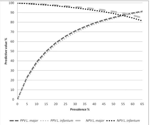
The accuracy of the ELISA tests usingL. majorandL. infantum antigens in are shown in Table 1. Briefly, sensitivity was 91.84% and 89.80% and specificity was 83.57% and 82.59% forL. major andL. infantumantigens, respectively. Figure 1 shows the results of the sensitivity analysis for the calculation of NPV and PPV based on seroprevalence rates ranged from of 1 to 65%.
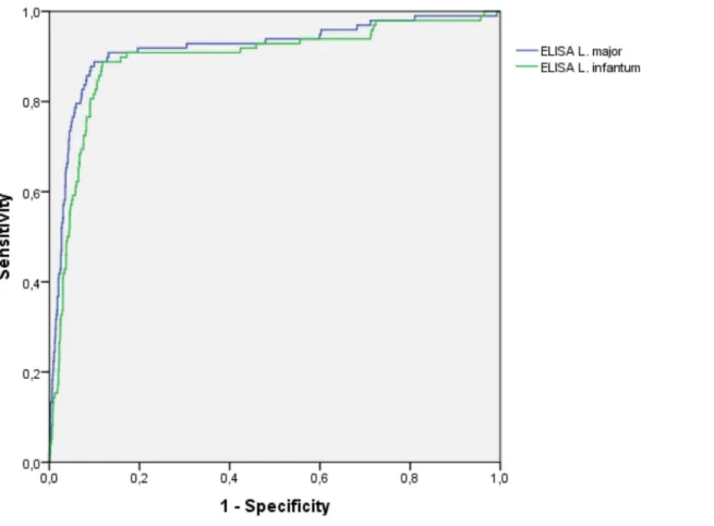
The area under the ROC curve (AUROC) was 0.920 and 0.898 forL. majorandL. infantumantigens, respectively. Figure 2.
The mean ICC of the tests ranged from 0.890 to 0.948 whenL. major was used as antigen, and from 0.818 to 0.879 when L. infantumwas used. Table 2.
Discussion
The relationship between human cases of VL and the prevalence of canine zoonotic disease has resulted in multiple efforts to reduce the risk of transmission. An important corner-stone of the national control program of the Brazilian Ministry of
Health is the monitoring of reservoirs based on the identification and euthanasia of infected dogs. The development of accurate diagnostic tests that meet the needs of both public health services and owners of dogs living in endemic areas in terms of the reliability of a valid diagnosis is a major challenge for researchers. The study of canine seroprevalence in endemic areas can generate much doubt depending on the sensitivity and specificity
Figure 1. Sensitivity analysis of positive and negative predictive values of ELISA tests usingLeishmania majororLeishmania infantum
antigen (Bio-ManguinhosH) according to variations in the prevalence of canine visceral leishmaniasis.PPV: positive predictive value; NPV: negative predictive value.
doi:10.1371/journal.pone.0069988.g001
Table 1.Accuracy results of ELISA tests usingLeishmania majororLeishmania infantumantigens (Bio-ManguinhosH) for the detection of visceral leishmaniasis in serum samples of dogs from endemic regions in Brazil (2011).
Leishmania majortest Leishmania infantumtest
Sensitivity 91.84% (86.42 to 97.26) 89.80% (83.80 to 95.79)
Specificity 83.75% (81.76 to 85.74) 82.69% (80.64 to 84.73)
AUROCa 0.917 (0.881 to 0.953) 0.893 (0.854 to 0.933)
PPVb 29.61% (24.47 to 34.74) 27.85% (22.91 to 32.79)
NPVc 99.28% (98.78 to 99.78) 99.09% (98.53 to 99.65)
The 95% confidence interval is given in parentheses.
aU
ROC: area under the ROC curve. bPositive predictive value. cNegative predictive value.
of the tests used, which vary between different validation studies. These variations are related mainly to differences in the reference population and sampling strategies used for the validation process, as well as to technical characteristics of the test, competence of the laboratory, choice of the gold standard, and cut-off value used for interpretation [26].Furthermore, biological factors can affect the accuracy of serological tests. Sensitivity varies according to the state of infection and immune status of the host. In addition, lower
specificity is due to cross-reactions with other agents or when this parameter is estimated in dogs that are truly infected but are not detected adequately by the gold standard [26]. Diagnostic tests based on recombinant more specific antigens have been devel-oped, however the lack of sensitivity precludes their use as the first choice tools for epidemiological surveys or control intervention programs.
In view of these considerations, the present study evaluated the accuracy and reproducibility of Bio-ManguinhosH ELISA tests usingL. majorand L. infantumantigens for the detection of VL in serum samples of a random sample of dogs including the whole spectrum ofLeishmaniainfection, from asymptomatic to seriously ill animals which represents the reality of canine VL in Brazil. The random sampling of dogs contributed to reduce selection bias, which is commonly seen in validation studies due to an unbalanced representation of symptomatic dogs. Another strong point of this study is the technical rigor and completeness of gold standard methods. However, our gold standard could fail in asymptomatic infected dogs with lower parasite burden producing a classification bias which would underestimate the true specificity value for both tests.
Comparison of the sensitivity between both antigens showed similar performance. Barbosa-De-Deus et al. [27] reported 98% sensitivity and 95% specificity of an ELISA test using antigen prepared from L. major-like promastigotes in a sample of 1,741 animals (1,582 negative and 159 positive). Similar results (97% sensitivity and 98% specificity) have been reported by Scalone et al. [28] for a sample of 415 animals (258 negative and 157 positive ) using recombinant rK39 antigen. Carvalho et al. [29] observed
Figure 2. Receiver operating curve (ROC) comparing the results of the optic density indexes obtained with ELISA tests using
Leishmania majororLeishmania infantumantigen (Bio-ManguinhosH).
doi:10.1371/journal.pone.0069988.g002
Table 2.Mean intraclass correlation coefficients between laboratories for the optic density indexes of ELISA tests using Leishmania majororLeishmania infantumantigen (Bio-ManguinhosH) for the detection of visceral leishmaniasis in serum samples of dogs from endemic regions in Brazil (2011).
Laboratory Leishmania majortest
Leishmania infantum test
ICCa ICC
CCZ-CGbx IAL-SPc 0.904 (0.884 to 0.919) 0.818 (0.791 to 0.841)
IAL-SP x FUNED-MGd 0.948 (0.942 to 0.953) 0.879 (0.865 to 0.891)
CCZ-CG x FUNED-MG 0.890 (0.876 to 0.902) 0.875 (0.834 to 0.903)
The 95% confidence interval is given in parentheses.
aIntraclass correlation coefficient.
bCentro de Controle de Zoonoses, Campo Grande. cInstituto Adolfo Lutz, Sa˜o Paulo.
dFundac¸a˜o Ezequiel Dias, Minas Gerais.
100% sensitivity and specificity of an in-house ELISA using L. infantum antigen in 125 animals (15 negative and 110 positive ). The fact that another serological test was used as the gold standard in those studies might be a disadvantage since the tests evaluated would detect the same phenomenon of antibody elevation identified by the gold standard and the chance of agreement would therefore be higher, overestimating sensitivity and specific-ity.
Studying ELISA tests that employedL. infantum/chagasiantigen, Oliveira et al. [30] reported 90% sensitivity and 100% specificity of the test for a sample of 101 dogs, including 30 animals with a confirmed parasitological diagnosis and 71 negative animals. The authors used exclusively sera from dogs with a confirmed parasitological diagnosis for the calculation of sensitivity and exclusively sera from dogs defined as negative for the calculation of specificity, an approach that improves artificially the accuracy of the tests. Rosa´rio et al. [13] compared ELISA tests employing crude antigens of L. amazonensis and L. chagasi/infantum and the recombinant antigens rK39 and rK26. A total of 131 samples were tested (25 negative and 106 positive) demonstrating sensitivity of 100% (95% CI: 95.6 to 100) forL. amazonensis, 98% (92.7 to 99.7) forL. chagasi/infantum, 98.1% (92.7 to 99.7) for antigen rK39, and 99.1% (94.1 to 100) for antigen rK26. Specificity was 100% (83.4 to100) for L. amazonensis, 100% (83.4 to 99.7) for L. chagasi/ infantum, 100% (83.4 to 100) for antigen rK39, and 96% (77.7 to 99.8%) for antigen rK26. Lira et al. [31] evaluated ELISA withL. major-like antigen (Bio-ManguinhosH), which is currently used by the Brazilian visceral leishmaniasis control program, in a sample of 41 animals (25 positive and 16 negative) and observed sensitivity of 72% (50.4 to 87.1) and specificity of 87.5% (60.4 to 97.8%). In addition to the imprecise estimates of that study, the authors used animals from unaffected areas as negative controls, a fact that may have favorably influenced the specificity results. Pinheiro et al. [32] compared ELISA tests using a recombinant cysteine proteinase (rLdccys1) and lysates ofL. chagasiamastigotes and promastigotes as antigens. In that study, sensitivity was 98% (rLdccys1), 89% (amastigotes) and 86% (promastigotes), and specificity was 96%, 69% and 68%, respectively. Like Oliveira et al. [30], the authors used sera from dogs with a confirmed parasitological diagnosis (209 animals) for the calculation of sensitivity and sera from 68 animals classified as negative for the calculation of specificity, including 46 samples from dogs with other diseases. However, the dog sera were obtained by convenience sampling which is prone to selection bias.
With respect to the predictive values shown in Table 1, the NPV were high (99,28 and 99,09%) indicating excellent sensitivity of the tests. On the other hand, the PPV was 29,61% when theL. major antigen was used and 27.85% with theL. chagasiantigen. These
results are a matter of concern since in cases in which the prevalence of infection is similar to that of the sample studied (6.9%), at least three dogs with a false-positive result would be eliminated per each truly infected animal. In this respect, although designed to improve the capacity for detection of infection, the gold standard used probably continues to be imperfect and does not identify some truly infected animals, with a consequent impact on specificity and PPV.
As can be seen in Figure 1, the predictive values of the ELISA tests are closely related to the prevalence of the disease. As already mention above prevalence of canine visceral leishmaniasis is variable and health decision-makers need to be aware of the expected predictive values using serological tests for control purposes. In this respect, sensitive tests are fundamental for surveillance and control programs of leishmaniasis since they permit the culling of a larger number of truly infected animals, whereas specific tests are more important for the confirmation of suspected clinical cases, being more relevant for veterinarians dedicated to individual animal care [14]. Barbosa-De-Deus et al. [27] studied an important convenience sample of dogs in which the ‘‘prevalence’’ of VL was 9.13% (159/1741), obtained good predictive values (100% NPV and 66% PPV). However, as discussed earlier the selection bias that may occur as a result of the use of another serological test as the gold standard should be taken into account when interpreting these results.
In our study the ICC indicated almost perfect agreement between tests (.0.81) in labs with different levels of complexity. This is a very relevant result because adequate reproducibility is essential to avoid unnecessary dog culling and optimize lab costs. The lack of significant differences in the accuracy and reliability of tests usingL. infantumorL. majorantigen indicates that there is no need to change the antigen composition of the enzyme immuno-assay currently used in Brazil for the diagnosis of canine VL.
Acknowledgments
We thank the Municipal and State Secretary Offices of Health that participated in the study for their help and commitment. Professor Jose´ Ue´leres Braga for critical review of the manuscript and useful suggestions for improving data analysis.
Author Contributions
Conceived and designed the experiments: MMA FBF FAC RMH JCMB MRFO EFN GASR. Performed the experiments: MMA FBF FAC RMH JCMB. Analyzed the data: MMA FBF MRFO EFN GASR. Contributed reagents/materials/analysis tools: MMA FBF FAC RMH JCMB. Wrote the paper: MMA FBF FAC RMH JCMB MRFO EFN GASR.
References
1. Schonian G, Mauricio I, Cupolillo E (2010) Is it time to revise the nomenclature ofLeishmania? Trends Parasitol. 26: 466–469.
2. Shaw JJ (2006) Further thoughts on the use of the nameLeishmania (Leishmania) infantumchagasi for the aetiological agent of American visceral leishmaniasis. Mem. Inst. Oswaldo Cruz. 101: 577–579.
3. Deane LM, Deane MP (1962) Visceral Leishmaniasis in Brazil: Geographical distribution and transmission. Rev. Inst.Med.Trop.Sa˜o Paulo 4: 198–212. 4. Rangel EF, ML V (2008)Lutzomyia longipalpis(Diptera, Psychodidae,
Phleboto-minae) and urbanization of visceral leishmaniasis in Brazil. Cad. Saude Publica. 24: 2948–2952.
5. Cerbino NJ, Werneck GL, Costa CH (2009 ) Factors associated with the incidence of urban visceral leishmaniasis: an ecological study in Teresina, Piaui State, Brazil. Cad. Saude Publica.25: 1543–1551.
6. Harhay MO, Olliaro PL, Costa DL, Costa CH (2011) Urban parasitology: visceral leishmaniasis in Brazil. Trends Parasitol. 27: 403–409.
7. Ministe´rio da Sau´de (2006) Manual de Vigilaˆncia e Controle da Leishmaniose Visceral. Brası´lia-DF. Ministe´rio da Sau´de - MS (ed), Brası´lia: Available: http://
portal.saude.gov.br/portal/arquivos/pdf/manual_leish_visceral2006.pdf Ac-cessed 25 January 2011.
8. Romero GA, Boelaert M (2010) Control of visceral leishmaniasis in latin america-a systematic review. PLoS Negl. Trop. Dis. 4: e584.
9. Costa CH (2011) How effective is dog culling in controlling zoonotic visceral leishmaniasis? A critical evaluation of the science, politics and ethics behind this public health policy. Rev. Soc. Bras. Med. Trop. 44: 232–242.
10. Silva AV, Paula AA, Cabrera MA, Carreira JC (2005) Leishmaniasis in domestic dogs: epidemiological aspects. Cad. Saude Publica. 21: 324–328.
11. Troncarelli MZ, Camargo JB, Machado JG, Lucheis SB, Langoni H (2009) Leishmaniaspp. and/orTrypanosoma cruzidiagnosis in dogs from endemic and nonendemic areas for canine visceral leishmaniasis. Vet. Parasitol. 164: 118– 123.
12. Souza AI, Oliveira TMFS, Machado RZ, Camacho AA (2009) Soroprevaleˆncia da infecc¸a˜o por Trypanosoma cruzi em ca˜es de uma a´rea rural do Estado de Mato Grosso do Sul. Pesq. Vet. Bras. 29: 150–152.
recombinant antigens as a diagnostic marker for canine visceral leishmaniasis. Mem. Inst. Oswaldo Cruz. 100: 197–203.
14. Mettler M, Grimm F, Capelli G, Camp H, Deplazes P (2005) Evaluation of enzyme-linked immunosorbent assays, an immunofluorescent-antibody test, and two rapid tests (immunochromatographic-dipstick and gel tests) for serological diagnosis of symptomatic and asymptomatic Leishmania infections in dogs. J. Clin. Microbiol. 43: 5515–5519.
15. Kar K (1995) Serodiagnosis of leishmaniasis. Crit. Rev. Microbiol. 21: 123–152. 16. Boarino A, Bollo E, Prunotto L, Canale L, Uslenghi F, et al. (2008) Application of a recombinant protein for the serological diagnosis of canine leishmaniasis. Comp. Immunol. Microbiol. Infect. Dis. 31: 527–536.
17. Figueiredo FB, Madeira MF, Menezes RC, Pacheco RS, Pires MQ, et al. (2010) Efficacy of an indirect immunofluorescence test in the diagnosis of canine leishmaniosis. Vet. J. 186: 123–124.
18. Madeira MF, Figueiredo FB, Pinto AG, Nascimento LD, Furtado M, et al. (2009) Parasitological diagnosis of canine visceral leishmaniasis: is intact skin a good target? Res. Vet. Sci. 87: 260–262.
19. Coutinho SG, Nunes MP, Marzochi MC, Tramontano N (1985) A survey for American cutaneous and visceral leishmaniasis among 1,342 dogs from areas in Rio de Janeiro (Brazil) where the human diseases occur. Mem. Inst. Oswaldo. Cruz. 80: 17–22.
20. Paranhos-Silva M, Freitas LA, Santos WC, Grimaldi GJ, Pontes-de-Carvalho LC, et al. (1996) A cross-sectional serodiagnostic survey of canine leishmaniasis due toLeishmania chagasi. Am. J. Trop. Med. Hyg. 55: 39–44.
21. Bernadina WE, De Luna R, Oliva G, Ciaramella P (1997) An immunodiffusion assay for the detection of canine leishmaniasis due to infection withLeishmania infantum. Vet. Parasitol. 73: 207–213.
22. Ashford DA, David JR, Freire M, David R, Sherlock I, et al. (1998) Studies on control of visceral leishmaniasis: impact of dog control on canine and human visceral leishmaniasis in Jacobina, Bahia, Brazil. Am. J. Trop. Med.Hyg. 59: 53– 57.
23. Moura ST, Fernandes CGN, Pandolpho VC, Rodrigues e Silva R (1999) Diagno´stico de leishmaniose canina na a´rea urbana do municı´pio de Cuiaba´, Estado de Mato Grosso, Brasil/Diagnosis of canine leishmaniasis in the urban
area of the District of Cuiaba´, State of Mato Grosso, Brazil. Braz. J. Vet. Res. Anim. Sci. 36: 101–102.
24. Franca-Silva JC, da Costa RT, Siqueira AM, Machado-Coelho GL, da Costa CA, et al. (2003) Epidemiology of canine visceral leishmaniosis in the endemic area of Montes Claros Municipality, Minas Gerais State, Brazil. Vet. Parasitol. 111: 161–173.
25. Queiroz PV, Monteiro GR, Macedo VP, Rocha MA, Batista LM, et al. (2009) Canine visceral leishmaniasis in urban and rural areas of Northeast Brazil. Res. Vet. Sci. 86: 267–273.
26. Greiner M, Gardner IA (2000) Epidemiologic issues in the validation of veterinary diagnostic tests. Prev. Vet. Med. 45: 3–22.
27. Barbosa-De-Deus R, Dos Mares-Guia ML, Nunes AZ, Costa KM, Junqueira RG, et al. (2002) Leishmania major-like antigen for specific and sensitive serodiagnosis of human and canine visceral leishmaniasis. Clin. Diagn. Lab. Immuno.l 9: 1361–1366.
28. Scalone A, De Luna R, Oliva G, Baldi L, Satta G, et al. (2002) Evaluation of the Leishmania recombinant K39 antigen as a diagnostic marker for canine leishmaniasis and validation of a standardized enzyme-linked immunosorbent assay. Vet. Parasitol. 104: 275–285.
29. Carvalho D, Oliveira TMFS, Baldani CD, Machado RZ (2009) An enzyme-linked immunosorbent assay (ELISA) for the detection of IgM antibodies against Leishmania chagasiin dogs. Pesq. Vet. Bras. 29: 120–124.
30. Oliveira LS, Julia˜o FS, Souza VMM, Freitas DS, Souza BMPS, et al. (2005) A utilizac¸a˜o da imunofluoreˆscencia indireta no Diagno´stico de rotina da leishmaniose visceral canina e suas implicac¸o˜es no controle da doenc¸a. Ci.Anim.Bras.6: 41–47.
31. Lira RA, Cavalcanti MP, Nakazawa M, Ferreira AGP, Silva ED, et al. (2006) Canine visceral leishmaniosis: a comparative analysis of the EIE-leishmaniose-visceral-canina-Bio-Manguinhos and the IFI-leishmaniose-visceral-canina-Bio-Manguinhos kits. Vet. Parasitol. 137: 11–16.