J Bras Pneumol. 2013;39(1):63-68
Histoplasmosis mimicking primary lung
cancer or pulmonary metastases*
,**
Histoplasmose simulando neoplasia primária de pulmão ou metástases pulmonares
Aline Gehlen Dall Bello, Cecilia Bittencourt Severo, Luciana Silva Guazzelli, Flavio Mattos Oliveira, Bruno Hochhegger, Luiz Carlos Severo
Abstract
Objective: To describe the main clinical and radiological characteristics of patients with histoplasmosis mimicking lung cancer. Methods: This was a retrospective descriptive study based on the analysis of the medical records of the 294 patients diagnosed with histoplasmosis between 1977 and 2011 at the Mycology Laboratory of the Santa Casa Sisters of Mercy Hospital of Porto Alegre in the city of Porto Alegre, Brazil. The diagnosis of histoplasmosis was established by culture, histopathological examination, or immunodiffusion testing (identification of M or H precipitation bands). After identifying the patients with macroscopic lesions, as well as radiological and CT findings consistent with malignancy, we divided the patients into two groups: those with a history of cancer and presenting with lesions mimicking metastases (HC group); and those with no such history but also presenting with lesions mimicking metastases (NHC group). Results: Of the 294 patients diagnosed with histoplasmosis, 15 had presented with lesions mimicking primary neoplasia or metastases (9 and 6 in the HC and NHC groups, respectively). The age of the patients ranged from 13 to 67 years (median, 44 years). Of the 15 patients, 14 (93%) presented with pulmonary lesions at the time of hospitalization. Conclusions: The clinical and radiological syndrome of neoplastic disease is not confined to malignancy, and granulomatous infectious diseases must therefore be considered in the differential diagnosis.
Keywords: Histoplasmosis; Multiple pulmonary nodules; Solitary pulmonary nodule.
Resumo
Objetivo: Descrever as principais características clínico-radiológicas de pacientes com histoplasmose simulando câncer de pulmão. Métodos: Estudo descritivo e retrospectivo baseado na análise dos prontuários médicos de 294 pacientes diagnosticados com histoplasmose no Laboratório de Micologia da Irmandade Santa Casa de Misericórdia de Porto Alegre, em Porto Alegre (RS) entre 1977 e 2011. O diagnóstico de histoplasmose foi estabelecido por cultura, exame histopatológico ou identificação de bandas M ou H por imunodifusão. Após identificar os pacientes com lesões macroscópicas e com achados compatíveis de malignidade em radiografia ou TC de tórax, os pacientes foram divididos em dois grupos: pacientes com história de câncer e lesões simulando metástases (grupo HC) e pacientes sem história de câncer com lesão simulando neoplasia primária (SHC). Resultados: Dos 294 pacientes com histoplasmose, 15 apresentaram lesões simulando neoplasia primária ou metástases (9 e 6 nos grupos HC e SHC, respectivamente). A idade dos pacientes variou de 13 a 67 anos (mediana, 44 anos) Dos 15 pacientes, 14 (93%) apresentaram lesões pulmonares no momento da internação. Conclusões: A síndrome clínica e radiológica da doença neoplásica não se limita a malignidade, e, portanto, as doenças infecciosas granulomatosas devem ser consideradas no diagnóstico diferencial.
Descritores: Histoplasmose; Nódulos pulmonares múltiplos; Nódulo pulmonar solitário.
* Study carried out at the Santa Casa Sisters of Mercy Hospital of Porto Alegre, Porto Alegre, Brazil.
Correspondence to: Aline Gehlen Dall Bello. Avenida Independência, 75, Centro, CEP 90050-070, Porto Alegre, RS, Brasil. Tel. 55 51 3214-8009. E-mail: [email protected]
Financial support: None.
Submitted: 23 July 2012. Accepted, after review: 2 October 2012.
lesion with homogenous soft-tissue attenuation and without benign calcification or spiculated margins.
Results
Of the 294 patients diagnosed with histoplasmosis, 15 presented with lesions mimicking primary neoplasia or simulating metastases. The most common clinical symptoms were fever, cough, weight loss, and chest pain. Five patients were asymptomatic. Of the 15 patients, 9 had a history of cancer and presented with lesions simulating metastases (HC group; Figure 1), and 6 patients had no history of cancer and presented with at least one lesion mimicking primary malignancy (NHC group; Figure 2).
Clinical, demographic, and biochemical findings are shown in Table 1. The age of the patients ranged from 13 to 67 years (median, 44 years). Of the 15 patients, 14 (93%) presented with pulmonary lesions at the time of hospitalization. In all 15 patients, the diagnosis of histoplasmosis was made by demonstrating oval budding yeasts, typical of H. capsulatum, on biopsy specimens stained with methenamine silver. The diagnosis was confirmed by culture in only 4 cases, although culture was requested in only 5. Seven patients were tested for the presence of specific antibodies to H. capsulatum, and 6 of those 7 tested negative.
Discussion
The results of the present study underscore the fact that the clinical syndrome of neoplastic disease is not confined to malignancy and that granulomatous infectious diseases must be considered in the differential diagnosis. Excluding histoplasmosis from the differential diagnosis of pulmonary lesions can delay the diagnostic process.
There are sporadic reports of patients with histoplasmosis that have been misdiagnosed as having head and neck cancer,(2,12) primary lung
cancer,(13-15) or lymphoma.(16,17) In such cases, the
cytological examination of the pleural fluid can suggest neoplasia, because giant cells in which H. capsulatum is present could be mistaken for of clinical manifestations, ranging from
self-limiting respiratory complaints to progressive, life-threatening infections. Primary pulmonary infection results from the inhalation of airborne microconidia, and the vast majority of infections are self-limiting—in most cases, the infection is limited to the lungs—however, since the onset of the AIDS epidemic, disseminated histoplasmosis has been reported with greater frequency.(1,2)
Air currents can carry the microconidia for miles, exposing unsuspecting individuals to indirect contact with a contaminated site. In addition, the fungus can be present at environmental sites that are not visibly contaminated with droppings from birds or bats, thus reducing the chance that histoplasmosis will be suspected.(3) In Brazil,
histoplasmosis is a recurrent topic in pulmonology.
(4-8) A review of the literature revealed that there
have been cases of histoplasmosis mimicking malignancy.(9-11) The rarity of this manifestation
prompted our study, the objective of which was to describe the main clinical and radiological characteristics of patients with histoplasmosis mimicking cancer. We also review the literature on the topic.
Methods
This was a retrospective descriptive study based on the analysis of the medical records of the 294 patients diagnosed with histoplasmosis between 1977 and 2011 at the Mycology Laboratory of the Santa Casa Sisters of Mercy Hospital of Porto Alegre in the city of Porto Alegre, Brazil. The study was approved by the Human Research Ethics Committee of the Hospital (Protocol no. 306/11). The diagnosis of histoplasmosis was established by culture, histopathological examination, or immunodiffusion testing (identification of M or H precipitation bands).(6)
Histoplasmosis mimicking primary lung cancer or pulmonary metastases
J Bras Pneumol. 2013;39(1):63-68
65
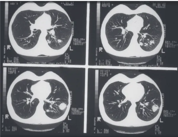
Figure 1 - Pulmonary histoplasmosis in a 29-year-old female patient with a history of melanoma and treatment with antineoplastic agents (patient 5). The CT scans reveal multiple, sharply circumscribed, randomly distributed nodules, predominantly in the left lung.
malignant lymphoblasts.(18) It is of interest that
histoplasmosis granuloma might be associated with a type of lung carcinoma.(14) On CT scans,
as well as on scans made with more advanced imaging techniques, such as positron emission tomography, histoplasmosis can mimic malignant lesions.
Immunocompromised patients, such as those in our HC group, are more susceptible to disseminated disease. In the literature, approximately two-thirds of patients with chronic disseminated histoplasmosis present with oropharyngeal or laryngeal involvement, which is almost invariably the clinical feature that leads to the diagnosis,(19)
and one of our patients (patient 6) presented with this type of lesion. In addition, adrenal involvement has been found in over half of all patients with disseminated histoplasmosis, the lesions being more commonly found in the zona reticularis; this could be due to the presence of higher downstream concentrations of cortisol, en route from secretion to the medullary central
venous system.(19) Three of the patients in the
HC group had disseminated disease. Of those 3, only 1 was submitted to autopsy (case 1), which showed adrenal involvement. Cutaneous lesions of disseminated histoplasmosis are infrequent, being most commonly found in individuals who are infected with HIV,(20) which nearly none of
our patients were.
The patients in the NHC group demonstrated that histoplasmosis could occur in individuals who are not apparently immunocompromised. Because imaging findings often mimic other granulomatous infections and neoplastic processes, they are not considered diagnostic.(17) Histoplasmosis in the
mediastinum is an uncommon diagnosis that has a presentation similar to that of other benign and neoplastic conditions encountered in the chest.(21) The principle that a solitary circumscribed
eira FM, Hochhegger B, Sev
ero LC
histoplasmoma
3 32/M Lymphoma Disseminated Mass Lung, skin + + + Untreated Died 4 67/M Squamous cell
carcinoma
Pulmonary/ histoplasmoma
Mass, consolidation Lung + − ND Itraconazole Improved
5 29/F Melanoma Pulmonary Nodules Lung + − ND Not reported Not reported 6 13/F Lymphoma Disseminated Nodules Lung, larynx + ND ND Itraconazole Improved 7 50/F Pancreatic
adenocarcinoma
Disseminated Nodules Lung, pleura + ND + Untreated Died
8 58/M Lymphoma Disseminated Pulmonary infiltrates
Lung + ND + Not reported Not reported
9 54/F Breast carcinoma Pulmonary Mass Lung + ND ND Surgery Improved Patients without a history of cancer
Patient Age,
years/Gender Primary disease Clinical presentation
Chest radiographic
findings Site(s) of infection
Diagnosis
Treatment Outcome Hist IDh Cult
10 25/M AIDS Disseminated Normal Pharynx, skin + ND ND Amphotericin B Improved 11 66/M Arterial hypertension Pulmonary/
histoplasmoma
Mass Lung, lymph node + − ND Itraconazole Improved
12 63/F None Pulmonary/ histoplasmoma
Mass Lung + − ND Surgery Improved
13 25/F None Pulmonary/ histoplasmoma
Mass Lung + − + Itraconazole Improved
14 13/M None Disseminated Nodule Lung, mediastinal + ND ND Itraconazole Improved 15 67/M Liver transplant Pulmonary Mass Lung + ND − Not reported Not reported
Hist: histopathology, tissue section (H&E and Grocott-Gomori methenamine silver); IDh: immunodiffusion for Histoplasma capsulatum; Cult: culture on Sabouraud dextrose agar and
Histoplasmosis mimicking primary lung cancer or pulmonary metastases
J Bras Pneumol. 2013;39(1):63-68
67
and metastatic lung cancer, because delaying treatment can allow severe manifestations of the former to occur.
References
1. Kauffman CA. Histoplasmosis. Clin Chest Med. 2009;30(2):217-25, v. PMid:19375629. http:// dx.doi.org/10.1016/j.ccm.2009.02.002
2. Loh FC, Yeo JF, Tan WC, Kumarasinghe G. Histoplasmosis presenting as hyperplastic gingival lesion. J Oral Pathol Med. 1989;18(9):533-6. PMid:2607474. http://dx.doi. org/10.1111/j.1600-0714.1989.tb01358.x
3. Wheat J, Sarosi G, McKinsey D, Hamill R, Bradsher R, Johnson P, et al. Practice guidelines for the management of patients with histoplasmosis. Infectious Diseases Society of America. Clin Infect Dis. 2000;30(4):688-95. PMid:10770731. http://dx.doi.org/10.1086/313752 4. Unis G, Roesch EW, Severo LC. Acute pulmonary
histoplasmosis in the State of Rio Grande do Sul, Brazil. J Bras Pneumol. 2005;31(1):52-9. http://dx.doi. org/10.1590/S1806-37132005000100010
5. Oliveira Fde M, Unis G, Severo LC. An outbreak of histoplasmosis in the city of Blumenau, Santa Catarina. J Bras Pneumol. 2006;32(4):375-8. PMid:17268739. http://dx.doi.org/10.1590/S1806-37132006000400018 6. Aidé MA. Chapter 4--histoplasmosis. J Bras
Pneumol. 2009;35(11):1145-51. PMid:20011851. http:// dx.doi.org/10.1590/S1806-37132009001100013 7. Santos JW, Michel GT, Lazzarotto M, Figaro JK, Spilmann
D, Homrich GK. Chronic cavitary pulmonary histoplasmosis. J Bras Pneumol. 2009;35(11):1161-4. PMid:20011854. http://dx.doi.org/10.1590/S1806-37132009001100016 8. Fortaleza SC, Lopes SK, Bandeira TJ, Nogueira TN,
Holanda MA. Acute disseminated histoplasmosis in an immunocompetent patient. J Bras Pneumol. 2004;30(3):270-3.
9. Mukhopadhyay S, Katzenstein AL. Biopsy findings in acute pulmonary histoplasmosis: unusual histologic features in 4 cases mimicking lymphomatoid granulomatosis. Am J Surg Pathol. 2010;34(4):541-6. PMid:20351490. http://dx.doi.org/10.1097/PAS.0b013e3181d4388b 10. Paphitou NI, Barnett BJ. Solitary parietal lobe
histoplasmoma mimicking a brain tumor. Scand J Infect Dis. 2002;34(3):229-32. http://dx.doi. org/10.1080/00365540110077308
11. Rolston KV, Rodriguez S, Dholakia N, Whimbey E, Raad I. Pulmonary infections mimicking cancer: a retrospective, three-year review. Support Care Cancer. 1997;5(2):90-3. PMid:9069606. http://dx.doi.org/10.1007/BF01262563 12. Antonello VS, Zaltron VF, Vial M, Oliveira FM, Severo
LC. Oropharyngeal histoplasmosis: report of eleven cases and review of the literature. Rev Soc Bras Med Trop. 2011;44(1):26-9. PMid:21340403. http://dx.doi. org/10.1590/S0037-86822011000100007
13. Kneale B, Turton C. Bronchoscopic findings in a case of bronchopulmonary histoplasmosis. Thorax. 1995;50(3):314-5; discussion 317-8. PMid:7660349 PMCid:1021200. http://dx.doi.org/10.1136/thx.50.3.314 14. Yoneda K. Scar carcinomas of the lung in a histoplasmosis
endemic area. Cancer. 1990;65(1):164-8. http://dx.doi. org/10.1002/1097-0142(19900101)65:1<164::AID-CNCR2820650131>3.0.CO;2-R
15. Ross P Jr, Magro CM, King MA. Endobronchial histoplasmosis: a masquerade of primary endobronchial The diagnostic approach depends on the type
of infection and the amount of microconidia inhaled. A variety of tests, including culture, specific staining for fungal cells, antigen detection, and serologic tests for antibodies, are used for the diagnosis of histoplasmosis. The sensitivity of laboratory tests depends on the clinical manifestation of histoplasmosis (disseminated, chronic pulmonary, or self-limiting).(20) We observed
these differences and the importance of using more than one diagnostic method. In our cohort, the patients with histoplasmoma (patients 11 and 12) showed no specific antibodies to H. capsulatum; this also can happen in immunocompromised patients with positive cultures.(20) This information
can be helpful because the absence of specific antibodies is usually associated with the absence of infection in immunocompetent patients.
21. Shersher DD, Hong E, Breard J, Warren WH, Liptay MJ. Anterior mediastinal mass secondary to histoplasmosis. Ann Thorac Surg. 2012;93(1):e9-10. PMid:22186488. http://dx.doi.org/10.1016/j.athoracsur.2011.07.093 22. Goodwin RA Jr, Snell JD Jr The enlarging histoplasmoma.
Concept of a tumor-like phenomenon encompassing the tuberculoma and coccidioidoma. Am Rev Respir Dis. 1969;100(1):1-12. PMid:5796688.
17. McGraw EP, Kane JM, Kleiman MB, Scherer LR. Cervical abscess and mediastinal adenopathy: an unusual presentation of childhood histoplasmosis. Pediatr Radiol. 2002;32(12):862-4. PMid:12447591. http:// dx.doi.org/10.1007/s00247-002-0808-2
18. Brodeur GM, Wilber RB, Melvin SL, Murphy SB. Histoplasmosis mimicking childhood non-Hodgkin lymphoma. Med Pediatr Oncol. 1979;7(1):77-81. PMid:522825. http://dx.doi.org/10.1002/mpo.2950070111
About the authors
Aline Gehlen Dall Bello
Doctoral Student. Mycology Laboratory, Santa Casa Sisters of Mercy Hospital of Porto Alegre, Porto Alegre, Brazil.
Cecilia Bittencourt Severo
Biochemist. Mycology Laboratory, Santa Casa Sisters of Mercy Hospital of Porto Alegre, Porto Alegre, Brazil.
Luciana Silva Guazzelli
Biochemist. Mycology Laboratory, Santa Casa Sisters of Mercy Hospital of Porto Alegre, Porto Alegre, Brazil.
Flavio Mattos Oliveira
Biochemist. Mycology Laboratory, Santa Casa Sisters of Mercy Hospital of Porto Alegre, Porto Alegre, Brazil.
Bruno Hochhegger
Radiologist. Radiology Department, Santa Casa Sisters of Mercy Hospital of Porto Alegre, Porto Alegre, Brazil; Radiology Department, Federal University of Rio de Janeiro, Rio de Janeiro, Brazil.
Luiz Carlos Severo