Abstract
Submitted: February 19, 2018 Modification: May 27, 2018 Accepted: July 1, 2018
Surface and vertical dimensional
changes of mineral trioxide
aggregate and biodentine in different
environmental conditions
Surface changes in biological environments are critical for the evaluation of physical and biological activity of biomaterials. Objective: This study investigated surface alterations of calcium silicate-based cements after exposure to different environments. Material and Methods: Forty-eight
cylindrical cavities were prepared on root surfaces. The cavities were filled
using ProRoot MTA or Biodentine and assigned to four subgroups (n=6): dry, wet, acidic, and blood. Surface topographies were evaluated using an optical
profilometer for 28 days, and the roughness of the material surfaces was quantified. Vertical dimensional change was measured by determining the height difference between the material surface and the flat tooth surface. Data were compared with a two-way repeated measures ANOVA and Bonferroni
tests. Results: In dry condition, the surface roughness of MTA or Biodentine
was constant up to 3 days (p>0.05) but decreased after 28 days (p<0.05).
In dry condition, ProRoot MTA presented constant surface level through
time, while Biodentine showed decreased surface level after 28 days. In wet
condition, the roughness and the surface levels of both materials increased
after 1 day (p<0.05). Neither the surface roughness nor the levels of the materials showed significant changes in acidic conditions (p>0.05). Both
materials showed the highest roughness in blood conditions on the 1st day
(p<0.05), while the surface roughness in blood decreased dramatically after 28 days. The roughness of Biodentine was higher in wet conditions up to 3 days compared with ProRoot MTA (p<0.05). Likewise, in blood condition, Biodentine showed higher roughness on the 28th day than ProRoot MTA
(p<0.05). Conclusions: Dry, wet, and blood conditions had a time-dependent
effect on the surface roughness and vertical dimensional changes of the materials. However, acidic conditions did not affect the roughness and the surface level of the materials.
Keywords: Biodentine. Calcium silicate-based cement. MTA. Profilometer. Roughness.
Hacer AKSEL1
Selen KÜÇÜKKAYA EREN1
Sevinc ASKERBEYLİ ÖRS1
Eda KARAISMAILOĞLU2
1Hacettepe University, Faculty of Dentistry, Department of Endodontics, Ankara, Turkey. 2Kastamonu University, Faculty of Medicine, Department of Biostatistics, Kastamonu, Turkey. Corresponding address:
Hacer Aksel Department of Periodontics and Endodontics - University at Buffalo -School of Dental Medicine - NY - USA.
Introduction
Mineral trioxide aggregate (MTA) and Biodentine are mostly used calcium silicate-based cements (CSCs) in several endodontic procedures, such as
retrograde filling, coronal barrier, pulp capping agent,
and perforation repair material.1 Both materials consist
of tricalcium silicate and dicalcium silicate as main components. MTA also contains calcium sulfate and
bismuth oxide (radiopacifier), and Biodentine contains calcium carbonate and zirconium oxide (radiopacifier).
Hydration of the materials forms calcium silicate hydrate gel and calcium hydroxide.2 MTA completes the
initial setting after 40 min and solidifies completely in
140 min.3 Biodentine has a smaller particle size that
increases the surface area and density of the material compared with MTA.4Biodentine has a relatively short setting time and showed initial setting after 9–12 min and solidifies after 45 min.5
The local physicochemical environment determines
the suitability of the materials for clinical applications.6
MTA and Biodentine generally come into contact with body fluids and moisture when used for endodontic applications, and both materials can solidify in blood, plasma, and other fluids.7 However, when these
materials are used as cavity liners and bases under final adhesive restorations, they are placed in relatively dry conditions.8 In addition, the materials can be
exposed to blood during endodontic applications, such
as apical surgery and revascularization procedures.9
The pH of the environment becomes acidic during
endodontic treatment of necrotic teeth with periapical
lesions or the repair of teeth with perforating furcal
lesions, which may affect the properties of the
materials.10
Different oral conditions may affect the surface characteristics of the materials.11 Surface roughness
is a component of the surface texture and is also the
measure of vertical (positive or negative) deviations
of the surface from an ideal flat surface. The biocompatibility and bioactivity of these materials provide a microenvironment for cell attachment and odonto-/osteogenic activity during pulp capping procedures,12 regenerative endodontics,13 or periapical
healing after apical plug formation.14 According to previous studies, a rough surface may promote the attachment and proliferation of the cells by increasing material-cell interactions.15,16 In addition, the materials
can release calcium ions, and the accumulation of the
calcium on the surface increases surface roughness, which leads to the formation of hydroxyapatite on the surface.17 However, excessive surface roughness might
have a negative impact on the strength and sealing of materials.18 MTA and Biodentine have a similar surface roughness in wet conditions.15 However, no
comparable data on the effects of different conditions on the roughness of MTA or Biodentine were found in literature.
Setting conditions could also affect the dimensional
stability of these materials.19 The dimensional stability
of a material should be adequate to improve its adaptation and prevent leakage. Slight expansion might contribute to the adaptation of the material, but excessive shrinkage or expansion during setting may lead to leakage, lack of marginal integrity, or cracks in the root canal walls.20
Recently, an optical profilometer was used to
measure the physical properties of dentin or the
rotary instruments, as well as the adaptation of
root end-filling materials.21 The optical profilometer
acquires a computer-based three-dimensional
(3D) measurement of information on the surface
characteristics of materials without damaging them during measurement.18
This study attempted to reveal the surface
characteristics of ProRoot MTA and Biodentine in
different environmental conditions with a non-contact 3D optical profilometer.
Material and Methods
The crowns of 24 human maxillary canine teeth
were removed, and the root halves were obtained by sectioning the roots longitudinally. The middle part of each root half was used for the experiments. Each root half was embedded in a circular self-cure acrylic resin (Meliodent, Heraeus-Kulzer, Senden,
Germany), and standardized cylindrical cavities (2
mm in height × 3 mm in diameter) were created in the middle part of each root half. A polishing device
(Mecapol P230, Presi, Contamine-sur-Arve, France)
using 600- and 1000-grit abrasive discs (Buehler,
Lake Bluff, IL, USA) was used to polish the surface of the blocks.22 Irrigation was performed during 3 min
using 2.5% sodium hypochlorite (NaOCl), followed by 1 min of 17% ethylenediaminetetraacetic acid (EDTA)
The tooth halves were assigned to the MTA group
or to the Biodentine group (n=24). To prepare the
materials, 1 g of the powder of White ProRoot MTA
(Dentsply Maillefer, Ballaigues, Switzerland) was
mixed with 0.34 g liquid on a glass slab for 30 s, and Biodentine (Septodont, Saint Maur des Fausses,
France) was prepared by pouring five liquid drops
into the powder capsule and mixing for 30 s in a triturator. The materials were then inserted into the
cavities using an MTA Endo gun (Dentsply Maillefer) and compacted with an endodontic plugger (Dentsply Maillefer) that was ultrasonically activated for 2 s using an ultrasonic tip CPR-1 (Dentsply Tulsa Dental, Tulsa, OK, USA) connected to a piezoelectric unit (Pmax,
Satelec, Merignac, France) at the medium power setting.23 The material surface was straightened from
the surface of the specimens with a scalpel. The same operator prepared and placed the materials to prevent inter-operator discrepancies. After placement, the specimens in MTA or Biodentine groups were assigned to four subgroups according to the experimental condition (n=6): dry, wet (phosphate buffered saline: PBS; Biochrom, Berlin, Germany, pH 7.4), acidic (PBS, pH 5.0), or blood. To prepare the blood condition,
human blood was obtained from a healthy volunteer,
member of the research group, by phlebotomy using
a 27-gauge needle. The blood was kept in a mold containing 50 IU of heparin (Nevparin, Mustafa Nevzat İlaç San. A.Ş., Istanbul, Turkey) per 1 mL of
blood to prevent coagulation. The specimens were
then incubated in 24-well plates with the respective
environmental medium, and the wells were placed in
an incubator with 100% humidity at 37°C for 28 days.
The media were refreshed three times per week.
Measurement of surface roughness
The surface of each specimen was scanned using
a 3D optical profilometer (Zygo Corp., Middlefield, CT, USA). The image resolution was 640×480 pixels. The mean of the 3D surface roughness of each material was
analyzed using image analysis software (MetroPro;
Zygo Corp.). To do so, three images were taken of
different parts of each material. The surface of each material was divided into three sections from left to right using analysis software to standardize the measurement area. The average roughness (Ra), root
mean square (RMS), and maximum peak-to-valley height (PV) values were the roughness parameters
used to determine the surface characteristics of the
materials.24 Ra represents the average roughness, RMS indicates the arithmetic mean of the height
and depth of the surface from a mean line, and PV
indicates the height distribution between the lowest
and the highest peaks. The measurements for surface
roughness analysis were performed for each sample at
four different time points (45 min, 1, 3, and 28 days).
Determination of vertical dimensional changes
The vertical dimensional change (VDC) of thematerials was measured by determining the height
difference between the material surface and the flat tooth surface. VDC was determined by creating a
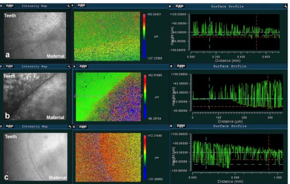
straight plane from the tooth surface to the material surface. Three straight planes were created for each sample to measure different areas of the surface. The height difference was also determined by the visual assessment of the color differences; the color change from red to blue indicates a height change from the highest to the lowest surface (Figure 1).
VDC measurements were performed for each
sample at three different time points (up to 1, 3, and
28 days) using the surface level of each material at
45 min as the baseline.
Statistical analysis
Changes in the surface roughness of ProRoot MTA and Biodentine in different conditions (dry, wet, acidic, and blood) at different time points (45 min, 1, 3 and
28 days) were compared using a two-way repeated measures analysis of variance (ANOVA) and Bonferroni tests. A p-value of <0.05 was considered significant.
The VDC changes in each condition were calculated
as a percentage change from the baseline value.
Results
Surface roughness
Figure 2 shows the representative 3D surface
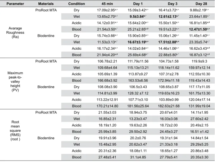
topographies of the groups. Table 1 shows the mean
± SD of Ra, RMS, and PV values of each group. Statistical analysis revealed no significant differences in RMS and PV values among the groups (p>0.05). However, significant differences were found in Ra
values depending on the experimental condition,
storage time, and material (p<0.05).
The surface roughness of ProRoot MTA and
Figure 1- Representative 3D images for the measurement of height differences between the tooth surface and the material surface showing (a) constant, (b) decreased (shrinkage), and (c) increased (expansion) surface level of materials relative to the tooth surface
Figure 2- Representative 3D images obtained using an optical profilometer showing surface topographies of ProRoot MTA and Biodentine at different environmental conditions over time. The color change from red to blue indicates a surface change from increased to decreased roughness. 3D surface images show that the surface roughness of both materials decreased in dry conditions after 28 days. In wet
conditions, the surface roughness of ProRoot MTA decreased on the 1st day; however, roughness increased for up to 28 days for both
materials. In acidic conditions, the surface roughness of both materials was similar at all time points. In blood conditions, the surface
days (p>0.05) but decreased markedly after 28 days
(p<0.05). In wet conditions, the roughness of MTA
decreased on the 1st day, while the roughness of both
materials increased after 1 day (p<0.05). In acidic
conditions, both materials showed similar roughness, and the roughness values of each material did not change through time (p>0.05). In blood conditions, the surface roughness of both materials had the highest roughness values on the 1st day, and they
showed decreased roughness after 28 days (p<0.05). The roughness of both materials was significantly
higher in wet conditions compared with those after
28 days (p<0.05). After incubation period of 28
days, the roughness of ProRoot MTA was similar in blood and dry conditions (p>0.05), while Biodentine exhibited increased roughness in blood compared with
dry conditions (p<0.05). Biodentine showed higher
roughness compared with ProRoot MTA on days 1
and 3, after storage in wet conditions (p<0.05). The
roughness of Biodentine was higher than ProRoot MTA
in blood conditions on 28 days (p<0.05).
Vertical dimensional changes
In dry conditions, the surface level of ProRoot
MTA was similar at all time points, while the surface
level of Biodentine showed 0.47% decrease after 28
days. In wet conditions, the surface levels of both materials remained constant up to the 1st day. ProRoot
MTA expanded 0.41% and 0.17%, while Biodentine expanded 0.85% and 1.44% on days 3 and 28,
respectively. Both materials presented similar surface levels to the tooth surface in the acidic environment.
The surface levels of both materials showed shrinkage
in blood conditions. The decrease in the surface level of ProRoot MTA was 0.60%, 0.69%, 0.25%, while for Biodentine, it was 0.22%, 0.56% and 0.79% on days 1, 3 and 28, respectively.
Parameter Materials Condition 45 min Day 1 Day 3 Day 28
Avarage Roughness
(Ra)
ProRoot MTA Dry 17.09±2.95a,+ 15.09±3.42a,+ 16.41±3.72a,+ 9.88±2.19b,+
Wet 13.65±2.70a,+ 9.5±3.84b,* 12.61±2.13a,* 23.64±1.85c,*
Acidic 14.12±0.91a,+ 15.64±2.00a,+ 15.50±1.50a,+ 16.81±1.85a,#
Blood 21.54±3.50a,* 25.21±2.85b,# 19.51±3.22a,+ 12.47±1.50c,+
Biodentine Dry 15.74±3.66a,+ 15.90±0.85a,+ 15.06±1.26a,+ 11.45±1.40b,+
Wet 11.53±3.13a,* 16.67±3.19b,+ 17.51±2.88b,+ 23.35±5.74c,*
Acidic 16.17±2.34a,+ 14.02±0.84a,+ 14.46±1.06a,+ 16.62±3.43a,#
Blood 21.94±4.20a,# 25.69±4.68b,* 22.88±5.80b,* 16.87±3.12c,#
Maximum peak-to- valley height (PV)
ProRoot MTA Dry 106.78±2.21 111.79±11.56 104.73±1.58 119.9±9.3
Wet 109.68±4.64 115.13±13.21 118.14±11.62 159.97±12.14
Acidic 105.69±1.39 113.87±9.27 107.31±2.78 112.55±10.30
Blood 166.68±3.92 163.53±6.56 172.94±11.18 119.43±14.43
Biodentine Dry 108.08±3.90 106.5±3.43 108.65±3.87 117.17±11.05
Wet 114.81±3.99 128.32 ±7.12 119.63±16.23 161.75±13.30
Acidic 113.22±12.91 107.71±3.10 103.89±0.99 120.04±17.14
Blood 170.21±14.80 181.56±25.64 182.63±21.68 131.99±19.04
Root mean square (RMS) (root )
ProRoot MTA Dry 21.55±3.03 18.94±3.75 20.67±4.01 14.11±1.95
Wet 16.85±2.31 13.23±3.47 16.03±3.08 27.60±2.42
Acidic 18.19±1.20 19.63±2.26 18.72±2.00 20.49±2.15
Blood 25.99±3.85 29.50±2.92 24.45±3.27 16.51 ±1.42
Biodentine Dry 19.91±3.96 20.2±0.76 19.31±1.94 14.84±1.54
Wet 15.48±2.95 20.62±3.47 21.33±3.18 29.29±5.25
Acidic 20.31±2.36 18.08±1.11 18.65±1.27 20.86±3.48
Blood 27.48±5.41 31.1±4.85 27.79±5.41 20.35±3.30
*In each row, different letters (a, b, c) indicate significant differences regarding storage time
*In each column, different symbols (+,*, #) show significant differences among the different test conditions *Bold values show significant differences between the materials (p<0.05)
Discussion
In this study, a 3D optical profilometer was used
to compare the effects of different environmental conditions on the surface roughness of ProRoot MTA and Biodentine over time. A 3D optical profilometer
can record a wide range of surface changes ranging
from <1 nm to 20000 µm in magnitude. In addition,
it allows repeatable analyses at different time points
without any sample preparation. It also allows the
measurement of height variations relative to a known
reference plane. The vertical dimensional change
of materials was measured in previous studies
using a dilatometer, a linear variable differential
transducer.7,22,25 This device contains a contact probe
that records the dimensional change according to the
probe position. However, the contact probe may limit
the expansion of the material due to the pressure it exerts, and the sensitivity (±1 µm) might not be high to detect the small changes in the material. For this
reason, a 3D optical profilometer was used to evaluate
the vertical dimensional changes in ProRoot MTA and
Biodentine in different conditions.
In clinical conditions, MTA and Biodentine are placed
in direct contact with dentin. The interaction of MTA or
Biodentine with dentin promotes a biomineralization
process due to the dissolution of bioactive dentin
matrix components and to the formation of carbonated
apatite precipitates.26 Dentin also has a high buffering
capacity that can change the pH of the environment
and the chemical and physical properties of the
materials.27 Therefore, the materials were placed into
dentin cavities to simulate clinical conditions.
In this study, the surface roughness of ProRoot MTA
and Biodentine in dry conditions was constant up to 3 days but decreased markedly after 28 days. When MTA sets completely in a dry condition, it has a thick microstructure that contains residual partially hydrated
grains.28 The surface roughness of the materials might
have decreased due to the loss of hydration over time.
In wet conditions, the surface roughness of ProRoot
MTA decreased on the 1st day, which may result from
washout of MTA due to its slow setting properties.29
However, a trend of increased surface roughness of
both materials in wet conditions was found. When
the material completed setting in 3 days, the excess
water might have prolonged the dissolution of the
biomaterials, leaving a more porous outer surface
on the remaining material. The increased roughness
in wet conditions might also be due to the material
properties. This can be related to the accumulation
of globular deposits on the surface of the materials
after storage in physiological solution.20 Regarding
the effects of acidic condition, Smith, et al.18 (2007) examined the surface characteristics of ProRoot MTA after exposure to EDTA (pH 7.4) and Biopure MTAD (pH 2) and reported increased surface roughness and higher calcium release in MTAD-treated specimens. However, in this study, the acidic pH did not affect
the surface roughness of the materials. In blood
conditions, ProRoot MTA and Biodentine showed
high roughness values up to 3 days but a significant decrease after 28 days. The high surface roughness
of the materials might be related to the accumulation of blood proteins on the surfaces of the materials.30
The initial roughness values of ProRoot MTA and
Biodentine were similar in this study. Similarly, a
previous study measured the surface roughness using
an atomic force microscopy and reported similar
values for ProRoot MTA and Biodentine.15 However,
the exposure of materials to different environmental
conditions can result in different morphological
changes on the surface.31 A previous study reported
that both materials formed white precipitates
consisting of hydroxyapatite crystals on their surfaces
after exposure to PBS.32 The formation of a white
precipitate in a different quantity on the surface of the
materials may explain the higher surface roughness
of Biodentine compared with ProRoot MTA on days 1
and 3 in wet conditions.33
In dry conditions, ProRoot MTA showed no dimensional change while Biodentine showed 0.47% shrinkage after 28 days. In agreement with our results, Camilleri, et al.20 (2014) reported structural changes,
crack formation and shrinkage of Biodentine when stored in dry conditions. In wet conditions, ProRoot
MTA expanded by 0.41% and 0.17% on days 3 and 28, while Biodentine expanded by 0.85% and 1.44%.
The greater expansion in Biodentine may stem from
the higher accumulation of hydroxyapatite on the
surface of the material due to its higher ability to release calcium in synthetic tissue fluids.34 In the acidic
environment, the surface level of both materials was
constant during all time periods. The different effects
of acidic and neutral pH levels on the properties of
the materials might be related to inhibition of the
setting reaction, which may lead to the fast dissolution
reason, the solubility of the material may impair the
dimensional expansion by preventing the accumulation
of hydroxyapatite on the material surface. In blood
conditions, both materials decreased in volume. The
crystalline phases of the materials during hydration are
also important to understand, as well as the cement
composition and changes that occur during the setting
process. By hydration, both materials showed poorly
crystallized amorphous calcium silicate hydrate gel
that contains globular particles and micro channels.10
This calcium silicate hydrate gel and the formation of
calcium hydroxide provide the setting and contribute
to the strength of these cements by the precipitation
of hydroxyapatite crystals.36 In a previous study,
the exposure to the blood conditions for four days
did not induce hydroxyapatite crystal formation on
the surface of white ProRoot MTA.37 This might be
related to the prohibition of the hydration process by
blood contamination, which prevents the formation of
this crystal structure and leads to unset materials.38
Additionally, exposure to blood reduced the formation
of calcium hydroxide by ProRoot MTA.39 For this reason,
the superficial reduction of the materials by blood contamination can be related to the inhibition of the
hydration and hydroxyapatite formation. Similarly, Grench, Mallia and Camilleri33 (2013) reported higher
wash-out tendency of Biodentine, with the loss of material upon contact with blood and other fluids. Unset materials might have been washed out after exposure to the blood conditions in this study.
Overall, the roughness of both materials decreased
after 28 days in dry conditions, which may be related
to the loss of hydration over time. Both materials increased surface roughness and expanded in wet conditions through time, which may improve the clinical performance of the materials when used in humid environments. In blood condition, both materials presented decreased surface roughness and
shrinkage through time and this can be a disadvantage
for apical surgery or revascularization treatments, as
it can disrupt adaptation and lead to microleakage.
In acidic conditions, the materials showed no
significant changes in terms of surface roughness
and dimensional change, which may be related to the increased solubility and reduced bioactivity properties of the materials. Further studies are required to clarify the clinical importance of these changes.
Conclusions
The environmental conditions tested had different effects on the surface of ProRoot MTA and Biodentine. The storage in dry, wet, and blood conditions had a time-dependent effect on the surface roughness and vertical dimensional changes of the materials. However, the roughness and the surface level of the materials were not affected by the storage in acidic conditions.
Acknowledgements
The authors deny any conflict of interests in the
subjects or materials related to this manuscript.
References
1- Torabinejad M, Watson T, Pitt Ford T. Sealing ability of a mineral trioxide aggregate when used as a root end filling material. J Endod. 1993;19(12):591-5.
2- Parirokh M, Torabinejad M. Mineral trioxide aggregate: a comprehensive literature review - part I: chemical, physical, and antibacterial properties. J Endod. 2010;36(1):16-27.
3- Islam I, Kheng Chng H, Jin Yap AU. Comparison of the physical and mechanical properties of MTA and Portland cement. J Endod. 2006;32(3):193-7.
4- Camilleri J, Sorrentino F, Damidot D. Investigation of the hydration and bioactivity of radiopacified tricalcium silicate cement, Biodentine and MTA Angelus. Dent Mater. 2013;29(5):580-93.
5- Grech L, Mallia B, Camilleri J. Investigation of the physical properties of tricalcium silicate cement-based root-end filling materials. Dent Mater. 2013;29(2):e20-8.
6- Lee YL, Lee BS, Lin FH, Lin AY, Lan WH, Lin CP. Effects of physiological environments on the hydration behavior of mineral trioxide aggregate. Biomaterials. 2004;25(5):787-93.
7- Gandolfi MG, Iacono F, Agee K, Siboni F, Tay F, Pashley DH, et al. Setting time and expansion in different soaking media of experimental accelerated calcium-silicate cements and ProRoot MTA. Oral Surg Oral Med Oral Pathol Oral Radiol Endod. 2009;108(6):E39-45.
8- Gandolfi MG, Siboni F, Prati C. Chemical–physical properties of TheraCal, a novel light-curable MTA-like material for pulp capping. Int Endod J. 2012;45(6):571-9.
9- Topcuoglu G, Topcuoglu HS. Regenerative endodontic therapy in a
single visit using platelet-rich plasma and Biodentine in necrotic and asymptomatic immature molar teeth: a report of 3 cases. J Endod. 2016;42(9):1344-6.
10- Elnaghy AM. Influence of acidic environment on properties of Biodentine and white mineral trioxide aggregate: a comparative study. J Endod. 2014;40(7):953-7.
11- Camilleri J, Formosa L, Damidot D. The setting characteristics of MTA Plus in different environmental conditions. Int Endod J. 2013;46(9):831-40.
12- Luo Z, Li D, Kohli MR, Yu Q, Kim S, He WX. Effect of Biodentine™ on the proliferation, migration and adhesion of human dental pulp stem cells. J Dent. 2014;42(4):490-7.
14- Felippe W, Felippe M, Rocha M. The effect of mineral trioxide aggregate on the apexification and periapical healing of teeth with incomplete root formation. Int Endod J. 2006;39(1):2-9.
15- Attik G, Villat C, Hallay F, Pradelle-Plasse N, Bonnet H, Moreau K, et al. In vitro biocompatibility of a dentine substitute cement on human MG63 osteoblasts cells: Biodentine™ versus MTA®. Int Endod J. 2014;47(12):1133-41.
16- Shi W, Mozumder MS, Zhang H, Zhu J, Perinpanayagam H. MTA-enriched nanocomposite TiO2-polymeric powder coatings support human mesenchymal cell attachment and growth. Biomed Mater. 2012;7(5):055006.
17- Formosa L, Mallia B, Bull T, Camilleri J. The microstructure and surface morphology of radiopaque tricalcium silicate cement exposed to different curing conditions. Dent Mater. 2012;28(5):584-95. 18- Smith JB, Loushine RJ, Weller RN, Rueggeberg FA, Whitford GM, Pashley DH, et al. Metrologic evaluation of the surface of white MTA after the use of two endodontic irrigants. J Endod. 2007;33(4):463-7. 19- Storm B, Eichmiller FC, Tordik PA, Goodell GG. Setting expansion of gray and white mineral trioxide aggregate and Portland cement. J Endod. 2008;34(1):80-2.
20- Camilleri J, Grech L, Galea K, Keir D, Fenech M, Formosa L, et al. Porosity and root dentine to material interface assessment of calcium silicate-based root-end filling materials. Clin Oral Investig. 2014;18(5):1437-46.
21- Munhoz MF, Marchesan MA, Cardoso DR, Silva SR, Silva-Sousa YT, Sousa-Neto MD. Quantitative 3D profilometry and SEM analysis of the adaptation of root-end filling materials placed under an optical microscope. Int Endod J. 2011;44(6):560-6.
22- Aksel H, Kücükkaya Eren S, Purali N, Serper A, Azim AA. Efficacy of different irrigant protocols and application systems on sealer penetration using a stepwise CLSM analysis. Microsc Res Tech. 2017;80(12):1323-7.
23- Küçükkaya Eren S, Aksel H, Serper A. Effect of placement technique on the push-out bond strength of calcium-silicate based cements. Dent Mater J. 2016;35(5):742-7.
24- Farshad FF, Pesacreta TC. Coated pipe interior surface roughness as measured by three scanning probe instruments. Anti Corros Method M. 2003;50(1):6-16.
25- Camilleri J, Mallia B. Evaluation of the dimensional changes of mineral trioxide aggregate sealer. Int Endod J. 2011;44(5):416-24. 26- Zhang H, Pappen FG, Haapasalo M. Dentin enhances the antibacterial effect of mineral trioxide aggregate and bioaggregate. J Endod. 2009;35(2):221-4.
27- Camps J, Pashley DH. Buffering action of human dentin in vitro. J
Adhes Dent. 2000;2(1):39-50.
28- Camilleri J. Characterization of hydration products of mineral trioxide aggregate. Int Endod J. 2008;41(5):408-17.
29- Jang GY, Park SJ, Heo SM, Yu MK, Lee KW, Min KS. Washout resistance of fast-setting pozzolan cement under various root canal irrigants. Restor Dent Endod. 2013;38(4):248-52.
30- Remadnia A, Dheilly RM, Laidoudi B, Quéneudec M. Use of animal proteins as foaming agent in cementitious concrete composites manufactured with recycled PET aggregates. Constr Build Mater. 2009;23(10):3118-23.
31- Agrafioti A, Tzimpoulas N, Chatzitheodoridis E, Kontakiotis EG. Comparative evaluation of sealing ability and microstructure of MTA and Biodentine after exposure to different environments. Clin Oral Investig. 2016;20(7):1535-40.
32- Asgary S, Parirokh M, Eghbal MJ, Brink F. Chemical differences between white and gray mineral trioxide aggregate. J Endod. 2005;31(2):101-3.
33- Grech L, Mallia B, Camilleri J. Characterization of set intermediate restorative material, Biodentine, Bioaggregate and a prototype calcium silicate cement for use as root-end filling materials. Int Endod J. 2013;46(7):632-41.
34- Han L, Okiji T. Bioactivity evaluation of three calcium silicate-based endodontic materials. Int Endod J. 2013;46(9):808-14.
35- Shie MY, Huang TH, Kao CT, Huang CH, Ding SJ. The effect of a physiologic solution pH on properties of white mineral trioxide aggregate. J Endod. 2009;35(1):98-101.
36- Lee YL, Lin FH, Wang WH, Ritchie HH, Lan WH, Lin CP. Effects of EDTA on the hydration mechanism of mineral trioxide aggregate. J Dent Res. 2007;86(6):534-8.
37- Nekoofar MH, Davies TE, Stone D, Basturk FB, Dummer PM. Microstructure and chemical analysis of blood-contaminated mineral trioxide aggregate. Int Endod J. 2011;44(11):1011-8.