Regulation of STEAP1 gene and its clinical
significance in human prostate cancer
Inês Margarida Amaral Santos Gomes
Tese para obtenção do Grau de Doutor em
Biomedicina
(3º Ciclo de Estudos)
Orientador: Prof. Doutor Cláudio Jorge Maia Baptista
Co-orientador: Prof.ª Doutora Cecília Reis Alves dos Santos
Regulation of STEAP1 gene and its clinical
significance in human prostate cancer
Inês Margarida Amaral Santos Gomes
Thesis for obtaining the Doctor Degree in
Biomedicine
(3rd Cycle of Studies)
Supervisor: Cláudio Jorge Maia Baptista, PhD
Co-supervisor: Cecília Reis Alves dos Santos, PhD
Depois de tantos altos e baixos, por entre momentos de sucesso e rasgados sorrisos e outros de tristeza e lágrimas, é com um grande sentimento de missão cumprida e grande felicidade que apresento este trabalho.
Aos meus orientadores, Prof. Doutor Cláudio Maia e Prof. Doutora Cecília Santos queria agradecer toda a ajuda disponibilizada e por terem sido meus mentores durante estes últimos cinco anos. O meu obrigado por me terem concedido o privilégio de trabalhar com os dois e por me terem sempre incentivado a olhar em frente sem desmotivar. Um agradecimento também ele especial à Prof. Doutora Sílvia Socorro por também ela fazer parte deste grupo de pessoas que sempre se demonstrou gentil e prestável.
Mas acima de tudo, tenho de agradecer a toda a minha família, especialmente aos meus pais, que sempre me apoiaram em todos os momentos, que foram o meu porto-seguro e os meus grandes exemplos de perseverança. A vocês devo a pessoa que sou e devo também mais esta conquista, que não é só minha, é também vossa.
Um bem-haja também muito especial ao meu avô, que é o melhor avô do mundo, e outro á minha estrelinha que me tem guiado sempre e a que com ela me faz querer brilhar sempre. Ao meu namorado, ao melhor namorado! que soube compreender-me e dar-me também ele o seu ombro de apoio, e que tanta força e vontade de sempre olhar em frente me deu! Sem ele certamente nada disto seria também possível. Fazes cada um dos meus dias ser especial! O teu beijinho ;)
Um beijinho muito especial ás minhas “manas”… Joana e Cláudia! Que venham mais 20 anos iguais aos que passaram!
Por último, a todos os meus amigos de laboratório, que com o tempo se tornaram muito mais do que colegas de bancada… Carina, Cátia, Margarida, Sofia, Ricardo, Carlinhos, Sara, Luís, Maria Inês, obrigado a todos vocês por cada momento único que partilhamos. Ficarão para sempre na memória os longos dias e algumas longas noites no CICS, todos aqueles momentos de parvoeira (habitualmente depois das 17h!), de alegria, trabalho árduo… terão sempre um lugar muito especial no meu coração! =)
Index
Thesis Overview xi
Resumo Alargado xiii
Abstract xvii List of Figures xxi
List of Tables xxv
List of Abbreviations xxvii
List of Scientific Publications xxxi
List of Scientific Communications xxxiii
CHAPTER I - General Introduction 1 1. Prostate Anatomy and Physiology 3
2. Prostate Pathology 5
2.1. Benign Prostatic Hyperplasia 5
2.2. Prostate Cancer 7 2.2.1. Epidemiology 12
2.2.2. Risk Factors 13
3. Molecular Pathways of Carcinogenesis: The role of Sex Steroid Hormones in Prostate Cancer 14 3.1. Androgens and Androgen Receptor 16
3.2. Estrogens and Estrogen Receptor 20
4. Biomarkers of Prostate Cancer 25
5. References 28 CHAPTER II - STEAP1 proteins: From structure to applications in cancer therapy 55
1. STEAP proteins 57
2. STEAP1: State of the Art 57
2.1. Structural Features of STEAP1 gene and protein 57
2.2. Tissue Expression and Cellular Localization 58
2.3. Physiologic Roles, Regulation and Implications in Cancer 60
2.4. STEAP1 as a Disease Biomarker 61
2.5. STEAP1 protein as Immunotherapeutic target 62
3. References 66 CHAPTER III – Aims of the Thesis 73
CHAPTER IV - Six transmembrane epithelial antigen of the prostate 1 is down-regulated by sex hormones in prostate cells (Original Paper 2)
77
Abstract 79
Introduction 79
Material and Methods 80
Results 82
Discussion 84
Conclusion 86
References 86
Supplemental Figure 89
CHAPTER V - Expression of STEAP1 and STEAP1B in prostate cell lines, and the putative regulation of STEAP1 by post-transcriptional and post-translational mechanisms (Original Paper 3)
91
Abstract 93
Introduction 93
Results 94
Discussion 97
Material and Methods 99
References 100
CHAPTER VI - Effect of down-regulation of STEAP1 by siRNA in cell cycle and apoptosis of human LNCaP prostate cells (Paper in Preparation)
105
Abstract 108
Introduction 108
Material and Methods 109
Results 112
Discussion 116
References 118
CHAPTER VII - STEAP1 is over-expressed in prostate cancer and prostatic intraepithelial neoplasia lesions, and it is positively associated with Gleason score (Original Paper 4)
121
Abstract 123
Introduction 123
Material and Methods 124
Results 125
Discussion 127
CHAPTER VIII- Conclusions and Future perspectives 131
Conclusions and Future perspectives 133
References 136
ATTACHMENTS Ai
Attachment I- Estrogens and prostate cancer: from biosynthesis to physiological effects(Original Book Chapter)
Aiii Attachment II- STEAP proteins: From structure to applications in cancer therapy
(Original paper 1)
Thesis Overview
This thesis is structured in eight main chapters. Chapter I presents a general introduction
that includes a short description of prostate anatomy, physiology and pathology. The molecular pathways that are involved in prostate carcinogenesis are also summarized, stressing the role of androgens and estrogens. A brief description of prostate cancer biomarkers and the potential of STEAP1 as biomarker and immunotherapeutic target is underlined. A small part of this chapter was adapted from the original book chapter (see attachment I). Chapter II is intended to put forward an extensive review on STEAP1 structural
features, its expression pattern both in normal and cancer tissues and cell lines. It is also summarized the role and regulation of STEAP1, as well as its physiologic roles, regulation and also STEAP1 implications in cancer. Additionally, the potential of STEAP1 as a biomarker and as an immunotherapeutic target are also stressed. This chapter is an adaption of the original paper 1 (see attachment II). In Chapter III, the main goals established for the development of
this thesis are described. The following chapters, Chapter IV, Chapter V, Chapter VI and Chapter VII present the results obtained during the course of the PhD, which were published
or submitted as original research papers in scientific international journals. These are organized as follows:
Chapter IV refers to the original paper 2 and describes the role of androgens and estrogens
on STEAP1 expression in LNCaP cells and rat prostate. STEAP1 expression was analyzed by qPCR, western blot and immunohistochemistry.
In Chapter V, STEAP1 and STEAP1B expression are characterized in malignant and non-malignant prostate cell lines. In addition, STEAP1 stability and post-translational mechanisms regulating STEAP1 expression are analyzed (Original paper 3). Evaluation of mRNA and protein expression was performed using qPCR and western blot, and putative post-translational mechanisms were evaluated using in silico tools.
Chapter VI outlines STEAP1 role in cell proliferation and apoptosis in LNCaP cells. LNCaP cells
were transfected with siRNA to induce the knockdown of STEAP1 gene. Cell proliferation and apoptosis were analyzed by MTS, flow cytometry and TUNEL assay. (Paper in Preparation)
Chapter VII describes STEAP1 expression and its clinical significance in prostate cancer. In
order to evaluate whether STEAP1 expression is associated with histologic diagnosis or clinical-pathological data of patients, STEAP1 immunoreactivity was evaluated in several samples of human prostatic tissues using immunohistochemistry. (Original paper 4)
The last and final chapter, Chapter VIII summarizes the general conclusions of the work and
Resumo Alargado
A próstata é uma glândula acessória do sistema reprodutor masculino e desempenha um papel fulcral na fertilidade masculina. A morfogénese, desenvolvimento e homeostase celular desta glândula são altamente dependentes das hormonas esteróides sexuais, nomeadamente androgénios e estrogénios. De um modo geral, o cancro surge de um desequilíbrio entre a proliferação celular e apoptose, conduzindo a um crescimento descontrolado. O cancro da próstata é uma doença multifatorial, afetada por fatores de risco exógenos e endógenos, de entre os quais se destacam a história familiar e a idade. O cancro da próstata é o segundo tipo de cancro mais frequente na população masculina, e em conjunto com a hiperplasia benigna da próstata, é encontrado na maioria dos homens com idade superior a 40 anos. A etiologia do cancro da próstata é complexa, mas acredita-se que processos inflamatórios estejam por detrás do aparecimento de lesões pré-cancerosas. A atrofia inflamatória proliferativa e a neoplasia intraepitelial prostática (PIN) são frequentemente referidas como percursoras do carcinoma prostático localizado, que por sua vez evolui tornando-se invasivo e metastizando para tecidos mais longínquos. O cancro da próstata é muitas vezes descrito como uma doença bi-etápica, uma vez que é inicialmente responsivo aos androgénios dependente), mas à medida que progride, torna-se não responsivo (hormono-independente). No final, o desenlace clínico é definido pelo potencial de crescimento, invasão e metástase do tumor. Disponíveis para a prática clínica, existem vários sistemas de classificação do grau e estadio tumorais que podem ser usados, dentro dos quais os mais frequentes são o sistema de Gleason e o sistema TNM.
As vias de sinalização envolvidas na carcinogénese da próstata são complexas. Neste trabalho destacam-se as vias de sinalização ativadas pelos androgénios e estrogénios. A 5 -dihidrotestosterona (DHT) apresenta-se como sendo o androgénio mais produzido, potente e o principal com ações fisiológicas na próstata. Também o 17-estradiol (E2) é produzido nesta
glândula a partir da aromatização da testosterona, e também ele é o estrogénio mais potente. Tanto os androgénios como os estrogénios têm um papel central na diferenciação, proliferação e sobrevivência de células tumorais prostáticas, contribuindo assim para a origem e desenvolvimento do carcinoma da próstata.
A identificação de biomarcadores que permitissem um diagnóstico e tratamento mais precisos revolucionou a prática clínica. Atualmente, o PSA é o biomarcador por excelência no diagnóstico do cancro da próstata. No entanto, apesar de ser um bom indicador clínico, o PSA apresenta várias limitações. Como tal, é necessário que novos biomarcadores sejam identificados.
O STEAP1 foi inicialmente identificado como sendo sobre-expresso no cancro da próstata. No entanto, é também encontrado como sobre-expresso em vários outros tipos de tumores. Em tecidos normais, a expressão do STEAP1 é quase que totalmente restrita à próstata, onde está
principalmente localizado na membrana plasmática das células epiteliais, especialmente nas junções celulares. A regulação e funções biológicas do STEAP1 estão ainda pouco definidas, mas crê-se que actue como um canal iónico ou proteína transportadora, regulando a comunicação inter- e intra-celular. Finalmente, apesar de o seu significado clínico não ser ainda claro, as características apresentadas pelo STEAP1 fazem dele um potencial biomarcador e alvo imunoterapêutico.
Deste modo, foi primeiramente investigado se a expressão do STEAP1 é regulada pelas hormonas sexuais esteróides in vitro e in vivo. Os resultados mostraram que a expressão do STEAP1 é inibida tanto pelo DHT como pelo E2 na linha celular maligna de cancro da próstata
LNCaP, assim como na próstata de rato, sugerindo que o STEAP1 poderá desempenhar funções relevantes na progressão do cancro da próstata de responsivo para não-responsivo aos androgénios. Subsequentemente, foi avaliado o padrão de expressão de um gene relacionado com o STEAP1, o STEAP1B, e se a expressão do STEAP1 está sujeita a mecanismos de regulação pós-transcrição e pós-tradução. A análise in silico revelou que o STEAP1 e o STEAP1B1 partilham grande homologia estrutural, e que o STEAP1 é mais estável em células tumorais da próstata do que em células não tumorais. Ainda, foi possível demonstrar in silico que a expressão do STEAP1 poderá ser regulada por várias modificações pós-tradução. Seguidamente, centrou-se o foco nas funções do STEAP1. Utilizando um siRNA específico para reprimir a expressão do STEAP1 na linha celular LNCaP, analisou-se o papel do STEAP1 na proliferação celular e apoptose, assim como o efeito do DHT nas células LNCaP com baixos níveis de expressão do STEAP1. Utilizando as técnicas de MTS, citometria de fluxo e TUNEL, verificou-se que o silenciamento do gene do STEAP1 reduz a viabilidade e crescimento celular, e aumenta o número de células em apoptose. Por outro lado, a ação do DHT parece estar dependente dos níveis de expressão do STEAP1, uma vez que o DHT não foi capaz de reverter os efeitos provocados pela sub-expressão do STEAP1.
Muitos esforços têm sido dirigidos para clarificar a potencial utilização do STEAP1 como biomarcador e alvo imunoterapêutico. Neste trabalho investigou-se a associação entre os níveis de expressão do STEAP1 com dados clínicos e histológicos provenientes de doentes com cancro da próstata. Os resultados demonstraram que o STEAP1 é sobre-expresso em casos de PIN e cancro da próstata, e que a sua expressão está positivamente associada com a escala de Gleason.
Como principais conclusões deste trabalho, os resultados apresentados demonstram que a expressão do STEAP1 é regulada negativamente pela presença das principais hormonas sexuais esteróides presentes na próstata. Também a elevada estabilidade do STEAP1 em células tumorais da próstata em comparação com células normais leva a crer que os mecanismos de regulação pós-transcrição e pós-tradução são dependentes do estado clínico do tumor. Mais ainda, o silenciamento do gene do STEAP1 inibe a viabilidade e proliferação celulares das células LNCaP, ao mesmo tempo que induz a sua apoptose, levando a crer que STEAP1 poderá desempenhar um papel relevante no cancro da próstata, nomeadamente na iniciação do tumor e no aparecimento de células tumorais hormono-independentes. O uso do STEAP1 per
se poderá não ser suficiente para ser usado com biomarcador na prática clínica diária, mas poderá abrir caminho para novas estratégias de diagnóstico e tratamento do cancro da próstata.
Abstract
The prostate gland is an accessory gland of the male reproductive system and displays a critical role in male fertility. This gland is dependent of sex steroid hormones, namely androgens and estrogens, for the gland morphogenesis, development and cellular homeostasis. Prostate cancer (PCa) is a multifactorial disease, affected by both endogenous and exogenous risk factors, especially family history, age and sex hormones. PCa is the second most common type of cancer among men, and along with BPH, is found in the majority of men over 40 years old. Although PCa etiology is complex, inflammatory processes are thought to be behind the appearance of pre-cancerous lesions. At molecular level, PCa is often described as a two step disease. Initially it is responsive to androgens, but usually, the PCa becomes androgen-independent. In the end, the clinical outcome of PCa is defined by the potential of the tumor to grow, invade and metastasize. Regardless of all the knowledge gathered from prostate cancer pathophysiology and clinical management, identification of genes associated with the pathology, the role of sex steroid hormones in their regulation, and their potential to be used as biomarkers or immunotherapeutic targets is urgent.
STEAP1 was firstly identified as being overexpressed in prostate cancer. However, it can also be found overexpressed in several other types of tumors. STEAP1 expression in normal tissues is almost restricted to the prostate, where it is primarily localized in the plasma membrane of epithelial cells, especially at cell-cell junctions. STEAP1 regulation and biological function are yet uncertain, but it is believed to act as an ion channel or transporter protein, regulating inter- and intra-cellular communications. Finally, although its clinical significance is not clear, STEAP1 features stress its potential as biomarker and immunotherapeutic target.
This way, it was evaluated if STEAP1 expression is regulated by sex steroid hormones, using both in vitro and in vivo assays. It was found that STEAP1 is down-regulated by DHT and E2 in
LNCaP prostate cancer cells and in rat prostate, suggesting that STEAP1 may have a role on prostate cancer progression from androgen-dependent to androgen-independent. Subsequently, it was also evaluated the expression pattern of a STEAP1 related gene (STEAP1B) in cancer cell lines, and whether STEAP1 expression is subjected to post-transcriptional or post-translational regulatory mechanisms. In silico analysis revealed that STEAP1 and STEAP1B1 share high homology, and STEAP1 is more stable in prostate cancer cells rather than in non-malignant ones. Furthermore, it was demonstrated by in silico analysis that STEAP1 expression may be regulated by several post-translational modifications. In order to clarify the role of STEAP1 in prostate cancer, it was used a specific siRNA to decrease the levels of STEAP1 in LNCaP cells. Then, it was evaluated the role of STEAP1 in cell proliferation and apoptosis, as well as the effect of dihydrotestosterone (DHT) in LNCaP cells with low levels of STEAP1. Using MTS assay, flow cytometry and TUNEL assay, it was
evident that STEAP1 gene silencing appears to reduce cell viability and growth, and to increase apoptosis of LNCaP cells. On the other hand, the DHT action seems to be dependent of STEAP1 levels as it could not revert the effects induced by STEAP1 knockdown.
Much effort has been done to clarify the potential of STEAP1 as biomarker and immunotherapeutic target. Here, it was investigated the association between STEAP1 expression with histologic and clinical data of patients. It was demonstrated that STEAP1 is overexpressed in PCa and PIN lesions, and it is positively associated with Gleason score. Taken together, the results here presented demonstrate that STEAP1 expression is negatively regulated by the two main sex steroid hormones presents on prostate. STEAP1 high stability in prostate tumor cells in comparison to normal ones lead us to believe that post-transcription and translational regulation mechanisms are dependent on the tumor stage. Moreover, STEAP1 gene silencing inhibits cell viability and proliferation, ate the same time that increases apoptosis in LNCaP cells, suggesting that STEAP1 may have an important role on PCa progression from androgen-dependent to hormone refractory. The use of STEAP1 per se may not be sufficient to be used as a biomarker in the daily clinical practice, but may open novel strategies for diagnosis and treatment of prostate cancer.
Keywords
List of Figures
[1linha de intervalo]Chapter I
Figure 1 – Representation of Prostate’s anatomy. Adapted from [66].
Figure 2- Human prostate histology. A – Human prostate H&E staining x30 magnification. B – Human prostate H&E staining x50 magnification. G- prostatic glands, CA- corpora amylacea, St- fibrous stroma. Adapted from [2].
Figure 3- Benign prostatic hyperplasia, H&E staining. Microscopic image of a small gland pattern area of benign nodular hyperplasia, composed by small to medium-sized and irregular acini, with rounded lumens and surrounded by stroma. Adapted from [97].
Figure 4- Global cellular homeostasis in normal prostate and BPH. As shown, androgens (testosterone and DHT) and growth factors (KGF, IGFs, EGF and TGFβ) can either have agonistic as well as antagonist effects on normal prostate, contributing to a rightful balance between cell proliferation and death. In BPH, this balance is disturbed and other hormones such estrogens may promote cell proliferation and inhibit cell death. DHT- 5α-dihydrotestosterone; KGF- Keratinocyte growth factor; IGF- Insulin-like growth factor; EGF- Epidermal growth factor; TGFβ- Transforming growth factor beta.Adapted from [40]. Figure 5- Schematic representation of prostate carcinogenesis. Prostate pathophysiology is a multistep process that comprises the appearance of pre-malignant lesions, proliferative inflammatory atrophy (PIA) and prostate intraepithelial neoplasia (PIN). This evolves into local carcinoma and locally invasive disease, and ultimately into metastasis with subsequent spread of tumor cells. Adapted from [396].
Figure 6- The distribution of inflammation, PIA, HGPIN and PCa in the human prostate H&E staining tissue sample, indicated, respectively, by red, white, green and black lines and arrows. PIA- proliferative inflammatory atrophy; HGPIN- High grade prostate intraepithelial neoplasia; PCa- Prostate cancer. Adapted from [397].
Figure 7- Gleason grading pattern system. A- Gleason pattern 1. Arrows indicate an individual acinus, and the lumen (L). 40x magnification. B- Gleason pattern 2, 40x magnification. C- Gleason pattern 3. Arrows indicate a small cell infiltration into the surrounding stroma. 100x magnification. D- Gleason pattern 4. Signs of increased stromal invasion. White arrows indicate areas of gland fusion and poorly defined lumens and green arrows indicate an area of
Gleason pattern 3. 40x magnification. E- Gleason pattern 5. Presence of solid sheets of cells with no glandular structure and poorly differentiated cells. 100x magnification. Adapted from [398].
Figure 8- Testosterone (T) is captured by prostatic epithelial and stromal cells, and can either bind to the androgen receptor or be converted into 5α- dihydrotestosterone (DHT) in the stromal cells. DHT can act in an autocrine manner inside the stromal cell or diffuse to the proximal epithelial cells acting in a paracrine manner. DHT produced by peripheral tissues can also diffuse into the prostate. Adapted from [40].
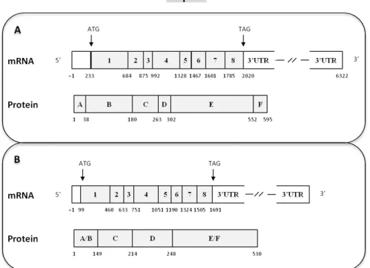
Figure 9- Structural organization of the androgen receptor (AR) mRNA and protein. Shading boxes indicate coding regions. Indicated below mRNA and protein structures are, respectively, exons size (bp) and aminoacid number. Adapted from [236].
Figure 10- Androgen Receptor (AR) genomic signaling pathway. AR is held inactive wild bound to Hsp. Upon ligand binding, Hsp is released and hormone/receptor homodimers translocate to the nucleus, where they bind DNA on AREs. AR transcriptional activity is regulated by chromatin remodeling complexes (purple), coactivators (light green), and RNA Polymerase II (PolII). AR shuttles between the chromatin-bound and free cytoplasmic state with a t1/2
around 5s. T- Testosterone; DHT- 5α-dihydrotestosterone; Hsp- Heat shock proteins; AREs- androgen responsive elements. Adapted from [230].
Figure 11- Structural organization of ERα (A) and ERβ (B) mRNA and protein. Shading boxes indicate coding regions. Indicated below mRNA and protein structures are, respectively, exons size (bp) and aminoacid number. ERα- Estrogen Receptor α; ERβ- Estrogen Receptor β. Adapted from [295].
Figure 12. Schematic diagram of estrogen signalling in prostate cancer. Binding of Estrogen to the Estrogen Receptor α (ERα) promotes the translocation of this complex to the nucleus and the transcription of ER target genes along with a set of independent genes, including Cyclin D1. Estrogen-ERα complex also activates Ras-MAPK and PI3K signalling pathways, which in turn triggers cell proliferation and anti-apoptotic routes. Estrogens may stimulate the transcription of anti-apoptotic genes, via PI3K-AKT pathway, upon activation of G protein-coupled receptor 30 (GPR30). In addition, Cyclin D1 and AIB1 potentiate the transcription of ER-target genes. Contrarily, the binding of estrogen to ERβ seems to inhibit cell growth and anti-apoptotic events. Adapted from [400].
Chapter II
Figure 1- Schematic representation of STEAP1 protein structure, cellular localization and physiologic functions. Similar on the structure, presenting a six transmembrane structure, intracellular C- and N- terminal and intramembrane heme group, STEAP1 lacks the innate metalloreductase activity conferred by the presence of FNO-like domain. STEAP1 actively increases intra- and intercellular communication through the modulation of Na+, Ca2+ and K+
concentration, as well as the concentration of small molecules. It stimulates cancer cell proliferation and tumor invasiveness capacity. Adapted from [54].
Chapter VIII
Figure 1- Schematic representation highlighting the proposed mechanisms of regulation, function and clinical significance of STEAP1 in human prostate cancer.
List of Tables
Chapter I
Table 1- TMN system for staging of prostate cancer. Adapted from [399].
Chapter II
Table 1– Characterization of STEAP1 and STEAP1b genes, mRNA transcripts and proteins. Adapted from [54].
Table 2- Expression of STEAP1 mRNA and proteins in normal and cancer tissues. Adapted from [54].
List of Abreviations
4-odione Androstenedioneaa Aminoacids
ACRATA Apoptosis, cancer and redox associated transmembrane
ADC Antibody-drug conjugates
ADIPOR1 Adiponectin receptor 1
AF Activation Function
Akt Protein kinase B
AP-1 Transcription factors activator protein 1
APC Antigen-presenting cells
AR Androgen receptor
ARE Androgen responsive elements
ArKO Aromatase deficiente mice
ATP Adenosine triphosphate
Bcl-2 B cell lymphoma 2
BPH Benign hyperplasia
BPSA BPH-associated Prostatic specific antigen
C Central domain CAG Polyglutamine CBX7 Chromobox homolog 7 Chr Chromosome COX-2 Clycooxygenase-2 CTL Cytotoxic T-lymphocyte D Hinge region DBD DNA-binding domain DES Diethylstilbesterol DHEA Dehydroepiandrosterone DHT 5- dihydrotestosterone
DNA Deoxyribonucleic acid
E1 Estrone
E2 17-estradiol
E3 Estriol
EGF Epidermal growth factor
EGFR Epidermal growth factor receptor
ER Estrogen receptor
ERE Estrogen-responsive elements
ERG-1 ETS related gene 1
ERK Extracelular signal regulated kinase ESR1 Estrogen receptor alfa encoding gene
ETV ETS variant
FNO F420H2:NADP+ oxidoreductase
fPSA Free Prostatic specific antigen
FRE Ferric reductase
GGC Polyglycine
GnRH Gonadotropin-releasing hormone
xxviii
GSTA1 Glutathione S-transferase alfa 1 GSTP1 Glutathione S-transferase-pi
HAT Histone acetyl transferase
HER Human epidermal growth factor receptor
HGPIN High-grade prostatic intraepithelial neoplasia
HHD HLA-A_0201 transgenic mice
HRPC Hormone refractory prostate cancer
Hsp Heat shock proteins
IGF-1 Insulin-like growth factor 1
IL-6 Interleukine 6
JNK c-jun N-terminal kinase
KGF Keratinocyte growth factor
KLK Kallikrein
LBD Ligand-binding domain
LEF Lymphoid enhancer factor
LH Luteinizing hormone
LUTS Lower urinary tract symptoms
MAPK Mitogen-activated protein kinase
MMAE Monomethyl auristatin E
MMP Matrix metalloproteinase
mPSCA Murine prostate stem cell antigen
MSC Mesenchymal stem cell
mSTEAP1 Murine six transmembrane epithelial antigen of the prostate 1
MVA Modified vaccinia Ankara
NADPH Nicotinamide adenine dinucleotide phosphate
NKX3.1 Neuromedin 3 homebox 1
NTD N-terminal domain
PAP Prostatic acid phosphatase
PCa Prostate cancer
PCA3 Prostate cancer antigen 3
PI3K Phosphatidylinositol-3 kinase
PIA Proliferative inflammatory atrophy
PIN Prostatic intraepithelial neoplasia
PR Progesterone receptor
PRC1 Polycomb repressive complex 1
PSA Prostatic specific antigen
PTEN Phosphate and tensine homolog
ROS Reactive oxygen species
Sp-1 specificity protein 1
STEAP1 Six transmembrane epithelial antigen of the prostate 1
TAA Tumor-associated antigens
TAF-1 Transcriptional activation function site 1 TAF-2 Transcriptional activation function site 2
TCF T-cell factor
Tf Transferrin
TfR1 Transferrin receptor 1
TGF Transforming growth factor
TMPRSS2 Type II transmembrane serine protease TNF- Tumor necrosis factor alpha
TNM Tumor Node and Metastasis
TRAMP-C Transgenic adenocarcinoma mouse model derived cell line TRPM2 Transient receptor potential cation channel M2
VEGF Vascular endothelial growth factor
ERKO Alpha estrogen receptor knockout
List of Scientific Publications
Papers related to this thesis
• Gomes IM, Santos CR, Gaspar C, Alvelos MI, Maia CJ, Effect of down-regulation of STEAP1 by siRNA in cell cycle and apoptosis of human LNCaP prostate cells, 2014 (Paper in
Preparation)
• Gomes IM, Arinto P, Lopes C, Santos CR, Maia CJ, STEAP1 is over-expressed in prostate cancer and prostatic intraepithelial neoplasia lesions, and it is positively associated with Gleason score, Urologic Oncology Seminars and Original Investigations 2014; 32(1): 53.e23–
53.e29 doi:10.1016/j.urolonc.2013.08.028
• Gomes IM, Santos CR, Maia CJ, Expression of STEAP1 and STEAP1B in prostate cell lines, and the putative regulation of STEAP1 by post-transcriptional and post-translational mechanisms, Genes & Cancer 2104; 5 (3-4): 142-151
• Gomes IM, Santos CR, Socorro S, Maia CJ, Six transmembrane epithelial antigen of the prostate 1 is down-regulated by sex hormones in prostate cells.
Prostate 2012; 73(6):605-13. doi: 10.1002/pros.22601
• Gomes IM, Maia CJ, Santos CR, STEAP proteins: From structure to applications in cancer therapy. Mol Cancer Res 2012; 10(5):573-87 doi: 10.1158/1541-7786.MCR-11-0281
Book Chapters related to this thesis
• Gomes IM, Vaz CV, Rodrigues D, Rocha SM, Socorro S, Santos CR, Maia CJ. Estrogens and prostate cancer: from biosynthesis to physiological effects. Book Title: Estradiol
synthesis: health effects and drug interactions. Nova Science Publishers, Inc, New York, USA. ISBN: 978-1-62808-962-2
Papers not related to this thesis
• Vaz CV, Maia CJ, Marques R, Gomes IM, Correia S, Alves M, Cavaco JE, Oliveira PF, Socorro S, Regucalcin is an androgen-target in the rat prostate associated with modulation of apoptotic pathways, Prostate 2013; doi: 10.1002/pros.22835
List of Scientific Communications
Poster communications related to this thesis
• Gomes IM, Santos CR, Alvelos MI, Gaspar C, Maia CJ. Effect of down-regulation of STEAP1 by siRNA in cell cycle and apoptosis of human LNCaP prostate cells. FEBS/EMBO Congress, 30 August- 4 September, Paris, France
• Gomes IM, Arinto P, Costa-Pinheiro P, Santos CR, Jerónimo C, Maia CJ. Expression and regulation of STEAP1 and STEAP1B in prostate cell lines through mRNA and protein stability and epigenetic mechanisms. 38th FEBS Congress- Mechanisms in Biology, 6-11 July
2013, Saint Petersburg, Russia
• Gomes IM, Arinto P, Santos CRA, Socorro S, Lopes C, Maia CJ. Regulation of STEAP1 expression in prostate by sex steroid hormones. 22nd Congress of European Association for
Cancer Research - from Basic Research to Personalized Cancer Treatment, 7-10 July 2012, Barcelona, Spain
• Gomes IM, Santos CR, Socorro S, Lopes C, Maia CJ. Regulation of STEAP1 expression in prostate by androgens. XX Porto Cancer Meeting, Porto, Portugal. 28-29 April 2011
• Gomes IM, Santos CR, Socorro S, Lopes C, Maia CJ. STEAP1 expression in prostate cancer and its regulation by androgens. 16th International Charles Heidelberg Symposium on
Cancer Research, Coimbra, Portugal. 26-28 September 2010
Poster communications not related to this thesis
• Marques R, Vaz CV, Peres CG, Gomes I, Santos CR, Maia, Socorro S. Effect of androgens on the expression of Ca2+ -binding protein, regucalcin, and Ca2+ -channels in MCF-7 cells.
22nd Congress of European Association for Cancer Research - from Basic Research to Personalized Cancer Treatment, 7-10 July 2012, Barcelona, Spain
1. Prostate Anatomy and Physiology
The human prostate gland is part of the male reproductive system, having the shape and size of a walnut. This gland is situated frontal to the rectum and immediately below the bladder, surrounding its neck and the first part of the urethra [1, 2]. The prostate is partially enclosed by a capsule on the posterior and lateral sides, and the anterior fibromuscular stroma binds the anterior and apical surfaces [2]. The prostate is divided in four main zones. These are the peripheral zone, central zone, transition zone and anterior fibromuscular zone, as shown in Figure 1 [2, 3]. The peripheral zone comprises the bulk, approximately 70% of the prostate gland, and the central and transition zones the remaining 30% [2]. With aging, the transition zone becomes the most prominent prostate zone due to hypertrophy development [4].
Figure 1- Representation of Prostate’s anatomy. Adapted from [66].
At a closer look, the gland has an irregular shape due to the folds formed by the epithelium (Figure 2). This is arranged into pseudostratified columnar to cuboidal cells, and composed by two cellular layers, the luminal one with tall columnar secretory cells with basal nuclei that express androgen receptor (AR), cytokeratin 8 and 18 and prostatic specific antigen (PSA), and the basal layer, often incomplete and consisting of flattened basal cells with the ability to produce keratin, p63, cytokeratin 5 and 14 [2, 5-7]. Although the precise role of basal cells has long been discussed, several functions are attributed to the basal layer, namely as a reservoir of stem cells with capacity to differentiate into columnar secretory cells, or as displaying an active part in the molecular trafficking between luminal cells and extracellular space [8, 9]. Basal cells selectively express the estrogen receptor alpha (ERα), the progesterone receptor (PR) and other non-hormonal receptors such as human epidermal
growth factor receptor (HER)-1 or HER-2, enabling them to maintain cell growth and proliferation [7].
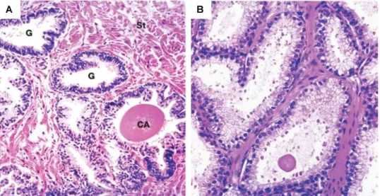
Figure 2- Human prostate histology. A – Human prostate H&E staining x30 magnification. B – Human prostate H&E staining x50 magnification. G- prostatic glands, CA- corpora amylacea, St- fibrous stroma. Adapted from [2].
A characteristic feature of aging is the appearance of corpora amylacea, a lamellated glycoprotein mass that later calcifies into prostatic concretions (Figure 2). The prostate stroma dwells on dense extracellular collagen matrix, fibroblasts, randomly arranged smooth muscle fibres, blood and lymphatic vessels, and nervous terminals [2, 10]. Prostate stroma is subdivided into two layers, the periacinar stroma that surrounds the epithelial ducts, and the interacinar stroma found between the adjacent periacinar sheaths [11]. Another type of cells found in the prostate are the neuroencodcrine cells, which are a rare type of cells located among epithelial cells and expressing chromogranin A [6, 7, 12].
Human prostate morphogenesis and growth takes place during fetal and puberty stages, while pathological modifications of this tissue begin around mid-forties [13]. Prostate growth period is characterized by increased rate of cell proliferation over cell death, whereas adult prostate displays comparable rates [14]. The initial epithelium differentiation is mediated by mesenchymal cells. The interaction between stroma and epithelial cells both in embryogenesis and adult differentiated prostate is imperative. The growth and development of the embryonic epithelium into differentiated prostate induced by androgens as well as the maintenance of the secretory epithelium in adult prostate is mediated by stroma cells [15, 16].
Physiologically, the maintenance of cellular homeostasis is not only attributed to androgens, but also to estrogens [17, 18]. The prostate gland secretes several factors, such as PSA, prostatic acid phosphatase (PAP), prostaglandins and citric acid, which play important roles in fertilization, sperm delivery and survival [19-23].
2. Prostate Pathology
In developed countries, prostate cancer (PCa) is the most frequently diagnosed malignancy in men [24, 25]. The etiology of this disease is complex and remains unclear. PCa is a gradual process that involves multiple genetic alterations in prostate cells, such as activation of oncogenes, inhibition of tumor suppressor genes and disturbance of stromal-epithelial balance [26, 27].
Prostate morphological zones display distinct architecture, histology and predisposition to disease development. Around 70 to 80% of the diagnosed prostatic adenocarcinomas emerge in the peripheral zone, while benign prostatic hyperplasia (BPH) commonly evolves in the transition zone [28].
The critical pathophysiological factor contributing to PCa development is the inhibition of apoptosis rather than enhanced cellular proliferation [29]. This event is characterized by a down-regulation of androgen responsive genes that inhibit proliferation, induce differentiation or mediate apoptosis [30-32]. There are three different stages involved in development of PCa. Initially, PCa progresses from precursor lesions, termed as prostatic intraepithelial neoplasia (PIN) and proliferative inflammatory atrophy (PIA), to carcinoma confined to the prostate, and finally, to metastatic and hormone-refractory carcinoma, a usually lethal prostate disease (Figure 5) [29].
2.1. Benign Prostatic Hyperplasia
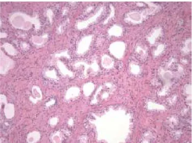
BPH is one of the most prevalent benign neoplasm and chronic disease associated with male aging [33]. BPH was first described by McNeal, and is defined by an enlargement of the prostate transition zone due to an increase of epithelial and stromal cells and consequently causing the effacement of the central zone [34-36]. Although BPH is an heterogeneous process, it encompasses the formation of miscellaneous nodules that differ in terms of glandular tissue and fibromuscular stroma percentage (Figure 3) [37].
Figure 3- Benign prostatic hyperplasia, H&E staining. Microscopic image of a small gland pattern area of benign nodular hyperplasia, composed by small to medium-sized and irregular acini, with rounded lumens and surrounded by stroma. Adapted from [97].
The nodules evolve in the transition zone or in the periurethral region, but prostate enlargement occurs independently of their formation [38]. The bulk of early developed periurethral nodules are entirely composed of stromal cells. On the other hand, the majority of nodules in the transition zone are due to glandular tissue proliferation, an event that may be subjacent to loss of overall stroma. The initial development of BPH is accompanied by an increased number of nodules, but with a slow growth rate. Later in disease progression, the nodules significantly increase in size, specially the glandular ones [38].
BPH etiology is uncertain, and the observed cellular proliferation may be due to several factors. Although androgens are not known to cause BPH, its development does require their presence [39]. Androgens may act either in an autocrine or paracrine manner, increasing the transcription of several androgen-responsive genes involved in cellular proliferation, or in some conditions, in cell death. The imbalance between cellular proliferation and cell death in response to growth factors and androgens may also be linked to the appearance of BPH (Figure 4) [40].
BPH development is often correlated with the appearance of lower urinary tract symptoms (LUTS), and when left untreated, bladder dysfunction and hypertrophy appear and may evolve to acute urinary retention [40-43].
Figure 4- Global cellular homeostasis in normal prostate and BPH. As shown, androgens (testosterone and DHT) and growth factors (KGF, IGFs, EGF and TGFβ) can either have agonistic as well as antagonist effects on normal prostate, contributing to a rightful balance between cell proliferation and death. In BPH, this balance is disturbed and other hormones such estrogens may promote cell proliferation and inhibit cell death. DHT- 5α- dihydrotestosterone; KGF- Keratinocyte growth factor; IGF- Insulin-like growth factor; EGF- Epidermal growth factor; TGFβ- Transforming growth factor beta. Adapted from [40].
2.2. Prostate Cancer
PCa is one of the most common types of cancer diagnosed in the male population over 40 years old. As previously mentioned, the majority of PCa arise in the peripheral zone of the prostate. These tumors initially start as small foci of intraductal dysplasia, and may stay silenced for a long period of time, to eventually differentiate and progress into an invasive state [35]. The tumor foci leads to a disruption of prostate tissue and a decrease on glandular activity and fluid production [35, 44, 45]. Only a small portion of PCa arise in the transition zone, and the central zone is rarely associated with the disease [45-47]. Histologically, PCa is characterized by an obliteration of the basal cell layer with consequent basal derangement of basal lamina, decreased epithelial polarity and a scarce glandular acini connection [48].
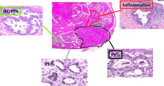
PCa is thought to arise from pre-cancerous lesions originated as a consequence of inflammatory processes (Figure 5 and 6) [49-51].
Figure 5- Schematic representation of prostate carcinogenesis. Prostate pathophysiology is a multistep process that comprises the appearance of pre-malignant lesions, proliferative inflammatory atrophy (PIA) and prostate intraepithelial neoplasia (PIN). This evolves into local carcinoma and locally invasive disease, and ultimately into metastasis with subsequent spread of tumor cells. Adapted from [396].
The stem cell population localized on the basal cell layer appears to have a preponderant role in pre-cancerous and PCa development. Occasionally, the population of adult basal stem cells may be restored, as in the case of prostatic tissue injury, and progresses into transit-amplifying/intermediate cells, which in turn can differentiate into luminal secretory epithelial cells or even neuroendocrine cells [49, 50, 52-54]. It has been proposed that a small number of these stem cells may represent the minority of epithelial cells that stand for PCa progenitor cells [49, 50, 52]. The continuous activation of signaling pathways by androgens, estrogens and other growth factors in progenitor epithelial cells may result in a heterogeneous population of progenitor cancer cells, provided with unlimited growth ability and possibility to trigger PIN-like lesions and PCa (Figure 6) [55].
Accordingly, the potential precursor of PCa is likely to be high-grade PIN (HGPIN) [56]. In general, PIN lesions are no more than an enhanced atypical cell number arising on the normal prostate gland architecture (Figure 6) [57]. According to the histomorphologic profile, PIN lesions were originally divided into three groups, namely Low-grade PIN1, grade 2 PIN and HGPIN (or grade 3 PIN) [58]. Later, PIN2 and 3 were fused and are mentioned as HGPIN [59]. Low-grade PIN architecture consists on crowding, stratified and irregular spacing epithelial cells, displaying anisonucleosis and rarely prominent and small nucleoli [58]. HGPIN lesion is characterized by architecturally benign prostatic acini and duct, lined by cytological atypical cells, with frequently prominent nucleoli, nuclear enlargement and crowding, increased cytoplasmatic density and multiple nucleolar size [58, 60]. HGPIN correlates with tumor stage, Gleason score and PCa risk [56, 61, 62]. Similar chromosomal alterations are found when comparing HGPIN with PCa, with loss of chromosome (chr) 8p, 10q and 13q and gain of chr 8q, 7 and Xq [63, 64]. In fact, the majority of the alterations that lead to PCa development appear to take place during benign epithelium to HGPIN transition [65].
PIA has also been pointed as a PCa precursor, possibly arising even before PIN-like lesions (Figure 6). PIA is thought to evolve in response to environmental factors that lead to cell damage and cause inflammation and oxidative stress. The resulting genetic modifications give rise to HGPIN lesions [66, 67]. PIA involvement on PCa etiology is somehow controversial. It is suggested that this type of lesion may only represent a primordial precursor, but is not related to PCa development [65]. Nonetheless, PIA displays molecular prints distinctive of
early neoplastic transformation, such as glutathione S-transferase-pi gene (GSTP1) hypermethylation, increased expression of glutathione S-transferase alpha 1 (GSTA1), cyclooxygenase-2 (COX-2) and B cell lymphoma 2 (Bcl-2), and reduced expression of NK3 homebox 1 (NKX3.1), early chromosomal abnormalities and shortening of telomere length [68-73]. In PIA lesions, discrete foci of proliferative glandular epithelium with the appearance of a simple atrophy or post atrophic hyperplasia in conjunction with inflammation can be found [74, 75]. PIA is characterized by the presence of two distinct cell layers. Among stromal and epithelial cells, a mononuclear and/or polymorphonuclear inflammatory cell layer can be found, and stromal atrophy with inconsistent fibrosis levels.
A number of different interactions between PCa cells and the stromal cell layer stimulate the stromal compartment originating a reactive microenvironment, where AR has a preponderant role promoting PCa cells growth and proliferation by activating distinct signaling pathways mentioned further ahead [76].
Figure 6- The distribution of inflammation, PIA, HGPIN and PCa in the human prostate H&E staining tissue sample, indicated, respectively, by red, white, green and black lines and arrows. PIA- proliferative inflammatory atrophy; HGPIN- High grade prostate intraepithelial neoplasia; PCa- Prostate cancer. Adapted from [397].
Hormone refractory prostate cancer (HRPC) is triggered after ablation therapies fail and tumor development is once again established. HRPC is considered a multifactorial and heterogeneous disease, and includes the activation of several signaling pathways in tumor cells. These induce a continuous AR-signaling because of an upregulation of AR expression, enhanced ligand-dependent activation by 5α-dihydrotestosterone (DHT) and testosterone and a broadened AR activation by non-androgenic and non-steroidogenic ligands, such as estrogens, progestins or growth factors. Furthermore, bypassing the AR signaling, cell growth and survival is also maintained by AR-independent mechanisms such as Bcl-2 activation, differentiation of neuroendocrine cells, ERα and PR signaling cascades, and others, and finally
by a continuous reposition of tumor cells through cancer stem cell regeneration [77-85]. Interestingly, much of the mechanisms culminating in HRPC are also essential for the growth and survival of the normal prostatic epithelium when exposed to adverse environments such limiting levels of androgens. The proper maintenance of the prostatic epithelium lies on basal cells and their particular resistance to androgen deprivation and invasive therapeutic approaches, much due to their androgen-independent phenotype and the harboring of a stem cells reservoir, as previously mentioned [7]. HRPC cells share great similarity to normal basal cells behavior, suggesting that HRPC cells mimic normal basal and stem cells in order to attain multidrug resistance, greatly improving their survival and proliferation ability [86].
The existing pseudocapsule surrounding the periphery and isolating it from the central zone may pose itself as a barrier for cancer cells to spread into the transition zone. When in the transition zone, tumors tend to be restricted to the prostate and have a better prognosis when comparing to peripheral tumors [47, 87]. Upon penetration of the capsule, PCa cells may spread and invade peri-prostatic tissues, firstly to the pelvic lymph nodes reaching as far as the distant lymph nodes, bones, brain, liver and lungs [88-91].
In the end, the clinical outcome associated with PCa is determined by the tumor growth, invasion, angiogenic and metastatic potential. PCa cells often metastasize to the bone tissue [92, 93]. The mechanism behind the high incidence of bone metastasis is poorly understood, but it may possibly be due to the tumor cell microenvironment and to the characteristics of the bone-specific matrix that facilitates the formation of metastasis [94].
Among the several grading systems available for tumor grading, the Gleason score system is the best known. This system is based on the tumor architecture pattern, glandular pattern, differentiation degree and interaction with stroma. Initially, the Gleason system comprised nine different patterns that were later compiled into five (Figure 7) [95, 96]. Briefly, Gleason patterns 1, 2 and 3 represent tumors with a normal resemblance and patterns 4 and 5 represent those with an increasing anaplastic appearance [95]. Gleason patterns 1,2, 3A and 3B indicate glands that are small in size, with separated acinis of various sizes. A large glandular pattern, with large simple acini, presence of papillary and cribiform structures and possible existence of central necrosis involving round duct-like structures, is attributed to Gleason 3A, C and 5A. Gleason patters 4A and 4B represent fused glandular adenocarcinomas. Finally, Gleason pattern 5B is attributed to solid adenocarcinomas, consisting on sheets, cords and single infiltrating cells [97]. A common finding among prostatic adenocarcinomas is the presence of heterogenic patterns within the tumor [98]. This way, the two most common patterns are selected and their sum accounts for a final Gleason score of the tumor. The Gleason system has been modified to better fit the modern needs, and nowadays the scale is restricted to patterns 3 to 5. Gleason patterns 1 and 2 are now considered as normal variants of prostate architecture [99].
Figure 7- Gleason grading pattern system. A- Gleason pattern 1. Arrows indicate an individual acinus, and the lumen (L). 40x magnification. B- Gleason pattern 2, 40x magnification. C- Gleason pattern 3. Arrows indicate a small cell infiltration into the surrounding stroma. 100x magnification. D- Gleason pattern 4. Signs of increased stromal invasion. White arrows indicate areas of gland fusion and poorly defined lumens and green arrows indicate an area of Gleason pattern 3. 40x magnification. E- Gleason pattern 5. Presence of solid sheets of cells with no glandular structure and poorly differentiated cells. 100x magnification. Adapted from [398].
Another system has also been introduced to classify the tumor stage and that could accurately correlate with the pathological stage and disease prognostic, the Tumor Node and Metastasis (TNM) system [100]. The TNM classification is based on the extent of the primary tumor (T stage), the absence or presence of migration to nearby lymph nodes (N stage) and the absence or presence of metastasis to distant organs (M stage) (Table 1) [101].
Table 1- TNM system for staging of prostate cancer. Adapted from [399]. Stage Sub-stage Definition
T1 Clinically unapparent tumor, not detected by digital rectal examination nor visible by imaging
T1a Incidental histological finding; ≤5% of tissue resected during TURP T1b Incidental histological finding; >5% of tissue resected during TURP T1c Tumor identified by needle biopsy
T2 Confined within the prostate
T2a Tumor involves half of the lobe or less
T2b Tumor involves more than one half of one lobe but not both lobes T2c Tumor involves both lobes
T3 Tumor extends through the prostate capsule but has not spread to other organs
T3a Extracapsular extension (unilateral or bilateral) T3b Tumor invades seminal vesicle(s)
T4 Tumor is fixed or invades adjacent structures other than seminal vesicles
Stage Sub-stage Definition
T4b Tumor invades levator muscles and/or is fixed to pelvic wall Node Regional lymph nodes
NX Regional lymph nodes can not be assessed N0 No regional lymph nodes metastasis N1 Regional lymph node metastasis Metastasis Systemic spread
MX Distant metastasis can not be assessed M0 No distant metastasis
M1a Non-regional lymph node(s) M1b Bone(s)
M1c Metastasis at other site(s)
2.2.1. Epidemiology
PCa is the most commonly diagnosed cancer and the second most common cause of cancer related death in men in the Western world. In general, PCa in mainly found in the elderly men, turning it into an issue in developed countries rather than developing ones [102]. In 2012, it was estimated that almost 240000 men would be diagnosed with PCa, and around 29.000 would die due to the disease [103].
In the United States of America, PCa alone will account for 27% (233.000) of all newly diagnosed cancers in men, and 29.480 estimated deaths for the year 2014. Possibly due to early diagnosis of PCa through PSA testing, the incidence rates of PCa appear to be declining since 2000 [25].
In the European continent, PCa is the most common non-skin solid tumor with an incidence of 214 cases per 1.000 individuals [104]. Across different countries, the incidence rate of PCa widely differs [24, 102]. An optimist trend appears to be setting, as from 2009 to 2013, all cancer age-standardized mortality rates are predicted to decrease by 6% (140,1 per 100.000 men). In the European Union only, the predicted number of deaths as a consequence of cancer is thought to affect 1.314.296 individuals, 737.747 of which are men, and 10,5 per 100.000 will die from PCa [105].
In Portugal, around 4000 new cases of PCa were diagnosed in 2002, and 2000 deaths were expected [106, 107]. According to the National Statistical Institute of Portugal, in 2008 PCa was the second most incident type of cancer, representing 12,3% of the total malignant tumors recorded [108]. During 2012, malignant tumors were the origin of 23,9% of all deaths recorded in Portugal, corresponding to a mortality rate of 245 deaths per 100.000 habitants, of which 1,7% were due to PCa, accounting for 1.814 deaths [109].
2.2.2. Risk Factors
The risk factors for a high prevalence of PCa can be classified as endogenous (age, family history, ethnicity, hormones and oxidative stress) or exogenous (dietary factors, physical inactivity, obesity, environmental factors, occupation, smoking). Of all these factors, family history and age are considered the strongest risk factors for PCa [110-112]. Epidemiologic data demonstrate that family history is often linked to PCa risk. The predisposition for PCa increases in individuals who have a genetic linkage to an affected relative, being even higher according to the number of relatives with PCa [113- 117]. Nevertheless, due to the fact that men with family history of PCa are being screened more thoroughly, it is more likely to obtain positive findings [118]. Increasing age leads invariably to a physiological decline, diminishing the ability to fight stress, damage and disease. It affects neuroendocrine and immunological responses, causes cellular senescence, and increases oxidative stress, predisposition for DNA damage and somatic mutations [119, 120]. Some studies demonstrated that increasing age decreases the vitamin D levels, p53 activity, expression of genes with anti-oxidant action and androgens levels. On the other hand, it increases inflammatory mechanisms and reactive oxygen species (ROS) levels. All together, these events turn prostate cells more susceptible to malignant transformation [121]. Ethnicity is also one of the risk factors behind PCa development. PCa is more incident among African-American males, and those in conjunction with Hispanic men are usually diagnosed at younger ages when compared to white males [122-124]. The mortality rate is rising worldwide, especially in Asian countries rather than in western countries, most likely because of earlier diagnosis [125]. The disparities between PCa risk associated with race are thought to be due to multiple factors, such as environmental exposure, diet and genetic background. Furthermore, the hormonal levels found between different races may also account for the distinct PCa risk incidence, although this is controversial [126-129]. The presence of specific nucleotide repeats of polyglutamine (CAG) and polyglycine (GGC) within AR gene has also been implicated in PCa development, and vary in number and length according to ethnicity [130, 131]. In fact, epidemiologic studies suggest that the high risk of PCa found in African-American men may be related to an higher frequency of shorter and fewer CAG and GGC repeats when compared to other populations [130, 132].
Deregulation of hormones metabolism, particularly that of androgens, may also be in the genesis of PCa [133, 134]. Analysis of several prospective studies are inconsistent and no clear association between circulating and intraprostatic levels of androgens and PCa has been established [135]. The role of estrogens is also unclear and the controversial data recorded demonstrate that no association between estrogen levels and PCa risk has yet been established [126, 136, 137]. To better understand the reason why androgens and estrogens are involved in PCa, the roles of both are described further ahead.
Consumption of food enriched in antioxidants has proven itself on preventing PCa progression. Inclusion of vitamins C, D and E, soy, lycopene and selenium on diet has a protective action over PCa etiology and incidence [138-142]. Although controversial, low fat intake has also been related with a lower tumor growth rate [143-146].
Both BPH and PCa epidemiologic studies suggest that their incidence and prevalence are linked to chronic prostatic inflammation [147, 148]. The persistence of inflammation arising from an infection or tissue injury origins an unbalanced repair versus inflammatory response [66, 149, 150]. This tends to increase cell proliferation and an increasing predisposition for malignant transformation due to cellular and DNA damage caused by a stable production of reactive species, cytokines, chemokines and growth factors [51, 151, 152].
3. Molecular Pathways of Carcinogenesis: The role of sex
steroid hormones in Prostate Cancer
Considering the several cellular and genetic alterations in PCa, the molecular pathways associated with carcinogenesis are poorly understood. The relationship between hormones and the pathogenesis of PCa has been extensively studied. PCa is generally considered a paradigm of androgen-dependent tumor. However, the role of estrogens appears to be equally important in both normal and malignant prostate. Recent epidemiologic and experimental data have clearly pinpointed the key roles of estrogens in PCa development and progression [153, 154].
The signaling pathways responsible for PCa carcinogenesis do not only include AR or ER genomic signaling. A number of other nongenomic pathways are involved on prostate pathogenesis and eventually crosstalk in between each other.
The cell surface receptor family of integrins has been thought to take part on cancer progression by modulating apoptosis, cell adhesion, cellular growth, gene expression and migration [155-158]. PCa cells display different extracellular matrix from the normal cells, suggesting that integrins display an important role in PCa progression and metastasis [159- 161]. Integrins act alongside other cell adhesion molecules, such as cadherins, which are crucial for activation of epithelial-mesenchymal transition and PCa metastasis [162]. The loss of expression of the tumor suppressor gene E-cadherin correlates with higher tumor grades, bone metastasis and an unfavorable diagnosis [163-166]. In contrast, the increased expression of N-cadherin correlates with advanced PCa and metastasis [167, 168].
Prostate carcinogenesis is also regulated by the activation of a number of other signaling pathways, some of which interacting with AR signaling. These include growth factor receptor signaling, mitogen-activated protein kinase (MAPK) signaling, cytokine signaling and
Wnt signaling. The insulin-like growth factor 1 (IGF-1) overexpression has been correlated with higher PCa risk, and potentially modulates AR signaling by interfering with its phosphorylation, translocation to the nucleus or by enhancing AR cofactors action [169-171]. The epidermal growth factor receptor (EGFR), and its ligand epidermal growth factor (EGF) are also associated with high Gleason scores and promotion of AR co-activators, respectively, thereby facilitating prostate tumor progression and metastasis [172-174]. EGF also stimulate the HER receptor tyrosine kinases 1-4, members of the EGFR family, highly expressed on PCa cells, leading to activation of downstream pathways involving MAPK and phosphatidylinositol- 3 kinase (PI3K)/ protein kinase B (Akt). Upon heregulin binding, HER2 and HER3 stimulate AR reporter genes transactivation [175]. Transforming growth factor alpha (TGFα) and EGFR act in a paracrine way influencing prostate growth and function [176]. Transforming growth factor beta (TGF-β) appears to have a dual effect, acting as a tumor suppressor and proliferation enhancer of PCa [177]. The phosphate and tensine homolog (PTEN) has also a vital role suppressing PCa initiation and progression, inhibiting AR and PI3K/Akt signaling [178, 179]. In PCa cells, PTEN is usually deleted, thus driving PCa progression [180]. The MAPK signaling cascade is a merging point of several pathways regulating prostate carcinogenesis. The primary endpoints of this cascade are the extracellular-signal regulated kinase (ERK), a protein involved on proliferation and usually activated by mitogens, c-jun N-terminal kinase (JNK) and p38 MAPK, both activated upon cellular stress and responsible for regulating the activity of nuclear transcription factors and proteins [181]. In PCa, ERK has either anti-apoptotic or proliferative activity, increasing PSA, and as p38, it correlates with increased cell proliferation and PCa initiation [182-185]. In opposite, PCa cells with activated JNK have increased apoptosis [186, 187].
PCa signaling pathways also include cytokine signaling, which are essential on the control of inflammation, cellular organization, apoptosis and cell survival. The tumor necrosis factor alpha (TNFα) is known to trigger the extrinsic apoptotic pathway, but contrarily to what happens in androgen-responsive PCa cells, normal and androgen-insensitive PCa cells are more resistant to apoptosis and growth arrest by TNFα suggesting that TNFα signaling differs according to AR expression in normal and PCa cells [188-190]. Another cytokine, interleukine-6 (IL-6) is prone to regulate inflammation, immune responses and tumor growth [191]. AR activity is also partially regulated by IL-6 by multiple pathways that include MAPKs and PI3K/Akt, and usually lead to AR expression and transcriptional activity enhancement, although mTor, a downstream target of Akt, suppress it [192-197]. Wnt signaling has also been implicated in PCa [198]. Binding of a Wnt ligand to the cell surface receptor frizzled family leads to nuclear translocation of β-catenin, where it complexes with lymphoid enhancer factor (LEF) and T-cell factor (TCF) activating several target genes transcription, such as c-myc and cyclin D1 [199, 200]. PCa is also characterized by an existing crosstalk between Wnt and AR signaling pathways. Both β-catenin and LEF-1/TCF complex induce AR upregulation and AR-regulated gene transcription [199]. β-catenin expression also correlates
with tumor grade and stage, leading to believe that altering Wnt pathway contributes to PCa progression into an androgen independent phase [201-206].
The Hedgehog pathway is directly implicated on embryonic development of the prostate tissue and the Hedgehog proteins expression in the human prostate decreases throughout prostate development until the adult stage, promoting cell growth and epithelial differentiation [207-212]. The downstream effects of this pathway include induction of proliferation and apoptosis repression, facilitating invasion and metastasis of tumor cells [213, 214]. Several studies have pointed out that the reactivation of this embryonic signaling pathway in PCa cells correlates with higher Gleason score [210, 215, 216].
Despite the high complexity of signaling crosstalk in PCa, it is clear that the signaling pathways triggered by AR and ER play an important role in PCa progression. Therefore, the comprehension of molecular mechanisms underlying the action sex steroid hormones is crucial to better understand the molecular mechanisms underlying PCa and to implement new therapeutic strategies.
3.1. Androgens and Androgen Receptor
The major circulating androgen, testosterone, is produced in the testis under hormonal control of the luteinizing hormone (LH) and gonadotropin-releasing hormone (GnRH). In several tissues, testosterone can be enzymatically converted into DHT by 5α-reductase [217]. Other androgens, such dehydroepiandrosterone (DHEA) and androstenedione (4-odione) are produced in small amounts in the adrenal glands [218]. DHT is the most potent androgen and is the main androgen on the prostate. As shown in Figure 8, both testosterone and DHT bind to AR. However, DHT is more potent and displays a higher affinity towards the receptor [219]. This way, the presence of DHT and a functional 5α-reductase enzyme is crucial for a complete prostate morphogenesis, as this organ is androgen-dependent [220, 221].

![Table 1- TNM system for staging of prostate cancer. Adapted from [399].](https://thumb-eu.123doks.com/thumbv2/123dok_br/18915425.936771/47.892.149.777.762.1151/table-tnm-staging-prostate-cancer-adapted.webp)