Brigham and Women’s Hospital, Harvard Medical School - Boston/MA. Children’s Hospital, Harvard Medical School - Boston/MA.
Email: [email protected]
Received for publication on January 23, 2006. Accepted for publication on February 22, 2006.
CLINICAL SCIENCES
MEASUREMENT OF PLASMA LEVELS OF VASCULAR
ENDOTHELIAL GROWTH FACTOR IN PROSTATE
CANCER PATIENTS: RELATIONSHIP WITH CLINICAL
STAGE, GLEASON SCORE, PROSTATE VOLUME, AND
SERUM PROSTATE-SPECIFIC ANTIGEN
José Luis Ferreira Duque, Kevin R. Loughlin, Rosalyn M. Adam, Philip Kantoff, Eduardo Mazzucchi, Michael R. Freeman
Duque JLF, Loughlin KR, Adam RM, Kantoff P, Mazzucchi E, Freeman MR. Measurement of plasma levels of vascular endothelial growth factor in prostate cancer patients: relationship with clinical stage, gleason score, prostate volume, and serum prostate-specific antigen. CLINICS. 2006;61(5):401-8.
PURPOSE: This study focused on circulating levels of vascular endothelial growth factor in patients with prostate cancer compared to a normal population.
METHODS: We analyzed 26 normal individuals and 80 patients with prostate cancer. Blood was drawn from all subjects, and plasma was extracted to determine the concentration of vascular endothelial growth factor using a quantitative immunoassay technique (ELISA—enzyme-linked immunosorbent assay).
RESULTS: The median plasma level of vascular endothelial growth factor was significantly elevated in patients with metastatic disease compared to patients with localized disease and with healthy controls. Patients with serum prostate-specific antigen > 20 ng/mL had significantly higher levels of plasma vascular endothelial growth factor than patients with serum prostate-specific antigen < 20 ng/mL. There was a trend for patients with a Gleason score of 8 to 10 to have higher levels of plasma vascular endothelial growth factor when compared to patients with lower Gleason scores. No relationship was found between plasma vascular endothelial growth factor and clinical staging, or between plasma vascular endothelial growth factor and prostate volume, in patients with localized prostate cancer.
CONCLUSION: This study indicates that patients with metastatic prostate cancer have higher plasma vascular endothelial growth factor levels than patients with localized disease or in healthy controls.
KEYWORDS: Cancer. Prostate. Growth factor. Cytokine. Metastasis.
INTRODUCTION
The incidence of prostate cancer in Brazil is high. There were 32,240 new cases and 8,230 deaths due to prostate cancer in Brazil according to the most recent data from the Instituto Nacional do Câncer (INCA) in 2003.1
Angiogen-esis is defined as the development of new blood vessels for
tissue formation. The development of a vascular supply is essential for organ formation and regeneration, tissue dif-ferentiation during embriogenesis, wound healing, repro-ductive functions in adults, and tumor tissue formation.2
VEGF in normal prostate tissue was first described in 1995 by Brown et al.3 After this study, several groups reported
the expression of VEGF in prostatic tissues, demonstrat-ing that VEGF is overexpressed in tumors and in poorly differentiated tissues when compared to normal tissues, proving the importance of VEGF in the pathogenesis of prostate cancer.4-6
The measurement of circulating levels of VEGF was initially described by Yamamoto et al,7 who reported that
serum VEGF levels were higher in cancer patients when compared to normal individuals. Subsequently, other arti-cles analyzing circulating levels of VEGF in many types of cancer have been published.8 Serum levels of VEGF have
been correlated with stage of disease in colorectal cancer. In patients with lung cancer, serum levels of VEGF have been correlated with poor clinical outcome (survival and treatment response). Patients with liver cancer, particularly those with metastatic disease, had higher circulating VEGF levels when compared to normal individuals. A strong cor-relation between circulating VEGF levels and advanced or metastatic breast cancer has been reported. There is con-troversy among different studies analyzing circulating VEGF levels in ovarian cancer.
Recently, several groups have reported that the analy-sis of circulating VEGF levels in serum is problematic be-cause VEGF is present in platelets and is released during the clotting process. Consequently, the quantitative meas-urement of VEGF by ELISA can be directly affected by number of platelets present in the serum sample. Most measurements of circulating VEGF levels in cancer patients published in the literature have been of serum VEGF lev-els. Banks et al claims that serum VEGF measurements are “totally unsuitable” and recommended the use of citrate or ethylenediaminetetraacetic acid (EDTA) plasma for VEGF quantitation in blood.9
The purpose of this study was to quantify plasma VEGF in patients with prostate cancer and in normal individuals, and to correlate plasma VEGF levels with clinical stage, serum PSA, histological grade, and prostate volume in pa-tients with prostate cancer.
METHODS
Subjects
A total of 80 patients with prostate cancer (from a sgle institution, diagnosed in 1998) and 26 cancer-free in-dividuals (control group) were retrospectively analyzed. Control subjects older than 50 years presented with a nor-mal digital rectal examination and nornor-mal prostate-specific antigen (PSA) levels (values between 0 and 4 ng/mL
us-ing the Hybritech® PSA test; Hybritech, Fullerton, CA). All
clinical information from the 80 patients with prostate can-cer was extracted from clinical records. Fifty-four patients were considered to have localized prostate cancer accord-ing to clinical parameters (digital rectal examination, PSA, and bone scan). Twenty-six patients were considered to have metastatic disease according to a positive bone scan or histologic confirmation of cancer metastasis to the pel-vic lymph node after node dissection. Patients with meta-static disease had not been hormonally manipulated before blood withdrawal. For clinical classification of patients with localized disease, the TNM classification system was adopted (1997). The clinical classification of patients with localized disease was performed by different urologists. We grouped them into T1c, T2, and T3 groups to avoid data dispersion. To analyze the correlation between VEGF and PSA, a PSA value of 20 ng/mL was adopted as a cutoff, since patients with PSA >20 ng/mL have a much higher incidence of metastatic disease.10 For the analysis of
his-tological grade, we divided cancer patients into 3 groups: patients with poorly differentiated tumors (Gleason score, 8, 9, and 10), patients with moderately differentiated tumors (Gleason score, 7) and well differentiated tumors (Gleason score, 5 and 6); because of their distinct behavior.11
Pros-tate volume was estimated by transrectal ultrasound in 51 out of 54 patients with localized disease.
Vascular endothelial growth factor (VEGF) measurements
Peripheral blood samples were drawn from the armfor all subjects, placed immediately in sterile tubes contain-ing 0.057 mL of 15% EDTA, taken immediately to the laboratory for processing of plasma (centrifugation at 1000 x g), and stored in a liquid nitrogen freezer at -70°C. One freeze-thaw cycle was used for all samples. The VEGF con-centrations were determined using an ELISA method (Quantikine, R&D Systems, Minneapolis, Minn), accord-ing to the manufacturer’s protocol. Values were calculated and converted to picograms per milliliter (pg/mL).
Statistical analysis
1-way layout procedure, followed by the Mann-Whitney U test. Vascular endothelial growth factor levels were also compared among Gleason score and clinical stage sub-groups with the same approach. A Bonferroni correction was used to adjust the type 1 error rate for multiple com-parisons, such that 2-tailed P < 0.017 was considered sig-nificant. Data analysis was performed using Statistical Analysis System version 6.12 statistical software.
RESULTS
Correlation between plasma VEGF levels and age
The median age of patients in the control group was 48.5 years (from 32 to 69 years), and 66 years in the pros-tate cancer group (45 to 84 years). The median age of the control group was significantly lower than the median age of the cancer group (P < 0.05). However, we found no re-lationship between age and plasma VEGF in patients with localized disease, metastatic disease, or controls (r = 0.10; P = 0.29) (Figure 1).
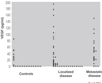
Comparison of plasma VEGF levels in patients with localized disease, metastatic disease, and control group
The median levels of VEGF in the 3 groups (controls, n = 26; localized disease, n = 54; and metastatic disease, n = 26) were compared. The Kruskal-Wallis test showed significant differences among the 3 groups (P < 0.001). The Mann-Whitney U test reveled a significant difference be-tween controls and patients with metastatic disease, and between patients with localized and metastatic disease (P
<0.001 and P <0.003, respectively). Table 1 and Figure 2 show the distribution of VEGF values among the 3 groups.
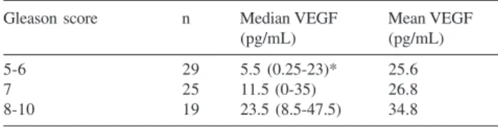
Correlation between Gleason score and plasma VEGF levels
The Gleason scores obtained from prostate needle bi-opsy were analyzed for an association with plasma VEGF levels. Six patients were excluded from the study because of lack of data. One patient with Gleason 2 + 2 was ex-cluded. Therefore, 73 patients were analyzed. There were no differences in plasma VEGF levels among the groups based on the Kruskal-Wallis test (P = 0.18). However, there was a trend for VEGF to be higher among patients with Gleason score 8 to 10 (Table 2).
Correlation between plasma VEGF levels and serum PSA in patients with prostate cancer
We did not observe a linear correlation between serum PSA and plasma VEGF levels (r = 0.14; P = 0.22). How-ever, patients with PSA levels greater than 20 ng/mL had
Table 1 - Plasma vascular endothelial growth factor (VEGF) concentrations in the 3 groups analyzed
Group n Median VEGF Mean VEGF
(pg/mL) (pg/mL)
Controls 26 0 (0-24)* 12.7
Localized disease 54 7.0 (0-26.5) 26.7
Metastatic disease 26 28.5 (19.3-57) 41.9 P < 0.001; *Number in parentheses = interquartile range
Figure 1 - Relationship between age and plasma vascular endothelial growth factor (VEGF).
significantly higher plasma VEGF values than patients with PSA levels less than 20 ng/mL (Table 3). Two patients were excluded for not having PSA levels determined at the time of the analysis.
Correlation between clinical stage and plasma VEGF in patients with localized disease
Regarding the 54 patients with localized prostate can-cer, VEGF levels were not related to clinical stage (P = 0.54). Six patients were excluded from the analysis for not having clinical stage described in the clinical records. Ta-ble 4 shows plasma VEGF levels according to clinical stage.
Correlation between prostate volume and plasma VEGF levels in patients with localized prostate cancer
No correlation between plasma VEGF levels and pros-tate volume in patients with localized disease were found (Table 5). Three patients were excluded from the analysis because they did not have measurements of prostate vol-ume when transrectal ultrasound was performed.
DISCUSSION
Angiogenesis, essential for tumor formation and the object of intense research, can be measured by microvessel density and by measurement of circulating levels of related growth factors, such as VEGF. Several studies suggest that the measurement of circulating levels of VEGF is a useful tool for analyzing prognosis and clinical outcome in many tumors. In the present study, plasma levels of VEGF was correlated with several parameters related to prostate can-cer, such as age, serum PSA, histological grade, clinical stage, and prostate volume.
No correlation was found between plasma VEGF and age of the patients. In the selection of the control group a younger population sample was deliberately collected in order to avoid inclusion of patients with occult prostate can-cer in this group. Accordingly, Yamamoto et al analyzed 184 normal individuals with ages varying from 21 to 59 years and found no correlation between age and circulat-ing VEGF levels.7 Kumar et al also found no correlation
between VEGF levels and age in a population of 136 indi-viduals with ages varying from 20 to 80 years.12
In this study, plasma VEGF levels were significantly higher in patients with metastatic prostate cancer in com-parison to patients with localized disease and healthy con-trols (P <0.001, Table 1). These findings are consistent with the study of Salven et al, which found elevated VEGF lev-els in patients with metastatic disease, although the fluid analyzed was serum in that study.13
Reported data regarding the measurement of circulat-ing VEGF levels in patients with prostate cancer is scarce. Jones et al measured VEGF in the serum of 78 individuals (21 controls, 9 with benign prostate hyperplasia, 16 with localized prostate cancer, and 32 with metastatic prostate cancer) and found elevated VEGF levels only in patients with hormonal-refractory metastatic prostate cancer. 14 They
found no differences in serum VEGF levels among controls, localized disease, and metastatic disease. They did not analyze VEGF levels in regard to clinical stage, Gleason score, or prostate volume. Their control group was also composed of younger individuals. In our study, patients
Table 4 - Plasma vascular endothelial growth factor (VEGF) levels according to clinical stage
Stage n Median VEGF Mean VEGF
(pg/mL) (pg/mL)
T1c 24 4.0 (0-18.8)* 24.4
T2 19 8.5 (4.7-20.5) 24.6
T3 5 4.5 (0-40) 16.9
P = 0.54; * interquartile range
Table 3 - Relationship between plasma vascular endothelial growth factor (VEGF) and prostate-specific antigen (PSA)
PSA n Median VEGF Mean VEGF
(pg/mL) (pg/mL)
PSA > 20 ng/mL 22 44.5 (44.5-68.5)* 53.9 PSA < 20 ng/mL 56 5.5 (5.5-21.5) 20.1 P < 0.001; * interquartile range
Table 2 - Relationship between plasma vascular endothelial growth factor (VEGF) and Gleason score
Gleason score n Median VEGF Mean VEGF
(pg/mL) (pg/mL)
5-6 29 5.5 (0.25-23)* 25.6
7 25 11.5 (0-35) 26.8
8-10 19 23.5 (8.5-47.5) 34.8
P = 0.18; * interquartile range
Table 5 - Plasma vascular endothelial growth factor (VEGF) according to prostate volume
Vol. n Median VEGF Mean VEGF
(pg/mL) (pg/mL)
< 20 10 8.5 (1.5-19)* 15.0
20-40 29 13.5 (2.5-58.5) 39.5
> 40 12 2.5 (0-7.5) 13.4
with metastatic disease were not subdivided into hormone-refractory and hormone-sensitive groups. Therefore, the data cannot be compared with regard to this issue. Addi-tionally, the 2 studies were not comparable because the fluid analyzed was different (plasma vs serum). Several studies in the literature analyzing circulating VEGF levels in patients with cancer, report high levels of this molecule in patients with advanced or metastatic cancer. 7,13 Such data
are compatible with both the study of Jones et al and our study. The fact that Jones et al14 did not find differences
between patients with localized and metastatic disease may reflect the small sample of their study (16 patients with lo-calized and 23 patients with metastatic disease).
Another study with 197 patients with hormone-refrac-tory prostate cancer reported plasma VEGF levels before and after treatment with suramin (patients in the protocol had already received hormones), and found an inverse cor-relation between plasma VEGF and survival. Yet, using a cutoff of 260 pg/mL, the plasma VEGF level was the strongest predictor of survival in a multivariate analysis, including other predictors such as serum PSA and alkaline phosphatase.15 The data suggests that plasma VEGF may
be clinically relevant in patients with hormone-refractory prostate cancer in terms of prognosis. However, plasma VEGF may be more than a disease marker, also reflecting a specific phenotype of a cancer that produces VEGF. Pro-spective studies including tumor histological analysis should be conducted to clarify this hypothesis.
Recently, Kohli et al reported a prospective study com-paring two groups of patients with advanced prostate can-cer in order to determine whether there is correlation be-tween plasma VEGF and tumor progression (from hor-mone-sensitive to hormone-refractory).16 The first group was
composed of patients with stable PSA during hormonal treatment, and the second group had patients with progres-sive PSA levels during treatment. There was no difference between the two groups in regard to plasma VEGF levels. One could argue that the development of metastatic spots would depend on angiogenesis and, therefore, would be accompanied by higher levels of related growth factors (like VEGF) in patients with disease progression. Alternatively, the increase in serum PSA levels in patients with disease progression may exert an anti-angiogenic effect, lowering circulating VEGF. The PSA anti-angiogenic effect on VEGF occurs due to its proteolytic action on plasminogen, creating an angiostatin-like protein with an anti-angiogenic effect.
In this study, we found not only a relationship between VEGF and advanced disease, but also a relationship be-tween VEGF and other clinical parameters associated with prostate cancer (PSA, clinical stage, and Gleason grade).
Patients with metastatic disease had significantly higher VEGF levels than patients with localized disease and con-trols. Although we did not find significant differences be-tween the control group and patients with localized disease, we observed a tendency of patients with localized disease to have higher VEGF levels than controls. The lack of sta-tistical significance could be attributed to the small sam-ple size; a larger series may or may not confirm this hy-pothesis. Yet, some patients with localized disease in this study had high VEGF levels. The reason for elevated plasma VEGF levels in patients with localized disease is unknown. Prospective study and long term follow-up of these patients should reveal whether patients with high plasma VEGF levels present worse prognoses than patients with low plasma VEGF levels. Even some patients in the control group were found to have high plasma VEGF, for unknown reasons. We should be aware that circulating VEGF may be elevated in patients with other, noncancerous disease.
We found no relationship between plasma VEGF and clinical stage in patients with localized disease. Two issues should be noted, ie, the limited sample size and the fact that different urologists performed the digital rectal exami-nation and clinical staging.
There was no relationship between plasma VEGF lev-els and Gleason score; however, we observed a tendency of patients with high Gleason scores (8 to 10) to have higher VEGF levels than patients with low Gleason score (5-6 or 7). Prostate cancer patients with high serum PSA levels and high Gleason scores are more likely to have metastatic disease.
No linear correlation between plasma VEGF and serum PSA levels were observed. However, patients with PSA lev-els greater than 20 ng/mL had significantly higher VEGF values than patients with PSA levels less than 20 ng/mL. This suggests that plasma VEGF is elevated in patients with disseminated disease. When prostate cancer presents as an organ-confined disease, plasma VEGF levels may not be elevated, regardless of clinical stage, Gleason grade, and PSA values.
In our study, there was no correlation between prostate volume and VEGF levels in patients with localized disease. Kohli et al, analyzing patients with localized prostate can-cer, found no differences between patients who had under-gone radical prostatectomy and patients with the intact gland (before treatment) with regard to plasma VEGF lev-els; this suggests that the prostate gland does not influence VEGF levels.16 Although the results of both studies are the
same, the data are not comparable, because the respective populations are totally different.
not affected by prostate size. However, George et al com-pared plasma VEGF levels in patients before and after radi-cal prostatectomy, and found a significant decrease in VEGF levels after surgery, suggesting that the prostate gland may be an important source of circulating VEGF.17
Additionally in this study, there was no correlation be-tween plasma VEGF and other prognostic factors related to prostate cancer before radical prostatectomy (PSA, clini-cal stage, Gleason grade). However, Shariat et al measured plasma VEGF in 215 patients with localized prostate can-cer before radical prostatectomy and found a correlation between plasma VEGF and prostate volume, Gleason grade obtained from prostate needle biopsy and definitive surgery, extra-prostatic disease, positive lymph nodes, and bio-chemical failure after surgery.18 The control group also had
lower VEGF values than patients with localized prostate cancer. Nine patients with metastatic disease had higher VEGF values when compared to the group of 215 patients with localized disease.
Based on all available published papers, controversies and unanswered questions persist about the role of circu-lating VEGF in prostate cancer patients. The role of VEGF remains unknown, in part due to its low specificity. Pro-spective studies with long-term follow-up may determine whether circulating VEGF can be used as a prognostic marker in patients with localized and metastatic disease, or as a tool in selecting patients who will benefit from cura-tive treatment. The value of VEGF in predicting lymph node disease remains uncertain. Yet, VEGF can be tested as a marker of response to treatment in patients with meta-static prostate cancer.
CONCLUSION
This study indicates that patients with metastatic pros-tate cancer have significantly higher plasma VEGF levels than patients with localized disease or healthy controls.
RESUMO
Duque JLF, Loughlin KR, Adam RM, Kantoff P, Mazzucchi E, Freeman MR. Medida da concentração plasmática do fator de crescimento do endotélio vascular em pacientes com câncer prostático: relação com estado clinico, gleason score, volume prostático e PSA sérico. CLINICS. 2006;61(5): 401-8.
OBJETIVO: Analisar os níveis circulantes do fator de cres-cimento do endotélio vascular em pacientes com câncer prostático comparados com uma população de indivíduos eutróficos.
MÉTODOS: Vinte e seis indivíduos eutróficos e oitenta
pacientes com câncer de próstata foram analisados nesse
estudo. A coleta sangüínea foi realizada da mesma manei-ra em todos os pacientes e o plasma foi extmanei-raído pamanei-ra a determinação dos níveis do fator de crescimento do endotélio vascular, utilizando-se o método quantitativo ELISA (enzyme-linked immunosorbent assay).
RESULTADOS: Os níveis de fator de crescimento do
Hou-ve uma tendência dos pacientes com escore de Gleason de 8 a 10 apresentarem níveis maiores do fator de crescimen-to do endotélio vascular plasmático em relação a pacien-tes com escores de Gleason menores que 8. Não houve re-lação entre fator de crescimento do endotélio vascular plasmático e estado clínico, ou entre fator de crescimento do endotélio vascular e volume prostático em pacientes com câncer de próstata localizado.
CONCLUSÃO: Os dados indicam que pacientes com
cân-cer de próstata metastático apresentam níveis significati-vamente mais elevados de fator de crescimento do endotélio vascular plasmático quando comparados com pacientes com câncer localizado e com indivíduos normais.
UNITERMOS: Câncer. Próstata. Fator de crescimento.
Metástases. Citocina.
REFERENCES
1. INCA – Instituto Nacional do Câncer. Câncer de próstata. Available at www.inca.gov.br/conteudo_view.asp?id=339
2. Ferrara N. Molecular and biological properties of vascular endothelial growth factor. J Mol Med. 1999;77:527-43.
3. Brown LF, Yeo KT, Berse B, Morgentaler A, Dvorak HF, Rosen F. Vascular permeability factor (vascular endothelial growth factor) is strongly expressed in the normal male genital tract and is present in substantial quantities in semen. J Urol. 1995;154:57-9.
4. Harper ME, Glynne-Jones E, Goddard L, Thurston VJ, and Griffiths K. Vascular endothalial growth factor (VEGF) expression in prostatic tumors and its relationship to neuroendocrine cells. Br J Cancer. 1996;74:910-6.
5. Jennbacker K, Vallbo C, Wang W, Damber JE. Expression of vascular endothelial growth factor C (VEGF-C) and VEGF receptor-3 in human prostate cancer is associated with regional lymph node metastasis. Prostate. 2005;65:10-6.
6. Kaushal V, Mukunyadzi P, Dennis RA, Siegel ER, Johnson DE, Kohli M. Stage-specific characterization of the vascular endothelial growth factor axis in prostate cancer: expression of lymphangiogenic markers is associated with advanced stage disease. Clin Cancer Res. 2005;11:584-93.
7. Yamamoto Y, Toi M, Kondo S, Matsumoto T, Suzuki H, Kitamura M, et al. Concentrations of vascular endothelial growth factor in the sera of normal controls and cancer patients. Clin Cancer Res. 1996;2:821-6. 8. Poon RT-P, Fan S-T, Wong J. Clinical implications of circulating
angiogenic factors in cancer patients. J Clin Oncol. 2001;19:1207-25. 9. Banks RE, Forbes MA, Kinsey SE, Stanley A, Ingham E, Walters C, et al. Release of the angiogenic cytokine vascular endothelial growth factor (VEGF) from platelets: significance for VEGF measurements and cancer biology. Br J Cancer. 1998;77:956-64.
11. Gleason DF, Mellinger GT. Prediction of prognosis for prostatic adenocarcinoma by combined histological grading and clinical staging. J Urol. 1974;111:58-64.
12. Kumar H, Heer K, Lee PWR, Duthie GS, Macdonald AW, Greenman J, et al. Preoperative serum vascular endothelial growth factor can predict stage in colorectal cancer. Clin Cancer Res. 1998;4:1279-85. 13. Salven P, Maenpaa H, Orpana A, Alitalo K, Joensuu H. Serum vascular
endothelial growth factor is often elevated in disseminated cancer. Clin Cancer Res. 1997;3:647-51.
14. Jones A, Fujiyama C, Turner K, Fuggle S, Cranston D, Bicknell R, et al. Elevated serum vascular endothelial growth factor in patients with hormone-escaped prostate cancer. Br J Urol. 2000;85:276-80. 15. George DJ, Halabi S, Shepard TF, Vogelzang NJ, Hayes DF, Small EJ,
et al. Prognostic significance of plasma vascular endothelial growth factor levels in patients with hormone-refractory prostate cancer treated on Cancer and Leukemia Group B 9480. Clin Cancer Res. 2001;7:1932-6.
16. Kohli M, Kaushal V, Spencer HJ, Mehta P. Prospective study of circulating angiogenic markers in prostate-specific antigen (PSA)-stable and PSA-progressive hormone-sensitive advanced prostate cancer. Urology. 2003;61:765-9.
17. George DJ, Regan MM, Oh WK, Tay M-H, Manola J, Decalo N, et al. Radical prostatectomy lowers plasma vascular endothelial growth factor levels in patients with prostate cancer. Urology. 2004;63:327-32. 18. Shariat SF, Anwuri VA, Lamb DJ, Shah NV, Wheeler TM, Slawin KM.