1
UNIVERSIDADE NOVA DE LISBOA
FACULDADE DE CIÊNCIAS E TECNOLOGIA
DEPARTAMENTO DE QUÍMICA
Joana Isabel Sobral Romão
“Development of cyclodextrin
-hydrogel polymeric systems
in scCO
2for drug delivery”
Thesis for the Degree of
Master of Science
in Bioorganic
Universidade Nova de Lisboa,
Faculdade de Ciências e Tecnologia
Supervisors:
Professora Maria Manuel Marques
Universidade Nova de Lisboa (UNL)
Faculdade de Ciências e Tecnologias (FCT)
Co-supervisors:
Professora Teresa Casimiro (UNL-FCT)
Jury:
Professor João Aires de Sousa (UNL-FCT)
Dr
aMarta Corvo (UNL-FCT)
Professora Maria Manuel Marques (UNL-FCT)
ii
UNIVERSIDADE NOVA DE LISBOA
FACULDADE DE CIÊNCIAS E TECNOLOGIA
DEPARTAMENTO DE QUÍMICA
Joana Isabel Sobral Romão
“Development of cyclodextrin
-hydrogel polymeric systems
in scCO
2for drug delivery”
Thesis for the Degree of
Master of Science
in Bioorganic
Universidade Nova de Lisboa,
Faculdade de Ciências e Tecnologia
Supervisors:
Professora Maria Manuel Marques
Universidade Nova de Lisboa (UNL)
Faculdade de Ciências e Tecnologias (FCT)
Co-supervisors:
Professora Teresa Casimiro (UNL-FCT)
Jury:
Professor João Aires de Sousa (UNL-FCT)
Dr
aMarta Corvo (UNL-FCT)
Professora Maria Manuel Marques (UNL-FCT)
iii
“Development of cyclodextrin
-hydrogel polymeric systems
in scCO
2for drug delivery”
Joana Isabel Sobral Romão,
Copyright
“
A Faculdade de Ciências e Tecnologia e a Universidade Nova de Lisboa têm o direito
perpétuo e sem limites geográficos, de arquivar e publicar esta dissertação através de exemplares
impressos reproduzidos em papel ou de forma digital, ou por qualquer outro meio conhecido ou
que venha a ser inventado, e de a divulgar através de repositórios científicos e de admitir a sua
cópia e distribuição com objectivos educacionais ou de investigação, não comerciais, desde que
v
Acknowledgements
In the first place I would like to thank Drª.Teresa Casimiro and Drª. Maria Manuel Marques for the opportunity they gave me, allowing me to understand what I truly like to achieve in my future career. This change proves that sometimes life, or destiny, decides what the best for us.
To Drª Maria Manuel Marques for the opportunity of constant learning, for all the scientific conversation at 8:00 am, for all the support and continuous motivation during the realization of this work.
To Drª Teresa Casimiro, I thank for the knowledge transfer and for the ongoing availability. To Mara Silva, I thank for the collaboration, the availability, help and support.
In the same way, I thank all my work colleagues of laboratory 202 for sharing the good and bad times of science.
I would also like to thank all my friends, especially those who accompanied me in my academic path: Ana Neto, Ana Fernandes, Daniel Silva, Marta Silva, João Domingos, Keno, Bruno for all the support, to Silvia for always being present and a big thanks to Alex for all the unconditional help and friendship, particularly this last year.
My parents and my sister, I thank for doing everything in their power for me and for always staying by my side.
Last but not least, I thank David Barata for all the support in my best and worst days, for always reminding me how lucky I truly am, for the smiles and good moments, for all the constructive discussions and opinions, for always pushing me one step ahead. I hope that life continue to smile at us.
vii
Abstract
This work describes the studies on the development of new cyclodextrin-hydrogel systems in supercritical carbon dioxide (scCO2) with potential application in drug delivery. Three β-cyclodextrin (CDs) derivatives were synthesized: 6-monoacryloyl-β-CD, 2-monoacryloyl-β-CD and 6-monoacryloly-heptakis-(2,3-di-O-benzyl)-β-CD. Their structures were assigned by nuclear magnetic resonance (NMR),
infrared (IR) and mass spectrometry (MS) using the technique of matrix-assisted laser desorption/ionisation-time-of-flight (MALDI-TOF). These functionalized β-CDs were co-polymerized in scCO2 and the resulting co-polymers were characterized by high resolution magnetic angle spinning (HR-MAS) NMR. Swelling tests were performed showing that the presence of CD decreases the swelling capacity of the corresponding co-polymers. The β-CD co-polymers were impregnated with a model drug, metronidazole, using a batch supercritical fluid impregnation process. Experiments in vitro were realized
ix
Resumo
Este trabalho descreve os estudos realizados no desenvolvimento de sistemas de ciclodextrinas (CDs)-hidrogel, em dióxido de carbono supercrítico com potencial aplicação para a libertação controlada de fármacos. Três derivados de β-CDs foram sintetizados: 6-monoacrilada-β-CD, 2-monoacrilada-β-CD e a 6-monoacrilada-hepta-(2,3-di-O-benzilo)-β-CD. As suas estruturas foram determinadas por ressonância
magnética nuclear, infravermelho e espectrometria de massa através da técnica de MALDI-TOF. As β -CDs funcionalizadas foram polimerizadas em dióxido de carbono supercrítico e os resultantes co-polímeros foram caracterizados por ressonância magnética nuclear usando a técnica de HR-MAS. Foram realizados testes de swelling que demonstraram que a presença da β-CD nos co-polímeros diminui o swelling. Os co-polímeros da β-CD foram impregnados com um fármaco, o metronidazol, usando um
processo de impregnação em dióxido de carbono supercrítico. Realizaram-se estudos in vitro com o intuito
xi
Index
Acknowledgements ... v
Abstract ... vii
Resumo ... ix
Index ... xi
List of Figures ... xv
List of Tables ... xix
Keywords ... xxi
List of Abbreviations ... xxiii
1. Introduction ... 1
1.1 Cyclodextrins ... 2
1.1.1 Fundamentals of CD chemistry ... 3
1.1.2 Chemical Modification of CDs ... 5
1.1.3 NMR Studies ... 10
1.1.4 CD inclusion complexes in aqueous solution ... 12
1.1.5 CD inclusion complexes in scCO2... 13
1.2 Polymers ... 15
1.2.1 scCO2 in polymer and impregnation process ... 15
1.2.2 Polymers in drug delivery ... 16
1.3 CD complexes and pharmaceutical applications. ... 20
1.3.1 Complexation and mechanism of drug release from CD complexes ... 20
1.3.2 Pharmaceutical applications of drug-CD complexes ... 23
2. Results Discussion ... 25
2.1 Objective ... 26
2.2 Synthesis of functionalized CDs ... 27
xii
2.4 Polymerization ... 46
2.4.1 Synthesis of CD-MAA-PNIPAAm co-polymer in scCO2 ... 46
2.4.2 Characterization of polymers ... 47
2.5 Final Conclusions ... 60
3. Experimental Part ... 63
3.1 Preamble ... 64
3.2 Synthesis ... 66
3.2.1 Synthesis of compound 10 (6-monoacryloyl-β-CD) ... 66
3.2.2 Synthesis of compound 11 (2-monoacryloyl-β-CD) ... 68
3.2.3 Synthesis of 12 (6-monoacryloly-heptakis-(2,3-di-O-benzyl)-β-CD) ... 69
3.2.4 Synthesis of acetylation β-CD ... 72
3.3 Studies in scCO2 ... 74
3.3.1 General methods for co-polymer synthesis in scCO2 ... 74
3.3.2 General methods for scCO2-assisted impregnation ... 75
3.3.3 General methods for in vitro drug release experiments ... 75
4. References ... 79
Appendix 1 ... 87
Appendix 2 ... 97
Appendix 3 ... 107
Appendix 4 ... 111
Appendix 5 ... 127
Appendix 6 ... 131
Appendix 7 ... 137
Appendix 8 ... 141
Appendix 9 ... 155
Appendix 10 ... 165
xiii
Appendix 12 ... 177
Appendix 13 ... 183
Appendix 14 ... 187
xv
List of Figures
Figure 1.1: Generic representation of the structure of the different types of CD. ... 2
Figure 1.2: CDs history, by József Szejtli. ... 3
Figure 1.3: Structure of α (1)-, β (2)- and γ-CD (3). ... 3
Figure 1.4: Schematic 3D representation of CD . ... 4
Figure 1.5: Conversion of a 6-substituted CD to a 3,6-anhydro CD. X is a good leaving group. ... 6
Figure 1.6: Strategies for pertosylation (I-IV) and peralkylation (I-VI). R1: TBDMS, R2: methyl or acetyl group, R3: alkyl group. ... 6
Figure 1.7: Strategy for synthesis of per-6-substituted CD via halogenated CD. X: Halogen atoms. R: Alkoxy, alkylamino, thio. ... 7
Figure 1.8: Summary of several pathways for the monosubstituion at 6-position of CDs. R1= I-, N3-, alkyl. ... 8
Figure 1.9: Strategies for modifications at the 2-positions of CDs. R: Sulfonly group, R: R1:TBDMS. ... 9
Figure 1.10: Representation of 1H-NMR spectra of β-CD: A- in D2O and B- in DMSO. C- Structure of the β-CD numbered. ... 11
Figure 1.11: Schematic representation of CD inclusion complex formation. The guest molecule is p -Xylene and small circles represent the water molecules. ... 12
Figure 1.12: Schematic illustration of the association of free CD and drug to form drug-CD complexes. 12 Figure 1.13: Representation of the equilibrium constant of inclusion complexes. ... 13
Figure 1.14: Schematic phase diagram for pure CO2. A- The critical point at the critical temperature and the pressure marks the end of the vapor-liquid equilibrium line and the beginning of the supercritical fluid region. B - Density of CO2 as a function of pressure at different temperatures (solid lines) and at the vapor-liquid equilibrium line (dashed line). ... 16
Figure 1.15: Graphical representations of A and B-type phase-solubility profiles. ... 21
Figure 1.16: Classes of CDs-containing polymers. ... 22
Figure 2.1: Functionalized β-CDs (10, 11 and 12) which were synthesized for application on polymerization reaction, to study the formation of inclusion complexes with drugs………..27
Figure 2.2: Mechanisms of mono-tosylation reaction with different reagents to obtain 5. ... 28
Figure 2.3: Structure of 5 and a representation of the corresponding 1H-NMR spectrum obtained. ... 29
Figure 2.4: Mechanism of reaction to obtain 10 and 13. ... 30
xvi
Figure 2.6: NOESY HR-MAS NMR spectrum and correlations between the protons of β-CD and acryloyl
group of 10. ... 31
Figure 2.7: Mechanism of preparation of acryloyl-β-CD. ... 32
Figure 2.8: Structure of 11 and a representation of the corresponding 1H-NMR spectrum obtained. ... 32
Figure 2.9: Plausible mechanism proposed to the formation of 11. a) Reaction of pyridine with acryloyl chloride. b) Mechanism of reaction forming an inclusion complex with pyridine salt. ... 33
Figure 2.10: Synthetic plan to obtain 12. ... 34
Figure 2.11: Mechanism of reaction with TBDMSCl to afford 4. ... 34
Figure 2.12: Structure of 4 and the corresponding 1H-NMR spectrum obtained. ... 35
Figure 2.13: Mechanism of reaction with benzyl bromide to fill the cavity of β-CD and obtain 14. ... 35
Figure 2.14: Structure of 14 and a representation of the corresponding 1H-NMR spectrum obtained. ... 36
Figure 2.15: Mechanism of deprotection of primary hydroxyl group to obtain 15. ... 36
Figure 2.16: Structure of compound 15 and 1H-NMR spectrum characterization. ... 37
Figure 2.17: Mechanism of reaction to obtain the final product (12) with acryloly chloride. ... 37
Figure 2.18: Structure of compound 12 and 1H-NMR spectrum characterization. ... 38
Figure 2.19: Mechanism of the acetylation reaction. ... 41
Figure 2.20: Examples of application of N-acylbenztriazole (Bt: benzotriazole) ... 42
Figure 2.21: a) Synthesis of N-acetylbenztriazole (17), b) Mechanism of acetylation reaction with benzatriazole as transfer agent of the acetyl group. ... 43
Figure 2.22: Structure of compound 19 and 1H-NMR spectrum characterization. ... 44
Figure 2.23: a) Scheme of membrane for UF. 1-Ultrafiltrate, 2-UF equipment, 3-Pressure supply. b) UF equipment. ... 45
Figure 2.24: Synthesized β-CDs that were co-polymerized in scCO2. ... 46
Figure 2.25: Scheme of the co-polymerization reaction... 46
Figure 2.26: Schematic representation of equipment used in polymerization reaction and impregnation. 1- Nitrogen cylinder; 2-Gas regulator; 3-Rupture disc; 4- High-pressure manometer; 5- Check-valve; 6- Line filter; 7-Water bath; 8-Immersible stirrer; 9-High pressure cell; 10- Platinum resistance RTD probe; 11-Temperature controller; 12-Vent; 13- Pneumatic CO2 compressor; 14- CO2 cylinder; M1,M2- bourbon manometers; wp-water recirculation pump; V1 to V7- HIP valves. ... 47
Figure 2.27: Appearance of the co-polymer obtained. ... 47
Figure 2.28: SEM images of: A - compound 10; B - NIPAAm-MAA-EGDMA co-polymer 10; C- NIPAAm-MAA-EGDMA co-polymer 11; D - NIPAAm-MAA-EGDMA co-polymer 12. ... 48
Figure 2.29: NOESY HR-MAS NMR spectrum and representation of co-polymer of compound 10. ... 49
xvii
Figure 2.31: XRD of P(NIPPAm-MAA) (blue), P(NIPAAm-MAA-10) co-polymer (orange) and
compound 10 (gray). ... 51
Figure 2.32: XRD of P(NIPPAm-MAA) (blue), P(NIPAAm-MAA-11) co-polymer (green) and compound 11 (gray). ... 51
Figure 2.33: Swelling of co-polymers at two different pHs 2.2 and 7.4. P(NIPAAm-MAA) (blue), P(NIPAAm-MAA-10) co-polymer (orange), P(NIPAAm-MAA-11) co-polymer (green) and (NIPAAm-MAA-12)(purple). ... 52
Figure 2.34: Test of swelling. A-(pH2.2); B-(pH7.4). ... 52
Figure 2.35: Structure of metronidazole. ... 53
Figure 2.36: High-pressure cell used for impregnation. ... 53
Figure 2.37: Drug release from the synthesized P(NIPAAm-MAA) (blue), P(NIPAAm-MAA-10) co-polymer (orange), P(NIPAAm-MAA-11) co-co-polymer (green) and (NIPAAm-MAA-12) (purple) at pH 2.2 (A) and pH 7.4 (B). ... 56
Figure 2.38: Drug release from the synthesized P(NIPPAm-MAA) (blue), P(NIPAAm-MAA-11) co-polymer with 2.5% (green) and P(NIPAAm-MAA-11) co-co-polymer with 8.8% (gray) at pH 2.2 (A) and pH 7.4 (B)... 58
Figure 2.39: Drug release from the synthesized P(NIPAAm-MAA-11) co-polymer with 8.8% at pH 2.2 (blue) and P(NIPAAm-MAA-11) co-polymer with 8.8% at pH 7.4 (gray). ... 58
Figure 2.40: Temperature test of co-polymers A- 23ºC, B- 45ºC. ... 59
Figure 3.1: Synthetic scheme to obtain products 10 and 13………66
Figure 3.2: Structure of product 11. ... 68
Figure 3.3: Synthetic scheme to obtain compound 12. ... 69
Figure 3.4: Structure of product 4. ... 70
Figure 3.5: Structure of product 14. ... 70
Figure 3.6: Structure of product 15. ... 71
Figure 3.7: Structure of product 12. ... 71
Figure 3.8: Structure of product 16. ... 72
Figure 3.9: Reaction between the acid anhydride and benzotriazole to obtain product 17. ... 72
Figure 3.10: Structure of product 18. ... 73
xix
List of Tables
Table 1.1: Properties of α-, β- and γ-CD. ... 4
Table 1.2: Examples of CDs pendent polymers. ... 22
Table 1.3: Approved and marketed drug-CD complexes. ... 23
Table 2.1: Yields of mono-tosylation reactions with different reagents. ... 28
Table 2. 2: Summary of structural characterization and yields of all compounds involved in synthesis of 10, 11 and 12. ... 40
Table 2.3: Number of equivalents of reagent used in the reactions of peracetylation and the products obtained. ... 42
Table 2.4: Conditions of the reactions with N-acylbenztriazole and products obtained ... 44
Table 2.5: Polymer loading during scCO2-assisted impregnation with Metronidazole. ... 54
Table 3.1: Results of metronidazole release of each co-polymer at pH 2.2. ... 76
xxi
Keywords
β-Cyclodextrin
Functionalization of β-Cyclodextrin
Supercritical carbon dioxide
Polymerization
xxiii
List of Abbreviations
13C-NMR Carbon Nuclear Magnetic Resonance Spectroscopy 1H-NMR Proton Nuclear Magnetic Resonance Spectroscopy
AIBN Azobisisobutyronitrile
Bt Benzotriazole
CD Cyclodextrin
DCC Carbodiimide
DMF N,N’-Dimethylformamide
DMP Dess-Martin periodinane
DMSO Dimethyl sulfoxide
DMSO-d6 Deuterated Dimethyl sulfoxide EGDMA Ethylene glycol dimethylacrylate
h Hours
HR-MAS High Resolution Magnetic Angle Spinning IR Infrared Spectroscopy
Kc Stability Constant
MAA Methacrylic acid
MALDI-TOF Matrix-assisted laser desorption/ionisation-time-of-flight mass spectrometer
min Minutes
MS Mass Spectrometry
NIPAAm N-isopropylacrylamide
NMR Nuclear Magnetic Resonance Spectroscopy NOESY Nuclear Overhauser effect spectroscopy
PGE Prostaglandins
RP Reverse Phase
RTD Resistence Temperature Detector scCO2 Supercritical carbon dioxide SEM Scanning Electron Microscope TBAF Tetra-n-butylamonium fluoride
TBDMS Tert-Butyldimethylsilyl
THF Tetrahydrofuran
TLC Thin layer chromatography
xxiv Ts p-Toluenesulfonyl
UF Ultrafiltration
UV Ultraviolet
1
2
1.1
Cyclodextrins
In 1981, A. Villiers, a French scientist, reported the formation of a crystalline
substance by fermentation of starch and determined its composition as (C
6H
10O
5)
2.3H
2O
naming
it as “cellulosine”.
[1-3]In 1903, an Austrian microbiologist, Franz Schardinger, isolated from the
microorganism
Bacillus macerans
, two distinct crystalline substances and identified them
as a cyclic structure of glucose oligomers
, called α
-cyclodextrin (CD)
and β
-CD. In 1935,
γ
-CD was discovered by Freudenberg and Jacobi.
[1, 2, 4]The corrected
chemical structure of α
-
, β
-
, γ
-CD was elucidated by Freudenberg and
co-workers in 1938, featuring them as cyclic structures composed of
α
-1,4-linked glucose
units (Figure 1.1). In the following years their molecular weight was determined.
[1, 5, 6]Figure 1.1: Generic representation of the structure of the different types of CD.
In 1953 the first patent on CD and their inclusion complex ability was registered by
Freudenberg, Cramer and Plieninger, who recognize the potential of these compounds to
form complexes. Later, in the 70s, only a small amount of CD could be produced with a
high production cost. However, advances in the biotechnology field allowed the
improvement of CDs production in terms of costs and purity.
[2]3
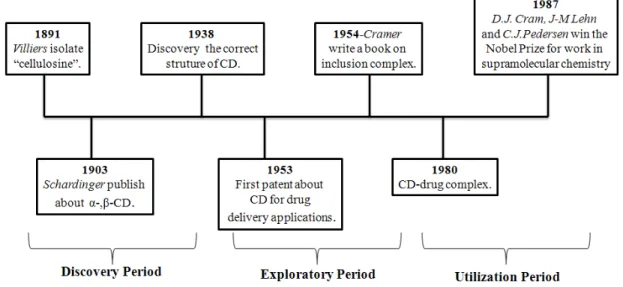
Figure 1.2 summarizes the main CDs development phases: the discovery period, the exploratory period, and the utilization period. [2, 7-9]
Figure 1.2: CDs history, by József Szejtli.[9]
1.1.1
Fundamentals of CD chemistry
CDs also known as cycloamyloses, cyclomaltoses or Schardinger dextrins, are macrocyclic oligosaccharides, most commonly composed by 6, 7 or 8 D-glucose units, known as α-, β- and γ
-CD, respectively (Figure 1.3).[8, 9] These CDs are crystalline and homogeneous substances, built up from glucopyranose units.[8, 9]
4
The most important properties of CDs are summarized in Table 1.1.[1, 5, 8-10]
Table 1.1: Properties of α-, β- and γ-CD.[5]
α-CD β-CD γ-CD
Number of glucose units 6 7 8
Molecular weight 972 1134 1296
Approximate inner cavity diameter (pm) 50 620 800
Approximate outer diameter (pm) 1460 1540 1750
Approximate volume of cavity (106 pm3) 174 262 427
[α]D at 25ºC 150.0 ± 0.5 162.5 ± 0.5 177.4 ± 0.5
Solubility in water (room temperature, g/100
ml) 14.5 1.85 23.2
Melting temperature range (ºC) 255-260 255-265 240-245
Water molecules in cavity 6 11 17
The units of D-glucose are attached by α-1,4-linkages, and the rigid 4C1-chair conformation
gives to the macrocycle structure the shape of a hollow truncated cone.[5] The hydroxyl groups located at the outer surface of the CD molecule are the primary hydroxyl groups that are located at the narrow face of the cone, while the secondary hydroxyls are located at the wide face, Figure 1.4. The cone is formed by the carbon skeletons of the glucose units and the glycosidic oxygen atoms in between.[1, 6, 8]
5
The C2-OH group of glucopyranoside unit can form a hydrogen bond with the C3-OH group of the adjacent glucopyranose unit, forming a complete secondary belt, which may explain the
rigid structure of β-CD and the lower water solubility comparing with the other CDs (Table 1.1).[5, 9, 11] In Table 1.1, it is possible to observe the different size and diameter of the cavities from the different CDs.[5, 8, 9]
The primary hydroxyl groups on the outside of the CDs cavity make them soluble in water, but simultaneously the secondary hydroxyl groups generate a cavity that is relatively hydrophobic[8, 9] or lipophilic[12, 13] (Figure 1.4).
1.1.2
Chemical Modification of CDs
The strategies for selective modification of CDs explored to date involve the exploration of the different reactivity of hydroxyl groups in CDs, and the selective modification is often adverted by regioselective protection and deprotection sequential steps.[6, 11, 14]
All modifications of CDs take place at the hydroxyl groups (C2-OH, C3-OH and C6-OH), which nucleophilic nature directs the regioselectivity and extension of modifications on all subsequent reactions.[14, 15]
The glucose units of CDs contain three different types of hydroxyl groups: primary hydroxyl groups at position C6, and secondary hydroxyl groups at C2 and C3. The primary side C6-OH groups are the most basic and the most nucleophilic, the C2-OH groups are the most acidic and the C3-OH groups are the most inaccessible.[5, 14-16] The different reactivity between both secondary hydroxyl groups is due to the fact that C2-OH is closer to the hemi-acetal than the C3-OH.[5, 14]
Some of the reported methods to functionalize CDs will be described next.
1.1.2.1
Modification at the primary face
6
Permodification at the C6-Position
Opposite to the mono-substitution of CDs, the persubstitution at C6 such as the persulfonates derivates are normally prepared directly from CDs by treatment with a large amount of sulfonyl chloride in pyridine. The 6-position tend to change to the 3,6-anhydro form, even in the absence of a base at room temperature (Figure 1.5).[14] This represents a limitation to this method, and in order to get reproducible results, the use of freshly prepared persulfonates is required.[14]
Figure 1.5: Conversion of a 6-substituted CD to a 3,6-anhydro CD. X is a good leaving group.[14]
Another approach reports for pertosylation or –mesylation involves silylation at the primary side of CDs with the TBDMS group (Figure 1.6, 4) as the first step, followed by esterification of the secondary side, subsequent desilylation and finally tosylation at the primary side (Figure 1.6, I-IV). [14, 17]
Figure 1.6: Strategies for pertosylation (I-IV) and peralkylation (I-VI). R1: TBDMS, R2: methyl or acetyl
group, R3: alkyl group. [14]
7
multi-step sequences, due to the good stability and easy removal of the silyl protecting groups. [14, 18]
Per 6-halogenocyclodextrins are important classes of compounds that, due to their greater stability compared to per-6-sulfonates, can be used for the selective functionalization of the primary face. However, these compounds are only soluble in polar solvents such as pyridine,
N,N’-dimethylformamide (DMF), dimethyl sulfoxide (DMSO), among others. [14] The secondary side of halogenated CDs can be acetylated in order to increase the solubility or allow a more selective functionalization (Figure 1.7). [14]
Figure 1.7: Strategy for synthesis of per-6-substituted CD via halogenated CD. X: Halogen atoms. R:
Alkoxy, alkylamino, thio. [14]
Monosubstitution at C-6-position
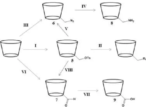
The most popular method for monomodifications at the 6-position of CDs is by nucleophilic attack to a properly functionalized CD, for example containing a good leaving group such as the tosyl group. [14, 16] The three main functional groups used for a variety of modification in CDs are the 6-tosyl (5), the 6-azide (6) and the 6-aldehydic (7) β-CDs (Figure 1.8). [5, 14] One of the most commonly used methods to perform mono-functionalizations at C6-OH is via the monotosyl CD
(Figure 1.8, I), which formation involves treatment of CD with tosyl chloride in pyridine or DMF. [14, 16, 19-21] However, this reaction produces a mixture of mono, di and tri-tosylated CDs. In order to obtain the desired product an extensive purification is required leading to low product yields.This compound is an important intermediate to achieve other desired modified products (Figure 1.8, II). [14, 16]
Many derivatives can be obtained from 6-monotosyl CD (5) by nucleophilic displacement of the tosyl group by a suitable nucleophile such as iodide, azide, thioacetate, hydroxylamine, alkyl, among others. This reaction allows the selective introduction of new functionalities at this position (Figure 1.8, II). [14, 15]
The 6-monoazide CD (6) can be obtained by two different methods: via the 6-monotosyl CD
(5) under heating with sodium or lithium azide salt in DMF (Figure 1.8, V); or via the
8
triphenylphosphine in DMF (Figure 1.8, III). The 6-monoazide CD (6) derivative is often used to obtain the 6-monoamine (8).[5, 14]
Figure 1.8: Summary of several pathways for the monosubstituion at 6-position of CDs. R1= I-, N3-,
alkyl.[14]
6-Monoaldehydic CDs (7) provide another route for further modifications (Figure 8, VI). The 6-monoaldehyde (7) has been synthesized by oxidizing 6-monotosyl-β-CD (5) using DMSO (Figure 1.8, VIII) or directly synthesized by reacting CDs with Dess-Martin periodinane (DMP), with a high yield (Figure 1.8, VI). Oxidation of 6-monoaldehyde (7) leads to the corresponding carboxylic acid (9) (Figure 1.8, VII). [5, 14]
1.1.2.2
Modification reaction at the secondary face
9
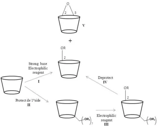
position, decreasing the selectivity. Positional isomerism in the secondary side, contributes to an increase in complexity. [5, 14]
Permodification at the C2-Position
Selective perfuncionalization of the secondary side is a difficult process due to the higher reactivity of the primary hydroxyl groups. A strong base selectively produces a 2-alcoxide ion which reacts with alkyl or sulfonyl chloride (Figure 1.8, I). This discriminatory behavior becomes less significant with increasing degree of substitution due to the steric crowding, where, as the reaction proceeds, the incoming electrophile finds the attack at the available primary hydroxyl groups more attractive. This problem can be overcomed by protecting the primary face of CDs with suitable group such as TBDMS (Figure 1.9, II). [5, 14]
Figure 1.9: Strategies for modifications at the 2-positions of CDs. R: Sulfonly group, R: R1:TBDMS. [14]
In order to prepare per-2-sulfonylation of CDs it is necessary to protect the primary hydroxyl groups due to their higher reactivity. The silyl group is usually selected as protecting group due to its easy removal with tetra-n-butylamonium fluoride (TBAF) (Figure 1.9, IV). After protection of
10
Due to the tendency to form 2,3-epoxy (Figure 1.9, V) derivatives in the basic medium,the yield of tosylation reaction is low. [14]
The direct alkylation of CDs at C2-OH is scarcely reported in literature, but an interesting approach has been described for the synthesis of per-2-alkylated CDs. This strategy involved the alkylation of per-2,6-di-O-TBDMS CD, due to the migratory property of the silyl group (from O
-2 to O-3). The per-2-alkylated compound was obtained after removal of the silyl groups from
positions O-3 and O-6. [14]
Per-modification at the C3-Position
The hydroxyl groups at the C3 are the least reactive groups, probably because of the hydrogen bonding with C2 hydroxyl groups. This position is hardly accessed and less reactive; consequently the modification in this position is not easy. [5, 14]
Selective per-3-sulfonation has not yet been reported, since sulfonyl chlorides fail to react with C3-OH hydroxyl groups. Indeed, strategies that involve protection of O-2- and O-6 positions
with TBDMS, lead to a high steric hindrance due to the bulky silyl functionality.[17] Moreover, the TBDMS groups have a tendency to migrate from the O-2 to O-3-hydroxyl groups under strongly
basic conditions, which also limits the attack of O-3-hydroxyl groups to the sulfonyl reagent. [17]
The use of TMS group for protection at the O-2- and O-6 positions does not constitute a better
choice, since these groups are easily hydrolyzed under neutral or acidic conditions.[14] Per-2-sulfonated CDs, when under basic condition, lead to per-2,3-epoxy-CDs (Figure 1.9, V). [14]
1.1.3
NMR Studies
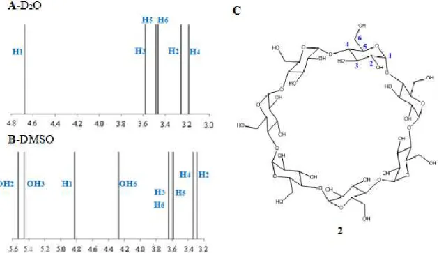
To perform an analysis and structural assignment of each monomer and polymer of CD, it is important to be familiar with NMR of non-functionalized β-CD.
The NMR spectroscopy has become the most important tool for structural elucidation of organic compounds. This technique allows structural assignment of compounds obtained by synthetic modifications in the different units of glucose of CD, by different 2D NMR experiments.[11, 22]
11
solvent. These different solvents allow a detailed insight into the intramecular CDs hydrogen-bond network.[22]
Figure 1.10: Representation of chemical shift spectra of β-CD: A- in D2O and B- in DMSO-d6. C- Structure
of the β-CD numbered.[22]
In protic solvents such as D2O, intermolecular exchange between solute and solvent is too fast on the NMR time scale for the observation of separate OH signals. The intramolecular hydrogen bond is visible by hydrogen-deuterium exchange rate constants in D2O for the equilibrium: [22]
In a solvent such as DMSO-d6 it is possible to observe separate signals for the OH groups and analyze the coupling of the vicinal C-H protons. The resonances of C3-OH and C2-OH were found to appear in DMSO-d6 between 5.4 and 5.6 ppm, clearly separated from the signals of the free, more shielded OH-6 group at 4.3 ppm (Figure 1.10, b). In DMSO-d6 as hydrogen bonding acceptor, the following hydrogen-bonding equilibrium is present: [22]
12
1.1.4
CD inclusion complexes in aqueous solution
Each CD has its own characteristics to form inclusion complexes with specific molecules, which is determined by the characteristics of the guest molecules, such as polarity, size and geometry that should be appropriate to the hydrophobicity and cavity size of a particular CD.[23]
In an aqueous solution, the slightly apolar CD cavity is occupied by water molecules which are energetically unfavored (polar-apolar interaction) and can be readily substituted by an appropriate guest molecule less polar than water (Figure 1.11).[7, 9, 10, 15] The dissolved CD is the
“host” molecule and the driving force for complex formation is the substitution of the high-enthalpy water molecules by an appropriate guest molecule (Figure 1.11).[7, 10, 24]
Figure 1.11: Schematic representation of CD inclusion complex formation. The guest molecule is p-Xylene
and small circles represent the water molecules.[7]
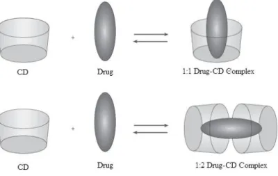
Frequently, the host:guest ratio is 1:1, however, 1:2, 2:1, 2:2 or other association can occur, sometimes simultaneously (Figure 1.12).[9, 24]
13
The cavity size of α-CD allows the formation of complexes with aliphatic chains and small molecules, whereas β-CD is appropriate for aromatic ring and γ-CD has a larger cavity so can form complexes with larger molecules, as steroids. [23, 25]
Upon dissolving these complexes, an equilibrium is established between dissociated and associated species, and this is expressed by the complex stability constant Kc. The association of
the CD and guest (D) molecules, and the dissociation of the formed CD/guest complex is governed by the thermodynamic equilibrium (Figure 1.13).[9, 15]
Figure 1.13: Representation of the equilibrium constant of inclusion complexes.[15]
This association and dissociation reactions of guest molecules from the CDs are dynamic processes that occur quite fast (in few milliseconds), even for complexes with high stability constants. [23] According to this properties, the kinetics of drug release from CDs will not be a limiting factor in absorption,other mechanisms may also be responsible for the release of the drug inclusion complex, as the effect of pH on CDs or ionizable drugs. [23] The change of pH can change the state of ionization of the drug or the CD and lead the dissociation of the complex by decreasing its Kc. The way all these factors mentioned will influence the dissociation of the
complex depends on the route of administration, the volume of distribution of the drug, the Kc
and the complex concentrations of the drug, the CD and other competitive entities. [23]
1.1.5
CD inclusion complexes in scCO
2Nowadays, the combinatorial chemistry and high throughput screening, allow the discovery of new drugs with good pharmacological activities, but many of them have low solubility.[26] The drug-CD complexes can bring substantial advantages like improvement of solubility, stability and bioavailability of drugs. The two main techniques to form complexes between CD and drugs in scCO2 are impregnation of CD matrixes with guest compounds and co-precipitation of CDs and guest molecules.[27]
14
solubilization of the drug in CO2 and the complexation of the solubilized drug with CD that would shift the equilibrium toward the dissolution of extra drug. A driving mechanism for the formation of CD inclusion complexes is the substitution of water of crystallization molecules in the CD cavity with hydrophobic guest. Since the removal of water of crystallization makes the hydrophobic cavity of the CD free to include other guest molecules, the extraction of the included water molecules in CO2 would favors the formation of the inclusion complex between the CD and the drug. The efficacy of CO2 in extracting water from the CD depends on the water solubility.[27]
The co-precipitation of CDs and guest compounds is another method that allows formation of complexes in CO2. The CD and the guest molecule are separately dissolved in different fluids and the two solutions were concomitantly contacted with the CO2 that expanded the two solvents, lowering their solvent power and causing the solutes precipitation. The solvents were removed from the systems by the CO2 and the product was collected in the form of dry powder. [27]
15
1.2
Polymers
1.2.1
scCO
2in polymer and impregnation process
In this work, scCO2 was used as an alternative green media of polymerization, once it offers numerous advantages compared with other traditional methods that use solvents.[28, 29] Conventional production routes involve an excessive use of organic solvents, either as a reaction medium in the polymerization step or as a processing medium for shaping, extraction, impregnation, and viscosity reduction. In each of these steps, most of the effort of the process is put into solvent recovery.[30] Therefore, it is highly desirable from environmental, safety and economical point of view, to develop alternative routes to reduce the use of solvents in polymerization processes. [28, 29, 31]
The scCO2 allows the preparations of hydrogels completely dry and free-flowing powders, with no solvent residues and with no need for intensive drying steps before processing.[32] Additionally, more attractive features are available: an easily accessible critical point (31.1 ºC and 73.8 bar), abundance, non-flammable, non-toxic and relatively inexpensive.[29, 31, 33] Due to the favorable practical physical and chemical properties of CO2, it is a solvent for monomers and a non-solvent for polymers, which facilitates the separation. [30]
A supercritical fluid is defined as a substance for which the temperature and pressure are above their critical values and which has a density close to or higher than its critical density.[29, 30] Above the critical temperature, the vapor-liquid coexistence line no longer exists. The supercritical fluids are versatile because their properties can be tuned from liquid to gas without crossing a phase boundary by simply changing the pressure or the temperature. [30]
16
Figure 1.14: Schematic phase diagram for pure CO2. A- The critical point at the critical
temperature and the pressure marks the end of the vapor-liquid equilibrium line and the beginning of the supercritical fluid region. B - Density of CO2 as a function of pressure at different temperatures (solid lines)
and at the vapor-liquid equilibrium line (dashed line).[30]
Supercritical carbon dioxide is a non-toxic and non-flammable solvent with a low viscosity and high diffusion rate and no surface tension.[28, 29] The disadvantage of CO2 is that only volatile or relatively non-polar compounds are soluble, [28, 29] however, the polarity can be turned by adding some amount of co-solvents and also several polymerizations approaches can be followed depending on the initial reaction phase behavior.[29, 33]
1.2.2
Polymers in drug delivery
Controlled drug delivery has emerged as a truly interdisciplinary science that aims to improve
human health. The basic goal of controlled drug delivery systems is to deliver biologically active molecules at a desired rate and for a desired period, maintaining the drug levels in the body within the therapeutic window.[35]
Most of the initially developed drug delivery systems were based on nondegradable polymers. However, the advent of synthetic biodegradable polymers coupled with the fact that macromolecules can be effectively delivered from these matrices, gave a new impetus to this branch of science.[35]
17
than the active ingredient.[36] Micro- and nano-electromechanical have many potentialities in drug release, like control the time and the doses administered. Micro-electromechanical has been used to construct microreservoirs, micropumps, nanoporous membranes, nanoparticles, valves, sensors, and other structures using biocompatible materials appropriate for drug administration. The focus of this section is polymeric drug delivery systems.[35]
In polymeric drug delivery systems, the drugs are incorporated in a polymer matrix. The rate of release of drugs from such a system depends on a multitude of parameters such as nature of the polymer matrix, matrix geometry, properties of the drug, initial drug loading, and drug-matrix interaction.[35] Therefore, controlled drug delivery can be used to achieve:[37]
Sustained constant concentration of therapeutically active compounds in the blood with minimum fluctuations;
Predictable and reproducible release rates over a long period time;
Protection of bioactive compounds having a very short half-life;
Elimination of side-effects waste of drug and frequent dosing;
Optimized therapy and better patient compliance
Solution to drug stability problems.
Furthermore, the mechanisms for drug delivery systems can be classified into diffusion controlled systems, chemically controlled systems, and solvent-activated systems.[38]
18
to formulate and gives higher initial release rate than a reservoir device and can be made to release at a nearly constant rate.[37]
In chemically controlled drug delivery systems, the release of a pharmacologically active agent usually takes place in the aqueous environment by one or more of the following mechanism:[37]
Gradual biodegradation of a drug containing polymer system;
Biodegradation of unstable bonds by which the drug is coupled to the polymer system;
Diffusion of a drug from injectable and biodegradable microbeads.
In biodegradable systems, the main advantages are elimination of the need for surgical removal, their small size and potential low cost. On the other hand, all biodegradable products as well as their metabolites must be non-toxic and non-carcinogenic, these requirements are not easily met and must be subject to careful scrutiny.[37]
In pendent chain systems, the drug molecule is chemically bonded to a polymer backbone and the drug is released by hydrolytic or enzyme cleavage. The rate of drug release is controlled by the rate of hydrolysis. This approach provides an opportunity to target the drug to a particular cell type or tissue.[37]
1.2.2.1
Aspects to approve a new drug
A number of factors that create hurdles for ultimate approval of the drug-loaded system in polymer medicine must be considered. Some of this factors are biocompatibility of the device, cytotoxicity, efficiency, inconvenience caused to patients, cost effectiveness, etc.[37]
For the design of drug delivery systems with optimum performance in specific circumstances, the drug delivery systems has to confront the following challenges:[37]
1. Improved efficacy;
2. Targeted delivery and reduced side effects; 3. Optimum performance;
4. Interfacing and pacing with modern methodologies; 5. Guarantees of safe environment;
19
The polymer hydrogels of different chemical architecture with novel physic-chemical properties have shown potential to find applications as promising drug carrying vehicles in several drug delivery platforms. Although their synthesis and in vivo study seems to be simple,
from the point of view in vivo applications the polymers systems need to be judged with extreme
20
1.3
CD complexes and pharmaceutical applications.
The potential uses of CDs in pharmaceutical industry have been increasing in the last few decades and may explain their main application in drug delivery field. The CDs have the ability to form inclusion complexes with some drugs and improve some of their features like solubility, stability, safety and bioavailability.[12, 39] However the most desirable attribute for a drug carrier is the ability to deliver a drug to targeted site.[40]
CDs with hydrophobic liner cavities and hydrophilic outer surfaces are capable of interacting with a large variety of guest molecule to form noncovalent inclusion complexes. The cavity size
of α-CD is insufficient for many drugs and γ-CD is expensive. β-CD has been widely used in the early stages of pharmaceutical studies because of its availability and cavity size, which gives a character suitable to interact with the widest range of drugs.[12]
The effect of CDs on chemical stability of drugs is a useful property and has been extensively studied.[41] CDs can be used to reduce or prevent gastrointestinal and ocular irritations, reduce/eliminate unpleasant smell or taste, prevent drug-drug or drug-additive interactions, or even to convert oils and liquid drugs into microcrystalline or amorphous powder.[13, 39] Besides this the CDs can improve the stability of several labile drugs against dehydration, hydrolysis, oxidation and photodecomposition and thus increase the shelf life of drugs.[12]
1.3.1
Complexation and mechanism of drug release from CD complexes
1.3.1.1
Aqueous Solution
In aqueous solutions, CDs are able to form inclusion complexes with many drugs by taking up the drug molecule or some hydrophobic moiety of the molecule into the central cavity. No covalent bonds are formed or broken during the complex formation and the drug molecules in complex find rapid equilibrium (with free molecules in the solution).[3, 39, 42]
Phase-solubility analysis about the effect of complexing agents on the compound being solubilized is a traditional approach to determine not only the value of the stability constant but also to give insight into the stoichiometry of the equilibrium.[3] The phase-solubility profiles as shown in Figure 1.15. In A systems, the apparent solubility of substrate increases as a function of CD concentration. Three subtypes have been defined: AL, when the complex is first order with
respect to ligand and first or higher order with respect to substrate (drug); AP, systems indicate an
21
order with respect to the substrate, but second or higher order with respect to the ligand and AN
relationships indicate a negative deviation from linearity, i.e the CD proportionally less effective at higher concentrations. The A-type phase-solubility profiles indicate that water-soluble complexes are being formed with solubility higher than that of uncomplexed substrate. [3, 39, 42] Type B phase-solubility profiles indicate formation of complexes with a limited solubility in the aqueous complexation medium. BS-type isotherms indicate that the CD concentration increases, a
soluble complex forms which increase the total solubility of the substrate. The BI systems are
similar in form to the BS profiles except that the complexes being formed are so insoluble that
they do not give rise to the initial ascending component of the isotherm.[3, 39, 42]
When the complexation of CD/drug is in aqueous solution two parameters are important, dilution and competitive displacement, because the complexation constant (K) and the lifetime of the complex are very importante for the drug release mechanism.[13]
1.3.1.2
Polymerization
There are different classes of CD containing polymers. One of the types of polymers are those possessing a crosslinked structure (Figure 1.16, a), one of the first agents used for crosslink in CDs was epichlorohydrin (1-chloro-2,3-epoxypropane).[11, 25] These were the initial CD-containing polymers investigated for drug delivery applications. Other agents used for crosslinking are the diepoxides and diisocyanates. Another class of polymers are those containing CDs as pendent moieties in the polymers backbone (Figure 1.16, b), however this type of
22
polymers are prepared by functionalized CDs. In Table 1.2, some examples of pendent polymers are referred.[15, 25]
Table 1.2: Examples of CDs pendent polymers.
Types of polymer CDs Preparation method
Polyacrylic esters α and β Polymerization of vinyl CD derivatives
Poly(allylamine)s β and γ Grafting of CD to preformed polymer
Polymethacrylates α, β and γ Polymerization of CD methacrylate monomers
Polyester β Gratting of CD to preformed polymer
A different type of CDs polymerized possesses a tubular structure, these polymers are then formed from the tubular configuration by crosslink between the CDs (Figure 1.16, c). The last class of polymers studied has a linear structure and contains CD as part of the backbone, as it can be seen in Figure 1.16, d.[15, 25]
23
1.3.2
Pharmaceutical applications of drug-CD complexes
Nowadays many drugs with CDs are available in the market with application in several areas of medicine, as shown in Table 1.3.[1, 25, 43]
Table 1.3: Approved and marketed drug-CD complexes. [1, 25, 43]
Drug Administration route Treatment Trade name Market
α-CD
Alprostadil (PGE1) Intravenous Vasodilation Prostavastin, Edex Europe, Japan,
USA
Cefotiam Hexetil HCl Oral Antibiotic Pansporin T Japan
β-CD
Dinoprostone (PGE2) Sublingual Labor induction Prostarmon E Japan
Benexate Oral Antiulcer Ulgut, Lonmiel Japan
Dexamethasone Dermal Analgesic Glymesason Japan
Nicotine Sublingual Reducing the symptoms of
absence
Nicorette Europe
Omeprazol Oral Antiulcer Omebeta Europe
2-Hydroxypropyl-β-CD
Hydrocortisone Buccal Anti-inflammatory Dexocort Europe
Indomethacin Eye drops Anti-inflammatory Indocid Europe
Random methylated β
-CD
17β-Oestradiol Nasal spray Hormone therapy Aerodiol Europe
Chloramphenicol Eye drops Antibiotic Clorocil Europe
Sulphobutylether β-CD
Voriconazole Intravenous Antimycotic Vfend Europe, USA
2-Hydroxypropyl γ-CD
Diclofenac sodium Eye drops Anti-inflammatory
24
The first studies about the application of CDs in drug delivery experiments were with the prostaglandins (PGE). CDs complexes of PGE1 and PGE2 resulted in a significant increase in their solid-state stability, and a product designed along these lines was approved for the first time in Japonese market in 1976. Prostaglandins are used to relax smooth muscles and increase blood flow, and were initially developed in the therapeutic of peripheral circulatory disorders. Just after, in 1979, alprostadil alphadex (Prostavasin) was approved for the treatment of peripheral vascular complications and showed activity against chronic arterial occlusions and asteriosclerosis. CDs were applied to this compound in an effort to improve several properties, including safety and drug dissolution rate. These improved characteristics reduced gastrointestinal irritation, and allowed faster drug absorption and faster onset of analgesic effect.[25]
The implementation of structural changes in the CD allowed different pharmaceutical applications. For instance, the randomly methylated β-CD avoids a number of issues related to oral or transdermal administration and allows administration strategies like eye drop and nasal.
The hydroxypropylated β-CD is available in registered oral, intravenous, buccal, rectal and
ophthalmic products. The sulphobutyl ether β-CD has products like intravenous formulation of the antifungal agent voriconazole and an intramuscular dosage form for the antipsychotic agent ziprasidone.[25]
25
26
2.1
Objective
From the point of view of pharmacotherapy optimization, drug release should be controlled in accordance to the therapeutic purpose and the pharmacological properties of active substances. There has been a growing interest in the development of a delivery system with rate- or time-controlled oral administration, because appropriate drug release from dosage forms have critical importance for efficient therapeutics.[44]
Cyclodextrins (CDs) are potential candidates for controlled drug delivery, once their cavities provide microenvironments where molecules can enter and form inclusion complexes. Supercritical CO2 (scCO2) is a sustainable alternative comparing to processes with organic solvents, and a very attractive medium for the preparation of these inclusion complexes.[45]
This work aims to develop a new pH responsive CDs-hydrogel system in scCO2 with potential application on controlled drug delivery. The first part of this study consists on functionalization of β-CD with a group, such as activated double bonds that allow polymerization in scCO2. In order to prepare the desired β-CDs, two different approaches were used: regioeselective functionalization in the most reactive hydroxyls groups (C6-OH) and functionalization of a complete set of hydroxyls groups.[3] The second part of this work envisages the polymerization of the functionalized β-CD in scCO2 and the investigation of the potential ability of polymer-CD to form inclusion complexes with the drug. In vitro experiments will be
performed in order to evaluate the performance of the CDs-hydrogel system as drug release device at different pHs, 2.2 and 7.4, which simulate the gastric and colon experiments. The intent is to study the most appropriate application of these complexes.1
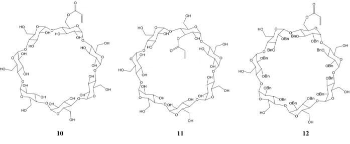
In order to investigate the importance and role of CD cavity, three different β-CD derivates need to be prepared: the first involves the synthesis the of a monoacrylate-β-CD via the
6-monotosyl-β-CD approach (Figure 2.1,1); the second consists of acrylation by a direct method (Figure 2.1, 2); and the third involves the exploration of a filled CD cavity to study its role in the polymerization and interaction with the drug (Figure 2.1,3).
1 The co-polymerization in scCO
2 and the in vitro drug release experiments were performed with the
27
Figure 2.1: Functionalized β-CDs (10, 11 and 12) which were synthesized for application on polymerization reaction, to study the formation of inclusion complexes with drugs.
2.2
Synthesis of functionalized CDs
The objective of synthesizing the compound 10 was to introduce a crosslink, a group with a double bond possessing an electron withdrawing group that allows subsequent polymerization in scCO2.
In the first chapter, the main approaches described in literature (Chapter 1.1.3.1) to selectively functionalize CDs were presented. From the reported approaches, a route for a selective modification at C6-OH consists on to the introduction of the tosyl group, a good leaving group and then substitution by the desired functional group, by tosyl group displacement (Figure 1.18).[14]
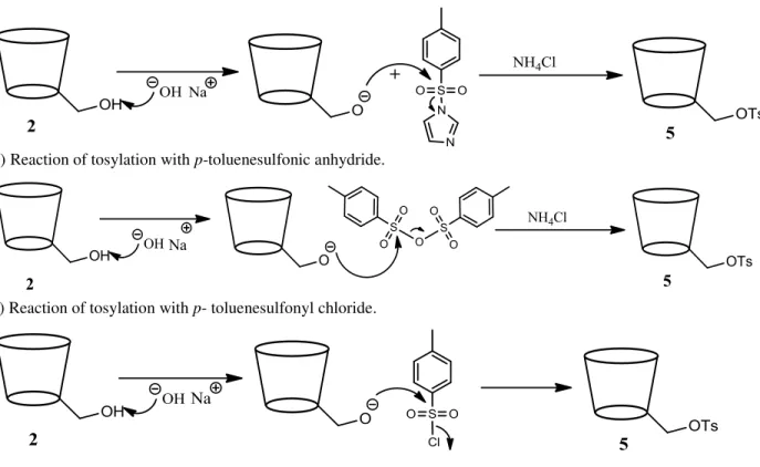
To synthesize the 6-monotosyl-β-CD (5) three procedures were tested in order to achieve high yields of conversion. The first attempts of mono-tosylation were performed by treatment with p-toluenesulfonly imidazole,[21] p-toluenosulfonic anhydride[20] and p-toluenesulfonyl
28
a) Reaction of tosylation with toluenesulfonly imidazole.
b)Reaction of tosylation with p-toluenesulfonic anhydride.
c) Reaction of tosylation with p- toluenesulfonyl chloride.
Figure 2.2: Mechanisms of mono-tosylation reaction with different reagents to obtain 5.
The yield of isolated and purified 5 varies depending on the tosylation reaction conditions (Table 2.1). Thus, a higher yield of 5 was obtained when p-TsCl was used as tosylation agent.
Table 2.1: Yields of mono-tosylation reactions with different reagents.
Reagents Yields
p-Toluenesulfonly Imidazole 19%
Toluenosulfonic Anhydride 10%
p-Toluenesulfonyl Chloride 21%
29
this compound: the aromatic protons (7.4-7.7 ppm) and the methylic protons (2.5 ppm) of tosyl group (Figure 2.3).[19-21] The NMR spectrum was in accordance with literature.[19-21, 46]
Figure 2.3: Structure of 5 and a representation of the chemical shift.
30
Figure 2.4: Mechanism of reaction to obtain 10 and 13.
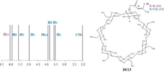
The NMR spectra of 10 and 13 were performed in D2O. In the case of
6-mono-methacryloyl-β-CD (13) two signals were expected in the 1H-NMR around 5.6-6.3 ppm, due to the presence of the double bond and 2 ppm due to the methyl group (Figure 2.5).[46, 48]
Figure 2.5: Structure of compounds 10 and 13 and a representation of the corresponding chemical shift.
31
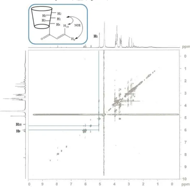
resonance signal correspondent to the carbon of carbonile carbon at 175.6 ppm and the signals of two carbons of the double bonds at 133.9 and 126.5 ppm were observed.[46] From all 2D spectra only in the NOESY experience was possible to see a correlation between the anomeric (H1) protons and the double bond protons (H9,10) (Figure 2.6).
Figure 2.6: NOESY HR-MAS NMR spectrum and correlations between the protons of β-CD and acryloyl group of 10.
32
In this second approach the β-CD (2) was slowly dissolved in pyridine to prevent the formation of a gel and subsequently the acryloly chloride in THF was added dropwise.[50] The mechanism of this reaction is basically the attack of the oxygen atom to the carbonyl group with the release of chloride ion, which is a good leaving group. In Figure 2.7, is represented a mechanism for the acrylation of β-CD (11).
Figure 2.7: Mechanism of preparation of acryloyl-β-CD.
The product 11 was obtained with 94 % yield and presented a yellow/brown color. To characterize 11 by NMR DMSO-d6 and D2O were used as solvents. In the 1H-NMR spectra performed in D2O it was possible to observe a signal correspondent to the resonance of double bound protons between 5.9- 6.4 ppm (Figure 2.8).[50]
Figure 2.8: Structure of 11 and a representation of the corresponding chemical shift.
33
experiments did not show evidence of a acryloly group covalently bonded connect to CD, in the 1H-NMR spectrum the integration of the number of protons for the hydroxyl groups (C2-OH,
C3-OH, C6-OH) suggests that the acryloly group is bounded to the C2-OH. The resonance signal of C2-OH and C3-OH appear in the same chemical shift while C6-OH appear at a higher field, thus integration suggest that the reaction occurred at C2-OH or C3-OH instead the most reactive hydroxyl group, C6-OH. However, due to the known higher reactivity of C2-OH it is proposed that the reaction occurred at this position.[5] Figure 2.9, shows a proposed mechanism to explain the fact that acrylation reaction occurred inside the cavity of the β-CD. In MALDI-TOF spectrum, the peak observed at 1268.6 m/z corresponds to monoacrylate-β-CD and in the IR spectrum a band at 1735 cm-1 confirm the presence of ester group.[47]
Figure 2.9:Plausible mechanism proposed to the formation of 11. a) Reaction of pyridine with acryloyl chloride. b) Mechanism of reaction forming an inclusion complex with pyridine salt.
The next synthesis envisaged the investigation of the importance of the free cavity of β-CD, in the process of polymerization and impregnation with a drug.
34
Figure 2.10: Synthetic plan to obtain 12.
The requirements for the choice of the protecting group are their stability in the following reaction steps and the conditions to remove them. The deprotection conditions should not affect the groups introduced in the secondary hydroxyl groups. From all the protective groups and procedures described in literature, the TBDMS was the group of choice due to its easy removal by treatment with TBAF and also due to its stability under the proposed synthetic sequence.[18] The synthesis of persilylated CD consisted on the use of TBDMSCl, that due to its bulkiness prevents the silylation at secondary hydroxyl groups.[50] However, it was necessary to purify the resulting crude in a chromatography column with a gradient of solvents (MeOH:CH2Cl2). The persilylate-β-CD (4) was collected as a white solid and a yield of 81% was obtained.
The mechanism of this reaction is depicted in Figure 2.11.
Figure 2.11: Mechanism of reaction with TBDMSCl to afford 4.
MALDI-35
TOF spectrum confirmed that the sylilation occurred at all C6-OH positions by the presence of the peak at 1956.0 m/z.[50]
Figure 2.12: Structure of 4 and the corresponding chemical shift.
The next step aimed to occupy the cavity of β-CD with benzyl groups, a large group that would fill the cavity and allow achievement of this study.[51]
One of the procedures reported, involved the use of benzyl bromide. β-CD was dissolved in DMF and sodium hydride was added, a base to remove protons from the secondary hydroxyl group forming the corresponding anion. This anion will act as nucleophile and when benzyl bromide is added, the attack to the carbon atom that is bound to bromine occurs since bromine is a good leaving group (Figure 2.13). [51] The product was collect as a white solid in 43% yield.
Figure 2.13: Mechanism of reaction with benzyl bromide to fill the cavity of β-CD and obtain 14.
36
that all the positions of secondary hydroxyl group are substituted by the presence of peak at 3217.8 m/z.[51]
Figure 2.14: Structure of 14 and a representation of the corresponding chemical shift.
The third step of this synthetic route consisted on the deprotection of primary hydroxyl group, removing the TBDMS group. This reaction was performed in tetrahydrofuran (THF) with the TBAF reagent, a quaternary ammonium salt (Figure 2.15).[18]
Figure 2.15: Mechanism of deprotection of primary hydroxyl group to obtain 15.
37
Figure 2.16: Structure of compound 15 and representation of chemical shift.
The last step consisted on the introduction of the cross-linking to polymerize the β-CD with filled cavity. The procedure for this step was the same used before for the product 11, by reaction with the acryloyl chloride (Figure 2.17).[49]
Figure 2.17: Mechanism of reaction to obtain the final product (12) with acryloly chloride.
38
Figure 2.18: Structure of compound 12 and representation of chemical shift.
In order to co-polymerize the compound 12 with functional monomers and study the interaction of the resulting polymer with a drug, a large amount was needed. With an overall yield of 21% it was necessary to
39
40
41
2.3
Exploration of novel approach towards the
functionalization of
β
-CD
The selective modifications of CDs are a challenge, due the hydrophobic cavity and the large number of hydroxyl groups. Due to the hydrophobic cavity there is a tendency to form inclusion complexes with reagents of the synthetic scheme.[14, 24] The hydroxyl groups C2-OH, C3-OH and C6-OH compete for the reagent and make the selective modification extremely difficult.[14, 24] In literature, the main methods to selectively functionalize the CDs at C6-OH are via the use of
6-monotosly-β-CD (5), but the synthesis of this compound has a very low yield.[5, 14] Due to the
importance of developing new methods that allow the regioselective functionalization of β-CD (2) with a good yield, synthetic studies were performed using the acetyl group as an alternative to protect the primary hydroxyl groups (C6-OH).
The first attempt consisted in the reproduction of a literature procedure using the acetyl chloride as acetylating agent (Figure 2.19).[52] Two assays were performed, one with seven equivalents of base and reagent and other with ten equivalents.
Figure 2.19: Mechanism of the acetylation reaction.
42
Table 2.3: Number of equivalents of reagent used in the reactions of peracetylation and the products obtained.
Nº equivalents of Acetyl Chloride
Mass Spectrum (m/z)
7 1241.7 (2 acetyl groups)
10 1283.7 (3 acetyl groups) and
1325.7 (4 acetyl groups)
As the previous procedure did not result in the expected product and revealed to be a non-selective approach, another approach was explored – use of benzotriazole as acetyl group transfer agent.[53] The most used acylating agents are acetic anhydride, acetyl chloride, between others, which are small compounds, can easily access the CD cavity and thus the regioselective functionalization of the β-CD is difficult.[16] The N-acylbenztriazole reagent is reported in the literature as a general reagent for N-acylation of amines and amides, the O-acylation of
aldehydes, and the C-acylation of ketones and heteroaromatics, alksulfones, alkylcyanides and
alkylazines (Figure 2.20).[54-56] Acylation with the stable and crystalline
N-acetylbenztriazole
(17)[56] has a great potential to selectively functionalize the β-CD, due to the size of the benzotriazole unit, the access to the cavity hydroxyls group C2-OH and C3-OH, will be more difficult.