(Annals of the Brazilian Academy of Sciences)
Printed version ISSN 0001-3765 / Online version ISSN 1678-2690 www.scielo.br/aabc
http://dx.doi.org/10.1590/0001-3765201520130298
Simplified three-dimensional model provides anatomical insights in lizards’ caudal autotomy as printed illustration
JOANA D.C.G. DE AMORIM1, ISADORA TRAVNIK2 and BERNADETE M. DE SOUSA1,2 1
Programa de Pós-Graduação em Comportamento e Biologia Animal, Universidade Federal de Juiz de Fora, Martelos, 36036-900 Juiz de Fora, MG, Brasil
2Universidade Federal de Juiz de Fora, Departamento de Zoologia, Laboratório de Herpetologia, Martelos, 36036-900 Juiz de Fora, MG, Brasil
Manuscript received on August 29, 2013; accepted for publication on January 8, 2014
ABSTRACT
Lizards’ caudal autotomy is a complex and vastly employed antipredator mechanism, with thorough anatomic adaptations involved. Due to its diminished size and intricate structures, vertebral anatomy is hard to be clearly conveyed to students and researchers of other areas. Three-dimensional models are prodigious tools in unveiling anatomical nuances. Some of the techniques used to create them can produce irregular and complicated forms, which despite being very accurate, lack didactical uniformity and simplicity. Since both
are considered fundamental characteristics for comprehension, a simplified model could be the key to improve
learning. The model here presented depicts the caudal osteology of Tropidurus itambere, and was designed to be concise, in order to be easily assimilated, yet complete, not to compromise the informative aspect. The creation process requires only basic skills in manipulating polygons in 3D modeling softwares, in addition to the appropriate knowledge of the structure to be modeled. As reference for the modeling, we used microscopic observation and a photograph database of the caudal structures. This way, no advanced laboratory equipment was needed and all biological materials were preserved for future research. Therefore, we propose a wider
usage of simplified 3D models both in the classroom and as illustrations for scientific publications. Key words: anatomy, 3D modeling, teaching, learning, Tropidurus itambere.
Correspondence to: Joana de Dornellas C.G. de Amorim E-mail: [email protected]
INTRODUCTION
Modern science is highlighting the importance of integrative and interdisciplinary approaches to undergraduate biology teaching (as pointed out by several reports such as the Bio2010), in the aim to strengthen the neural pathways that consolidate true
and deep learning. Identifying difficulties in the
learning process or in the information conveyance is of great importance to improve the effectiveness of
communication within the scientific community. We
personally observed that students, and even teachers
from other fields, usually found it hard to comprehend
the anatomical features involved in autotomy, but
that images of a simplified three-dimensional model
greatly facilitated their understanding.
noteworthy for their particularly complex ability to shed and regenerate their tails (Alibardi 2010). The saxicolous lizard Tropidurus itambere Rodrigues, 1987 inhabits rocky outcrops throughout South America (Rodrigues 1988), resorting to autotomy as an anti-predator tactic (Van Sluys et al. 2002).
Volume visualization is the method of comprehension of a volumetric object or situation through interactive graphics or imaging techniques (Kaufmann 2000). Since anatomical structures usually present complex spatial organization in three dimensions, visual representation with the conversion of the structure in a simpler pattern of geometric forms can facilitate the creation of a mental model that can be easily recalled, promoting and enhancing the learning process (Miller 2000, Garg et al. 2001).
This paper will address two main issues concerning 3D modeling in the context of
propagating knowledge: 1) the benefits of developing a simplified tridimensional model,
regarding the creation process aspects and the easiness of assimilation; and 2) the possibility of presenting volumetric solids as printed images
without significant prejudice to the informative
contents, and the advantages of using those images
as scientific illustrations.
THE PROCESS OF AUTOTOMY AND ITS ANATOMICAL
PECULIARITIES
To ensure the success and easiness of autotomy, the tissues involved in this process present several adaptations, such as musculature arranged in segmented display, with cartilaginous septum preventing tissue damage when tail is severed, and muscular sphincters along the main caudal artery,
which constrict its flow, avoiding unnecessary
blood loss during autotomy (Arnold 1984, Bellairs and Bryant 1985, Payne 2012).
One of the most remarkable features involved in the occurrence of autotomy is the intervertebral breakage, which literally splits the vertebrae in two,
resulting in the derived, yet most common form
of autotomy (Woodland 1920, Arnold 1984). The
presence or absence of the intravertebral fracture plane in a vertebra is associated not only with the capacity to release that portion of the tail, but also with its subsequent regeneration (Etheridge 1967). That is because from the exposed bone surface of the broken vertebra, a cartilaginous hollow tube will originate, which will serve as the backbone for the regenerated tail, providing mechanical support
and fixation points to the growing tissues (Barber
1944, Hughes and New 1959, Alibardi 2010). The fracture plane location within the vertebrae is associated with a number of anatomical structures, such as the transverse processes, the neural arch and the hemal process (Arnold 1984), in a complex arrangement. Therefore, lizards’ caudal and vertebral osteology have been fairly explored since autotomy has drawn the attention of researchers (e.g. Woodland 1920, Etheridge 1967, Ritzman et al. 2012, Sanggaard et al. 2012), in an attempt to enlighten the mechanisms underlying tail severing and regeneration.
For undergraduate students and even for
scholars from other fields, one of the most critical
impairments to fully understanding the subtleties of this process concerns the structural features of lizards’ vertebrae. Because of its particularly complex set of structures, vertebrae’s anatomy is not very easily assimilated through regular means
of scientific illustration and written description
(J.D. Amorim, personal observation), due to the fact that these means of communication do not facilitate the development of a mental model of volumetric forms. It has been shown that self-directed examination of an object, from multiple perspectives and different views can greatly improve spatial learning (Garg et al. 2001).
However, the diminished size of the vertebral structures can make proper handling tricky and impair detailed observation, compromising the
under-graduate students frequently found it difficult to
form a mental model, and thus were usually unable to accurately place and correlate the structures
described in the textbooks, finding it arduous to
comprehend the peculiarities of the autotomy.
VIRTUAL MODELS AS CONSOLIDATED TEACHING TOOLS
Three-dimensional modeling is proving to be a powerful tool in unveiling anatomical particula-rities, especially within the context of functional anatomy, where morphological features interact in complex physiological mechanisms (El-Khalili 2005, Lu et al. 2010, Lee et al. 2010, Ruisoto et al. 2012). It has also solved a multitude of practical issues and inconveniences of working with the actual structures to be studied, which could result in being expensive and time consuming, not to mention ethically questionable (McLachlan and De Bere 2004, Peat and Taylor 2005, Oakley 2012).
A number of different techniques are available to digitalize a solid form, such as 3D-MRIs or CT-scans, maximum intensity projection (MIP), high-resolution digital photographs of cross-sections,
weighted distance transform (WDT), stereoscopic
view, inter alios (Johnson et al. 1998, Anastasi et al. 2007, Kerwin et al. 2010, Lu et al. 2010, Adams
and Wilson 2011, Moreno et al. 2012, Ribaupierre and Wilson 2012), most of which rely upon digital
imaging devices or resort to previously gathered medical records and images, vastly available in a number of databases for human beings, but not quite as much for animals.
Techniques such as the ones mentioned above, although very accurate and reliable in their representation of the concerned object, lack the didactic component of the schematic drawings. In that regard, Miller (2000) pointed out that gross anatomy fundamentals are better learned from simpler patterns that highlight the principles of collinearity and symmetry, rather than from analyzing the real anatomical parts, which could
lead to confusion in a first contact.
In scientific publications, where it is of utmost
importance to convey the intended message as clearly as possible for the greatest plausible range
of audience, it is important to find ways to simplify
complex systems and yet detail intricate structures.
That is why researchers usually rely on scientific drawings to expose their findings, and that is why simplified 3D models could better serve this purpose.
POTENTIAL APPLICATION OF 3DMODELS AS SCIENTIFIC
ILLUSTRATIONS
To the best of our knowledge, volumetric visua-lization has yet to be further explored in zoological and biological publications, especially regarding lizards caudal anatomy. Kuhn et al. (2008) presented some Micro-CT obtained 3D images of the caudal vertebras of two Australian lizards, evidencing the fracture plane and related structures. Despite their great breakthrough in offering an innovative method for illustrating anatomical features involved in autotomy, the images presented could appear confusing to the unfamiliarized observer, as image artefacts can confer a distorted aspect to structures and surfaces.
Most of the studies involving three-dimen-sional models try to validate them as a substitute for practical lessons, comparing the performance of students learning from a virtual reality simulation with that from students subjected to regular dissection/experimental practices (e.g. Quinn et al. 2009). Such comparisons not always allow for unanimous conclusions, leaving
questions as to the final results of such methods.
Our goal with this communication is to direct the use of 3D models (and perhaps expand it), not just as a computer based teaching tool, but as a means to illustrate academic papers, or to complement publication images.
Even though regular scientific illustrations
true comprehension, while 3D models can be
more easily assimilated at first contact (Hoyek et al. 2011). However, the majority of scientific
propagation is still 2D or print based (El-Khalili 2005), so combining the best of two worlds (turning 3D models into printed images) could be the key to facilitate learning and understanding, given the most common means of knowledge dissemination.
Moreover, it seems that printed versions of three-dimensional objects (photographs or rendered images) are as good as the interactive computer-based models in enhancing the spatial abilities required in the process of anatomical learning. Appelhof et al. (2008) tested students learning from a 3D model exhibited in a computer device with the ones exposed to 2D printed versions of the same model, and Hoyek et al. (2011) compared the performance of students learning from an interactive PDF 3D femur model with others who have learned from photographs of the same bone structure. Both studies found no differences in the acquired skills of the subject from experimental groups, although showing improvement in those experimental groups when compared to the control groups (learning without aid of any 3D imagery), showing that, although volumetric visualization is of great use to the learning process, it is not necessary to present the models in a computer-based system.
That being true, volumetric visualization could step out of computer platforms to be spread through-out regular dissemination tools such as textbooks and article papers, as a promising substitute for
scientific illustration when detailed and complex
forms are to be presented, or for introducing new structures to an unfamiliarized audience.
CREATING OUR SIMPLIFIED VIRTUAL MODEL
The model presented here is a volumetric repre-sentation of the vertebral skeleton of Tropidurus itambere, a small to medium sized lizard, relatively common around the area of study (Van Sluys 2000, Nunes et al. 2007). Regarding autotomy’s mechanisms, the anatomical features of T. itambere can typify those of congener species, and even be associated with those of other iguanids with similar vertebral osteology (Etheridge 1967).
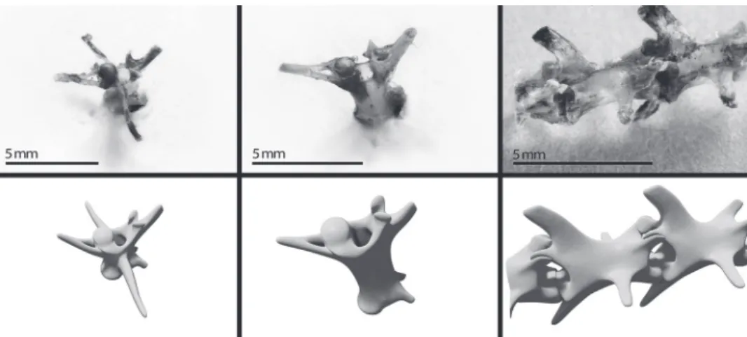
To serve as reference for the 3D modeling we used dissected full tails and autotomized caudal portions of T. itambere, all from the herpetological collection of the Federal University of Juiz de Fora (UFJF). Because the structures were so small and fragile, we analyzed them through microscopic observation, and compiled a database of high
definition macro photographs from a multitude of
different angles to use as guidance in the modeling process (Figure 1). The process did not destroy or
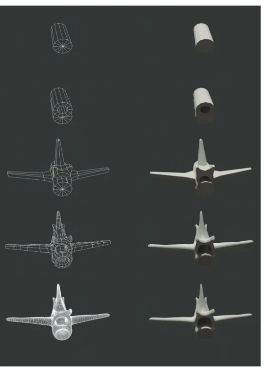
Figure 2 - Sequence of progress in the modeling process. The column on the left presents the polygonal grid visualization and the column on the right presents the solid view.
permanently damage the biological material, nor did it require any specialized laboratory equipment or rigorous procedures, as many of the methods which are usually employed to create digital models, do.
This model was created using the software Autodesk Maya 2012 for the initial structural
3D modelling is an artistic task, and therefore requires some degree of artistic skills combined with a minimum level of familiarity with one of a range of commercially available rendering
softwares. Scientific drawing also requires specific
tools and skill, and takes about the same amount of time and effort. Furthermore, digital modeling can
also be outsourced, as often occurs with scientific drawing, seeing that this is now an expanding field.
Therefore, using 3D models as illustration for papers is just a matter of changing the approach, and can be very advantageous for depicting detailed or complex structures.
CONCLUSIONS
Our digital model was initially meant to be rendered for printed illustration in a master’s degree thesis on ecological and morphological features involved in the intravertebral autotomy and posterior tail regeneration (Figure 3). The aim was to create a model simple enough to be easily understood without jeopardizing informative quality, to serve as illustration and to be presented to undergraduate students in classes focused on caudal anatomy and autotomy.
However, we did not anticipate the astonishing success of the images in facilitating the learning
Figure 3 - Original illustration rendering, showing the autotomy’s osteological peculiarities of Tropidurus itambere. A: Intravertebral fracture planes; B: The occurrence of autotomy in a vertebrae; C: Four stages of cartilage regeneration; and D: Fully regenerated tail with cartilaginous stick replacing the vertebrae.
process of virtually every student who was exposed to the images. That drew our attention to a window
of possibilities that simplified virtual models present both in science education and in scientific
publications, which are currently being overlooked within most of the biological areas.
With the methodology proposed here, the
process of creating a 3D model can be just as easy as
the process of creating any other scientific illustration,
and the results obtained by volumetric visualization, even when printed, are much more positive in terms of general comprehension of the forms presented than those of regular drawings. Therefore we strongly
encourage a more prominent use of simplified
three-dimensional models in articles, text books and any
ACKNOWLEDGMENTS
We sincerely thank the digital artists at SixHeads
Studios, and especially Alexandre Vieira for his patience and perfectionism in teaching and conducting
the three-dimensional modeling. We are grateful as well
to Tiago Cotrim for his excellence in photographing and post-editing of both the images here presented
and the photographs used as reference. We also thank
the Zoology Laboratory of Universidade Federal de Juiz de Fora (UFJF) for providing a great working
environment. The first author thanks Coordenação
de Aperfeiçoamento de Pessoal de Nível Superior (CAPES) for MSc scholarship and the last author thanks Conselho Nacional de Desenvolvimento
Científico e Tecnológico (CNPq) – Brazil, for the
productivity scholarship.
RESUMO
A autotomia caudal é um mecanismo anti-predação complexo e amplamente utilizado por lagartos, que envolve diversas adaptações anatômicas. Por seu tamanho diminuto e estruturas intrincadas, é difícil explicar claramente a anatomia vertebral para estudantes e pesquisadores de outras áreas. Modelos tridimensionais são ferramentas prodigiosas no esclarecimento de nuances anatômicas. Algumas das técnicas usadas para criá-los produzem formas complicadas e irregulares, que ainda que primem pela exatidão, não apresentam uniformidade e simplicidade. Como ambas são consideradas características fundamentais para a
compreensão, um modelo simplificado poderia ser a
chave para melhorar a aprendizagem. O modelo aqui apresentado ilustra a osteologia caudal de Tropidurus itambere, e foi desenvolvido para ser conciso, de maneira a ser facilmente compreendido, mas completo, não comprometendo o caráter informativo. O processo de criação requer apenas habilidades básicas de manipulação de polígonos em softwares de modelagem 3D, além do conhecimento apropriado da estrutura a ser modelada. Como referência para a modelagem usamos
observações microscópica e uma base de fotografias das
estruturas caudais. Assim, não se fez necessário ou uso de nenhum equipamento laboratorial avançado, e todos os materiais biológicos foram preservados para futuras pesquisas. Portanto, propõe-se maior utilização de
modelos 3D simplificados tanto em sala de aula quanto como ilustrações em publicações científicas.
Palavras-chave: anatomia, modelagem 3D, ensino, aprendizado, Tropidurus itambere.
REFERENCES
ADAMS CM AND WILSON TD. 2011. Virtual cerebral ventricular system: An MR-based three-dimensional computer model. Anat Sci Educ 4: 340-347.
ALIBARDI L. 2010. Regeneration in Reptiles and Its Position Among Vertebrates. In: Morphological and Cellular Aspects of Tail and Limb Regeneration in Lizards. Springer Berlin Heidelberg 207: 1-49.
ANASTASI G, BRAMANTI P, DI BELLA P, FAVALORO A,
TRIMARCHI F, MAGAUDDA L, GAETA M, SCRIBANO E,
BRUSCHETTA D AND MILARDI D. 2007. Volume rendering based on magnetic resonance imaging: advances in understanding the three-dimensional anatomy of the human knee. J Anat 211:399-406.
APPELHOF H, LANDMAN R AND VLIEGEN S. 2008. The effect of virtual anatomical learning. Available in: <http:// portfolio.io.utwente.nl/student/appelhofh/pdf/ALV_ virtuallearning.pdf> Web. 17 july 2013.
ARNOLD EN. 1984. Evolutionary aspects of tail shedding in lizards and their relatives. J Nat Hist 1: 127-169.
BARBER LW. 1944. Correlations between wound healing and regeneration in fore-limbs and tails of lizards. Anat Rec 89: 441-453.
BATEMAN PW AND FLEMING PA. 2009. To cut a long tail short: a review of lizard caudal autotomy studies carried out over the last 20 years. J Zool, London 277: 1-14.
BELLAIRS DA AND BRYANT SV. 1985. Autotomy and regeneration in reptiles. In: Gans C and Billet F (Eds), Biology of the reptilia, J Wiley & Sons, New York, USA, p. 301-410.
COOPER JR WE. 2003. Effect of risk on aspects of escape behavior by a lizard, Holbrookia propinqua, in relation to optimal escape theory. Ethology 109: 617-626.
EL-KHALILI N. 2005. 3D web-based anatomy computer-aided learning tools. Int Arab J Info Tech 2: 248-252.
ETHERIDGE R. 1967. Lizard Caudal Vertebrae. Copeia 4: 699-721. GARG AX, NORMAN G AND SPEROTABLE L. 2001. How medical
students learn spatial anatomy. Lancet 357: 363-364. HOYEK EN, COLLET C, GUILLOT A, THIRIET P AND SYLVESTRE
HUGHES A AND NEW D. 1959. Tail regeneration in the gecknonid lizard, Sphaerodactylus. J Ernbryol Exp Morph 7: 281-302.
JOHNSON PT, FISHMAN EK, DUCKWALL JR, CALHOUN PS AND
HEATH DG. 1998. Interactive three-dimensional volume rendering of spiral CT data: current applications in the thorax. Radiographics 18: 165-87.
KAUFMANN AE. 2000. Volume visualization in Medicine. In: Bankman IR (Ed), Handbook of Medical Imaging. Academic Press, San Diego, USA, p. 713-730.
KERWIN T, HITTLE B, SHEN HW, STREDNEY D AND WIET G.
2010. Anatomical Volume Visualization with Weighted
Distance Fields. VCBM 2010: 117-124.
KUHN PG, GRUBER RM AND RÜHLI F. 2008. Three-dimensional evaluation of structures in small bones by Micro- CT: tail fracture planes of autotomizing lizards (Scincidae and Gecconidae families). (The Internet Journal of Biological Anthropology).
LEE H, KIM J, CHO Y, KIM M, KIM N AND LEE K. 2010. Three-dimensional computed tomographic volume rendering imaging as a teaching tool in veterinary radiology instruction. Vet Med Czech 55: 603-609.
LU J, LI L AND SUN GP. 2010. A Multimodal Virtual Anatomy E-Learning Tool for Medical Education. In: Entertainment for Education. Digital Techniques and Systems, p. 278-287. MAGINNIS TL. 2006. Costs of regeneration and autotomy in
animals: a review and framework for future research. Behav Ecol 17: 857-872.
MCLACHLAN JC AND DE BERE SR. 2004. How we teach anatomy without cadavers. Clin Teach 1: 49-52.
MILLER R. 2000. Approaches to learning spatial relationships in gross anatomy: perspectives from wider principles of leaning. Clin Anat 13: 439-443.
MORENO AG ET AL. 2012. Application of 2D and 3D models for teaching of natural sciences. In: Proceedings of INTED2012 Conference, Valencia, Spain.
NUNES JV, ELISEI T, LOPES JFS, GOMIDES SC AND SOUSA BM.
2007. Aspectos da ecologia termal do lagarto Tropidurus itambere Rodrigues, 1987 (Squamata: Tropiduridae) no Parque Estadual do Ibitipoca, Minas Gerais (dados preliminares). In: VIII Congresso de Ecologia do Brasil, 2007, Caxambu, Brasil.
OAKLEY J. 2012. Science teachers and the dissection debate: Perspectives on animal dissection and alternatives. Int J Environ Sci Ed 7: 253-267.
PAYNE SL. 2012. Angiogenesis during Multi-Tissue Regeneration Following Tail Loss in the Leopard Gecko (Eublepharis macularius). Master’s thesis - The University of Guelph. (Unpublished).
PEAT M AND TAYLOR C. 2005. Virtual biology: how well can it replace authentic activities? CAL-laborate 13: 21-24. QUINN JG, KING K, ROBERTS D, CAREY L AND MOUSLEY A.
2009. Computer based learning packages have a role, but care needs to be given as to when they are delivered. (Bioscience Educacion Eletronic Journal).
RIBAUPIERRE S AND WILSON TD. 2012. Construction of a 3-D anatomical model for teaching temporal lobectomy. Comput Biol Med 42: 692-696.
RITZMAN TB, STROIK LK, JULIK E, HUTCHINS ED, LASKU E,
DENARDO DF, WILSON-RAWLS J, RAWLS JA, KUSUMI
K AND FISHER ER. 2012. The gross anatomy of the original and regenerated tail in the green anole (Anolis carolinensis). Anat Rec 295: 1596-1608.
RODRIGUES MT. 1987. Sistemática, ecologia e zoogeografia
dos Tropidurus do Grupo Torquatus ao sul do rio Amazonas (Sauria, Iguanidae). Arq Zool 3:105-230. RODRIGUES MT. 1988. Distribution of lizards of the genus
Tropidurus in Brazil (Sauria, Iguanidae), In: Vanzolini PE and Heyer WR (Eds), Proceedings of a Workshop on Neotropical Distribution, Academia Brasileira de Ciências, Rio de Janeiro, p. 413-425.
RUISOTO P, JUANES JA, MAYORAL P AND PRATS-GALINO A.
2012. Experimental evidence for improved neuroimaging interpretation using three-dimensional graphic models. Anat Sci Educ 5(3): 132-137.
SANGGAARD KW ET AL. 2012. Unique structural features facilitate lizard tail autotomy. PLoS ONE 7: e51803.
VAN SLUYS M. 2000. Population dynamics of the saxicolous lizard Tropidurus itambere (Tropiduridae) in a seasonal habitat of southeastern Brazil. Herpetologica 1: 55-62. VAN SLUYS M, VRCIBRADIC D AND ROCHA CFD. 2002. Tail loss
in the syntopic lizards Tropidurus itambere (Tropiduridae) and Mabuya frenata (Scincidae) in southeastern Brazil. Stud. Neotrop. Fauna Environ 37: 227-231.
WOODLAND WNF. 1920. Some observations on caudal autotomy and regeneration in the gecko (Hemidactylus
flaviviridis Rüppel), with notes on the tail of Sphenodon