Vol.49, n. 4 : pp. 605-609, July 2006
ISSN 1516-8913 Printed in Brazil BRAZILIAN ARCHIVES OF
BIOLOGY AND TECHNOLOGY
A N I N T E R N A T I O N A L J O U R N A L
Production of Specific Polyclonal Antibodies anti-Human
Factor IX
Daniel Gonçalves Chaves, Cibele Velloso Rodrigues*, Wanderley Almeida Ferreira, Pollyana Fantini Miranda Siqueira, Clara Guerra Duarte, Anna Bárbara de Freitas Carneiro-Proietti and Marcelo Matos Santoro
Fundação Centro de Hematologia e Hemoterapia de Minas Gerais - HEMOMINAS; Laboratório de Biotecnologia, Serviço de Pesquisa; Alameda Ezequiel Dias, 321; Santa Efigênia; 30130-110; Belo Horizonte - MG - Brasil
ABSTRACT
In this work, polyclonal antibodies anti-human Factor IX were produced in New Zealand rabbits by immunization with commercial pure human FIX (hFIX) (Octanyne®, Octapharma, USA). The serum containing immunoglobulins anti-hFIX was useful to detect hFIX antigen in human plasma fractions submitted to anionic exchange chromatographic process and with a large yield. Immunoassays (ELISA) using bovine serum albumin, trypsin and peptides generated by cleavage assays with trypsin as digestion enzyme was performed and revealed adequate specificity of the polyclonal antibodies produced.
Key words: Factor IX, hemophilia B, HPLC, purification, antibodies, ELISA
*
Author for correspondence
INTRODUCTION
The human blood coagulation factor IX (hFIX) is an essential glycoprotein for blood coagulation. The molecular weight of this protein is between 55.000 and 65.000 Da and its concentration in human plasma is about 5µg/mL (0.1µmol) in healthy individuals (Hoffer et al, 1999). The absence or a significant deficiency of hFIX causes hemophilia B, an X-linked recessive disorder of blood coagulation, also known as Christmas disease. The treatment of affected individuals consists of periodic intravenous infusions of purified hFIX concentrates.
The Brazilian blood center network generates a considerable volume of plasma of good quality (over 200 thousand liters per year) (Tanaka et al, 2002). This could be used for production of hFIX and other blood derivatives, reducing treatment
costs and providing autonomy to Brazil in the treatment of hemophilia and other disorders depending on plasma protein replacement. To accomplish this aim, in an industrial setting, it is
necessary to develop plasma fractionation
MATERIAL AND METHODS
Immunization
Two male New Zealand rabbits were immunized
with pure hFIX (Octanyne® 500, Human
Concentrate Factor IX for intravenous use, USA). The immunization plan (dose/day of protein load applied) was: (1) day “0” - 50µg; (2) 27th day - 100µg; (3) 41st day - 150µg; (4) 48th day - 200µg. The hFIX concentration injected in the rabbits was adjusted in PBS buffer 0.15M, 0.3M Al(OH)3 and pH 7.2. Aluminum hydroxide was used as adjuvant. The subcutaneous application of 500µL total solution containing hFIX was distributed in four dorsal points in the animals.
Three bleedings of each animal were performed along the experiment. The first on day 0 and the second on 41st day for antibodies titers checking, both from the central vessel of the ear. The last bleeding on 55th day was done utilizing cardiac punction. After collection, blood was immediately centrifuged at 2,500 rpm for 15 minutes at room temperature. The serum was stored at -30ºC.
Serum titers by ELISA
Calcium chloride was added in the solutions used in ELISA tests for recognition of activated hFIX by the antibodies, except in the coating buffer, as reported by Chang et al, (2003). The serum reactivity was revealed using a sheep anti-rabbit IgG conjugate marked with peroxidase, kindly provided by Olortegui, C.C. Titer curves of the collected serum were performed on 41st and 55th days to confirm the antibody production and determine the dilution to be used in the subsequent ELISA tests. Pure hFIX Octanyne (250ng) was coated in the wells of the plate and serial dilutions of the serum were analyzed.
Polyclonal antibodies specificity
ELISA tests were done by adding bovine serum albumin (BSA) or trypsin in the wells to verify the polyclonal antibodies specificity. Enzymatic digestions assays of hFIX (Octanyne®) were done using trypsin (Macarrone et al, 2001). The enzymatic digestion was performed in five defined times of 1, 5, 20 and 60 minutes plus 24. The reaction was stopped by adding 60% acetic acid solution stopped the reaction. The peptide pools
plates for ELISA test in the same conditions used for the undigested hFIX.
In addiction to these specificity tests with unrelated proteins, the polyclonal antibody produced was also tested in immunoassays for the recognition of partially purified hFIX from human plasma. Samples of human plasma poor for cryoprecipitate (PEX) (49mL) were divided into fractions by anionic exchange chromatography
using Sepharose DEAE FF resin (Pharmacia
Biotech, Sweden). In this process, the condition were: flux of 10mL/min in a 520mL column, equilibrium with 1.5 column volume (CV), unbound material elution with 1CV (citrate buffer 10µM, Gly 10µM, Lys 10µM, CaCl2 1µM, pH 7.0), salt linear gradient (0 to 1.0M NaCl) established with 1.5CV, washing with 1.0M NaCl solution (1 CV) and the column regeneration with citrate buffer (1 CV). To make the salt gradient citrate buffer 1.0M NaCl was used. The collected protein peaks were tested with ELISA assays using the polyclonal antibodies anti-hFIX previously produced.
Protein content of the collected samples was assayed using the Bradford method (Bradford, 1976) with BSA as a reference standard.
RESULTS AND DISCUSSION
Serum titers by ELISA
The serum titration graph is shown in Fig. 1. Observing the data obtained, we could establish satisfactory serum dilutions for all the realized ELISA tests in the standardization and in subsequent tests. For the serum collected after the second immunization, the chosen dilution was 1:16. The ideal dilution for the serum was 1:500 when it was collected after the 4th immunization.
Polyclonal antibodies specificity
Figure 1 - Serum titer of rabbits immunized with hFIX. Bars represent the medium obtained in duplicated assays and the vertical lines represent standard error. The solid bars represent the serum collected in 41st day and the spotted one represents the serum collected in 55th day.
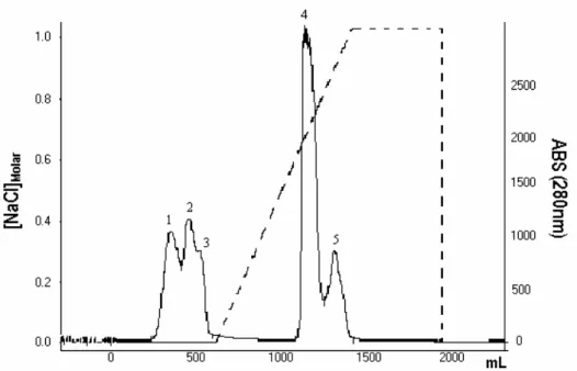
Figure 2 - Chromatographic profile obtained when PEX is applied in an anionic exchange
resin (Sepharose DEAE FF). The continuous line represents the 280nm absorbance and the dashed line represents the salt gradient. Five peaks are observed. Only peak number five is significantly recognized by the anti-hFIX antibodies.
0 0,2 0,4
1
:1
1
:2
1
:4
1
:8
1
:1
6
1
:3
2
1
:1
2
8
1
:2
5
6
1
:5
0
2
1
:1
0
0
4
1
:2
0
0
8
1
:4
0
1
6
1
:8
0
3
2
Serum dilutions
A
B
S
(
4
9
0
n
m
0,00 0,15 0,30
1 2 3 4 5 6
Peaks
A
B
S
(
4
9
0
n
m
)
Figure 3 - ELISA to verify the presence of hFIX in collected fractions in the chromatography
of PEX. The bars represent means obtained in duplicate assays and vertical lines represent the standard errors. Bar number 6 denotes the recognition of the anti-hFIX antibodies of pure anti-hFIX (positive control).
The immunoglobulins presented a significantly lower recognition of the peptides generated in the hFIX digestion by trypsin when compared to the pure commercial hFIX (Octanyne®). They were 68% lower with one-minute reaction and 88% lower with one-hour reaction. In the 24 digestion reaction there, was no reactivity.
As shown in Fig. 2, in the anionic exchange chromatography of the human plasma poor in cryoprecipitate, five protein peaks were obtained. Among these, only one was significantly recognized by the anti-hFIX antibodies in ELISA tests (Fig. 3). The low recognition of the other fractions by the anti-hFIX antibodies could be
caused by its premature elution in the
chromatographic process used, causing a
diminutive loss. The premature elution of hFIX could be caused by its high aggregation capacity with other plasmatic proteins, due to a Gla domain of this glycoprotein, which favored this type of interaction (Blostein et al, 2003).
These results showed that the polyclonal antibodies anti-hFIX could be readily produced in New Zealand rabbits and presented a good specificity in ELISA tests. Other immunoassays such as Western Blot could also be used to test for hFIX purification, as reported by Iberer et al
showing the specificity of the produced antibodies, which was reinforced by the control assay with BSA or trypsin. The polyclonal antibodies produced in rabbits presented a good specificity to human FIX as demonstrated in other works using primates, dogs and rats (Tomokiyo et al, 2001). In conclusion, it was possible to produce polyclonal antibodies of good specificity in New Zealand rabbits, useful for the monitoring of hFIX purification by chromatographic methods. There could be other uses of these antibodies for clinical laboratory tests, for example, in the diagnosis of FIX deficiency.
ACKNOWLEDGEMENTS
The authors would like to thank the technicians of the Laboratório de Hematologia of Fundação Hemominas, Ana Celi de Carvalho and the technicians of the Laboratório de Enzimologia e Físico-Química de Proteínas, UFMG. This work was supported by Finep and FAPEMIG.
comercial puro (Octanyne®, Octapharma, EUA). O soro contendo as imunoglobulinas anti-hFIX foi útil para a detecção do antígeno hFIX em frações do plasma humano submetido a cromatografia de troca iônica. Imunoensaios (ELISA) usando soro-albumina bovina, tripsina e peptídeos gerados por ensaios de clivagem com tripsina com enzima de
digestão foram realizados e revelaram
especificidade adequada dos anticorpos policlonais produzidos.
REFERENCES
Blostein, M. D.; Furie, B. C.; Rajotte, I. and Furie, B. (2003), The Gla domain of factor IXa binds to factor VIIIa in the tenase complex. J. Biol. Chem., 278 : (33), 31297-31.
Bradford, M. M. (1976), A rapid and sensitive method for the quantification of microgram quantities of protein utilizing the principle of protein-dye biding.
Anal Biochem, 72, 248.
Branovic, K.; Buchacher, A.; Barut, M.; Strancar, A. and Djuro Josic (2003), Application of semi-industrial monolithic columns for downstream processing of clotting factor IX. J. Chromat B, 790, 175-182.
Chang, Y.; Wu, H.; Hsu, Y.; Hamaguchi, N.; Shi, G.; Shen, M. and Lin, S. (2003), Discontinuous residues of factor IX constitute a surface for binding the anti-factor IX monoclonal antibody A-5. Thromb Res, 111, 293-299.
Hoffer, L.; Schwinn, H. and Djuro Josic (1999), Production of highly purified clotting factor IX by a combination of different chromatographic methods. J Chromatography A, 844, 119-128.
Iberer, I.; Schwinn, H.; Djuro Josic; Jungbauer, A. and Buchacher, A. (2002), Continuous purification of a clotting factor IX concentrate and continuous regeneration by preparative annular chromatography.
J Chromatogr A, 972, 115-129.
Macarrone, M.; Salucci, M. L.; Zadelhoff, G.; Malatesta, C.; Veldink, G.; Vliegenthart, J. F. G. and Finanzzi-Agro, A. (2001), Tryptic digestion of soybean lipoxygenase-1 generates a 60 kDa fragment with improved activity and membrane binding ability.
Biochemistry, 40, 6819-6827.
Ono, K. and Smith, K. J. (1984), Monoclonal antibodies to factor IX: characterization and use in immunoassays for factor IX. Thromb Res, 33 : (2), 211-224.
Tanaka, K.; Sawatani, E.; Arashiro, F.; Andrade Dias, G.; Shigueoka, E. M. and Fujita, C. S. (2002), Fracionamento de plasma humano pelo método de cromatografia líquida. LAES and HAES, 137, 98-114. Tomokiyo, K.; Teshima, K.; Nakatomi, Y.; Watanabe,
T.; Mizuguchi, J.; Nozaki, C.; Nakagaki, T.; Miyamoto, S.; Funatsu, A. and Iwanaga, S. (2001), Induction of acquired factor IX inhibitors in cynomolgus monkey (Macaca fascicularis): a new primate model of hemophilia B. Thrombosis Research, 102, 363-374.