Vol. 51, Special Number : pp. 121-126, December 2008
ISSN 1516-8913 Printed in Brazil BRAZILIAN ARCHIVES OF
BIOLOGY AND TECHNOLOGY
A N I N T E R N A T I O N A L J O U R N A L
Alkaline Gel Electrophoresis Assay to Detect DNA Strand
Breaks and Repair Mechanisms in
Escherichia coli
José Carlos Pelielo de Mattos
*, Ellen Serri da Motta, Márcia Betania Nunes de Oliveira,
Flávio José da Silva Dantas and Adriano Caldeira de Araujo
Laboratório de Radio e Fotobiologia; Departamento de Biofísica e Biometria; Universidade do Estado do Rio de Janeiro; Av. 28 de Setembro, 87; 20551030; jcmattos@uerj.br; Rio de Janeiro - RJ - Brasil
ABSTRACT
Reactive oxygen species (ROS) can induce lesions in different cellular targets, including DNA. Stannous chloride (SnCl2) is a ROS generator, leading to lethality in Escherichia coli (E. coli), with the base excision repair (BER)
mechanism playing a role in this process. Many techniques have been developed to detect genotoxicity, as comet assay, in eukaryotic cells, and plasmid DNA agarose gel electrophoresis. In this study, an adaptation of the alkaline gel electrophoresis method was carried out to ascertain the induction of strand breaks by SnCl2 in bacterial DNA,
from E. coli BER mutants, and its repair pathway. Results obtained show that SnCl2 was able to induce DNA strand
breaks in all strains tested. Moreover, endonuclease IV and exonuclease III play a role in DNA repair. On the whole, data has shown that the alkaline gel electrophoresis assay could be used both for studying DNA strand breaks induction and for associated repair mechanisms.
Keywords: Alkaline gel electrophoresis, Escherichia coli, base excision repair, stannous chloride, DNA strand breaks, reactive oxygen species
*
Author for correspondence
INTRODUCTION
Several studies have pointed out that reactive oxygen species (ROS) can generate lesions in many cellular structures, including DNA (Marnett, 2000; Cooke et al., 2003; Halliwell and Whiteman, 2004). Among the ROS, hydroxyl radical (OH•) has been described as a powerful oxidant, able to react with any molecule (Friedberg et al., 2006). Some authors have suggested that transition metals (Asad and Leitão, 1991; Kehrer, 2000), such as Fe(II), take part, as reducing agents, in OH•
formation through a Fenton reaction, a
phenomenon also observed, in a Fenton-like reaction, with Cu(I) ions. Dantas and collaborators
(1996) have described that stannous chloride (SnCl2)is also able to produce ROS in a
Fenton-like reaction. In the same article, the authors, by using ROS scavengers and antioxidant enzymes, have concluded that SnCl2 induces lethality in
Escherichia coli (E. coli), through ROS
generation.
technique was developed by Ostling and Johanson (1984) and Singh et al. (1988). It is a simple method for measuring DNA strand breaks in eukaryotic cells. Briefly, cells embedded in agarose on a microscope slide are lysed with detergent and high salt to form nucleoids containing supercoiled loops of DNA linked to the nuclear matrix. Electrophoresis at high pH results in structures resembling comets, observed by fluorescence microscopy; the intensity of the comet tail relative to the head reflects the number of DNA breaks. The likely basis for this is that loops containing a break lose their supercoiling and become free to extend toward the anode. The sensitivity and specificity of the assay could be greatly enhanced if the nucleoids are incubated with bacterial repair endonucleases that recognize specific kinds of damage in the DNA and convert lesions to DNA breaks, increasing the amount of DNA in the comet tail. On the other hand, DNA repair can be monitored by incubating cells after treatment with damaging agent and measuring the damage remaining at intervals (Tice et al., 2000). Relative to other genotoxicity tests, the advantages of the comet assay include its demonstrated sensitivity for detecting low levels of DNA damage, the requirement for small numbers of cells per sample, its flexibility, its low costs, its ease of application, and the short time needed to complete a study.
Agarose gel electrophoresis could also be used to observe genotoxic lesions, in vitro, through plasmid topology alteration. Plasmids are circular minichromosomes found in bacteria and in a number other organisms. The protocol assay is based on conformational differences between plasmid molecules. The supercoiled form (or form I) can be maintained only if both polynucleotide strands are intact, providing for a more compact topology that migrates faster in the gel. If one of the double strands is broken, the plasmid will take on another conformation, called open circle (or form II), and will display a slower migration
pattern on agarose gel electrophoresis.
Furthermore, the linear conformation (or form III) could appear if breaks occur in the same position on both strands, also leading to a variation in migration through agarose gel, which is slower than the supercoiled form and faster than the open circle form. The test is based on the plasmid
incubation with a known break-inducing agent such as SnCl2 as a positive control. Then, tested
products (artificial or natural) can be used alone or in combination (De Mattos et al., 2004).
Lesions leading to cell inactivation in prokaryotes could be, for instance, alkali-labile sites and strand breaks, which can be detected by alkaline sucrose gradient (MacGrath and Williams, 1966). This experimental procedure follows many detailed steps and requires special care and supplies, including radionuclide manipulation. Therefore, we have proposed, in the present paper, a faster and easier methodology, named bacterial alkaline gel electrophoresis, adapted from Zirkle and Krieg (1996). This assay allows visualizing not only strand breaks and alkali-labile sites, but also following DNA lesions repair kinetics. Thus, we decided to use this procedure, in order to investigate genotoxicity and repair activity, using SnCl2, as positive control, and E. coli base
excision repair.
Stannous chloride (SnCl2) is used in different areas
of human daily life, for example in nuclear medicine as a reducing agent of technetium-99m, with the aim to radiolabel pharmaceutical products (Saha, 1998; Santos-Filho et al., 2007). The SnCl2
biological effects have been studied by many authors in the last decade (Caldeira-de-Araujo et al., 1996; Cabral et al., 1998; Dantas et al., 1999; De Mattos et al., 2000; De Mattos et al., 2005).
MATERIALS AND METHODS
Chemicals
Stannous chloride (SnCl2.2H2O) and sodium
dodecyl sulfate (SDS) were acquired from Sigma Chemical Co. (USA). Sodium chloride and ethylene diaminetetracetic acid (EDTA) were obtained from Lafan (Brazil). Sodium hydroxide (NaOH) and Tris (hydroxymethyl) amino-methane were obtained from Nuclear (Brazil). Normal point agarose (NPA), low-melting point agarose (LMPA) and ethidium bromide were acquired from Invitrogen (Brazil).
Bacterial strains
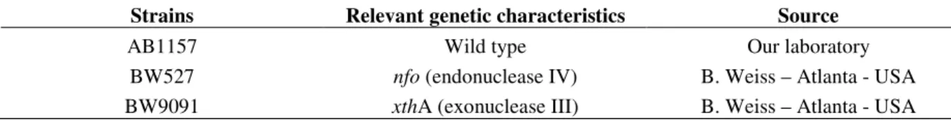
Table 1 -Escherichia coli K12 strains and their relevant genetic characteristics for this study.
Strains Relevant genetic characteristics Source
AB1157 Wild type Our laboratory
BW527 nfo (endonuclease IV) B. Weiss – Atlanta - USA
BW9091 xthA (exonuclease III) B. Weiss – Atlanta - USA
Growth medium
Bacterial cells were grown overnight at 37ºC, with shaking in LB medium (Luria and Burrous, 1957). A starting culture for experiments was taken from overnight samples, and the cells were grown in the same medium up to exponential phase (1-2 x 10⁸ cell/ml), harvested by centrifugation (2,940 xg; 15 min; 4ºC; washed twice) and suspended in 0.9 % NaCl.
Alkaline gel electrophoresis
Alkaline gel electrophoresis was done as described by Zirkle and Krieg (1996), with modifications. All E. coli cell cultures assayed, at exponential growth phase, were separated in two samples, each one containing 1.5 ml, with exception to AB1157 cells, whose samples contained 6 ml, on account of the repair kinetics study. One fraction, control, was incubated with NaCl (0.9 %) at 37ºC, by shaking, for 40 min; the second one was incubated with SnCl2 (25 µg/ml), under the same conditions.
After this period, 1.5 ml from each fraction was centrifuged (1 min; 12,000 xg; room temperature) and suspended in 10 l of cold TE (50 mM Tris•Cl pH 8.0; 5 mM EDTA pH 8.0). Following, 50 l of low melting point agarose (1.5 %, in TE), at 37ºC, were added to each 10 l sample. The mixture (60 l) was placed in an acrylic container holding small squared wells and incubated, at room temperature, for 30 min to form 0.5 cm2 squared blocks. Following gellification, the blocks were incubated for 2 h, at room temperature, in the dark, in a lysing solution (0.1 mM EDTA pH 8.0; 0.5 M NaOH; 0.05 % SDS). Then, the blocks were washed three times (5 min each) in cold TE (1 ml) and placed onto the teeth combs of an electrophoresis gel apparatus (Horizon 58 “GIBCO BRL - Life technologies” - EUA). TE excess was removed with filter paper to facilitate block adherence to the comb.
Normal agarose solution (0.76 %) was prepared in alkaline buffer (0.03 mM NaOH; 0.01 mM EDTA pH 8.0) and heated for complete homogenization. After cooling, the solution was deposited in the gel electrophoresis chamber and the comb, containing
the blocks, was fitted. Following gellification, the comb was removed, the blocks remaining in the gel, which was submitted to 7 V for 14 h. Then, this was neutralized, for 1 h, by soaking in a solution containing 30 mM NaCl and 50 mM Tris•Cl pH 6.0. Following this step, the gel was stained in ethidium bromide solution (0.5 g/ml), for 10 min, visualized in a transilluminator UVP (Model TM-10E-EUA), and then digitalized using Canon Power Shot S2IS. The image was analyzed using the software Image J, 1.33u version, in order to quantify the DNA strand breaks. The collected data were submitted to chi-square test (χ2), adopting 5 % as significance level.
To ascertain of SnCl2-induced lesion repair, after
40 min of incubation, the AB1157 residual fractions (control and treated, 4.5 ml each one) were centrifuged (2,940 xg; 15 min; 4ºC), suspended in fresh LB medium and incubated with shaking for 90 min, at 37ºC. Aliquot (1.5 ml) from each sample was taken every 30 min. Blocks were performed and alkaline electrophoresis was carried out as described above.
RESULTS AND DISCUSSION
Previous studies pointed to that base excision repair (BER) genes are involved in cell restoration after oxidative stress (Friedberg et al., 2006). As presented elsewhere, SnCl2 is able to promote
different inactivation levels in E. coli base excision repair mutants (Dantas et al., 1996). Stannous chloride can induce ROS (Dantas et al., 1996), in addition to a direct effect, as previously described (De Mattos et al., 2000; De Mattos et al., 2005). So, in the present study, the participation of xthA (exonuclease III) and nfo (endonuclease IV) genes in the repair of SnCl2-induced lesions was
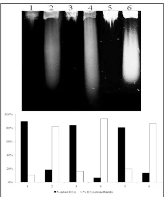
observed. Bacterial DNA was analyzed through alkaline gel electrophoresis; the results have shown that SnCl2 is able to induce DNA strand
Figure 1 - Alkaline gel electrophoresis of E. coli with different DNA repair capacities treated with 25 µg/ml SnCl2. Lanes: (1) AB1157; (2) AB1157 + SnCl2; (3) BW9091; (4) BW9091 +
SnCl2; (5) BW527; (6) BW527 + SnCl2.
Gel densitometrical analysis revealed that AB1157 was the strain displaying the lowest number of strand breaks, suggesting that this strain has an
advantage regarding other strains. The
performance of all base excision repair proteins would assure quick repair of SnCl2-induced
lesions, with few DNA strand breaks visualized in the gel.
BER mutant alkaline gel analysis has shown that the BW9091 strain exhibited a number of strand breaks higher than both strains wild type (p<0.0001) and BW527 mutant (p<0,0001). As demonstrated by Cabral and collaborators (1998), SnCl2 is able to generate 8-hydroxyguanine
lesions, and it is known that this lesion can be recognized and removed by a BER protein, called
formamidopyrimidine glycosylase (Fpg)
(Friedberg et al., 2006). This process generates apyrimidinic sites (Friedberg et al., 2006), which could be translated in DNA strand breaks through the present proposed methodology. Thus, these results suggest that SnCl2-induced lesions could be
recognized and removed by Fpg protein, but the repair process in the mutant strain would be slower, when compared with the wild type strain, because of the lack of other BER proteins.
According to these results, we could suggest that nfo gene product (endonuclease IV) seems to be more important than exonuclease III (xthA gene) in the repair of SnCl2- induced DNA lesions, since
BW527 was the strain which showed the lowest number of DNA strand breaks, when compared with all the others (p<0.0001). So, SnCl2 could be
inducing damage, recognized preferentialy by nfo gene product, as it occurs with the chemical agents
tert-butil hydroperoxide and bleomicin,
responsible for producing lesions not recognized by exonuclease III, but only by endonuclease IV (Friedberg et al., 2006). Experiments are carrying out, in order to confirm this hypothesis.
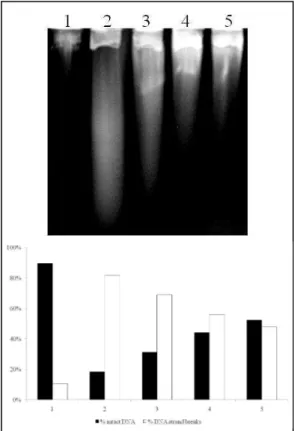
To demonstrate the occurrence of repair events, AB1157 strain aliquots, after SnCl2 treatment,
were submitted to incubation for 90 min, with the repair accompanied every 30 min (as described in material and methods) by a decrease of DNA strand break number (Fig. 2).
evident after 90 minutes (p<0.0001, when compared with all lanes - Fig. 2, lane 5). These results appear to be true, since AB1157 displays all the repair mechanisms active.
It is important to emphasize that the experimental approach used here seems to be appropriate to study DNA oxidative lesions and its repair in E.
coli.
Figure 2 - Repair of SnCl2-induced lesions in E. coli AB1157 by alkaline gel electrophoresis. The
culture was treated with 25 µg/ml of SnCl2, for 40 min. Afterwards, the culture was
centrifuged, suspended in LB medium and incubated for 90 min. Aliquots were collected after 40 min of treatment and following every 30 min of incubation time. Lanes: (1) control; (2) SnCl2 after 40 min of treatment; (3) 30 min of repair; (4) 60 min
of repair; (5) 90 min of repair.
Other experiments, using strains lacking one or more base excision repair enzymes, are being carried out in order to clarify the mechanism involved in SnCl2-induced lesions, as well as the
repair of the damage induced by this agent.
ACKNOWLEDGEMENTS
The authors are grateful to Carlos Brown Scavarda for English language support. This work received grants from CNPq, Capes and FAPERJ (E-26/171.332/2006 and E-26/171.162/2006) .
RESUMO
Espécies reativas de oxigênio (ERO) podem induzir lesões em diferentes alvos celulares, incluindo o DNA. O cloreto estanoso (SnCl2) é um
gerador de ERO que induz letalidade em E. coli, sendo o reparo por excisão de bases (BER) um mecanismo importante neste processo. Técnicas como o ensaio cometa (em eucariotos) e a eletroforese de DNA plasmidial em gel de agarose têm sido utilizadas para detectar genotoxicidade. No presente estudo, uma adaptação do método de eletroforese em gel alcalino de agarose foi usada para verificar a indução de quebras, pelo SnCl2, no
no DNA de todas as cepas testadas. Além disso, endonuclease IV e exonuclease III estão envolvidas na reparação dos danos. Em resumo, os dados obtidos indicam que a metodologia de eletroforese em gel alcalino de agarose pode ser empregada tanto para o estudo de quebras no DNA, quanto para avaliação dos mecanismos de reparação associados.
REFERENCES
Asad, N. R.; Leitão, A. C. (1991), Effects of metal ion chelators on DNA strand breaks and inactivation produced by hydrogen peroxide in Escherichia coli: detection of iron-independent lesions. J Bacteriol.,
173, 2562-2568.
Cabral, R. E. C.; Leitão, A. C.; Lage, C.; Caldeira-de-Araujo, A.; Bernardo-Filho, M.; Dantas, F. J. S.; Cabral-Neto, J. B. (1998), Mutational potentiality of stannous chloride: an important reducing agent in the Tc-99m-radiopharmaceuticals. Mutat Res., 408, 129-135.
Caldeira-de-Araujo, A.; Dantas, F. J. S.; Moraes, M. O.; Felzenszwalb, I.; Bernardo-Filho, M. (1996), Stannous chloride participates in the generation of reactive oxygen species. Free Radical Research in Latin America, 48, 109-113.
Cooke, M. S.; Evans, M. D.; Dizdaroglu, M.; Lunec, J. (2003), Oxidative DNA damage: mechanisms, mutation and disease. FASEB J., 17, 1195-1210. Dantas, F. J. S.; Moraes, M. O.; Carvalho, E. F.; Valsa,
J. O.; Bernardo-Filho, M.; Caldeira-de-Araujo, A. (1996), Lethality induced by stannous chloride on
Escherichia coli AB1157: participation of reactive oxygen species. Food Chem Toxicol., 34, 959-962. Dantas, F. J. S.; Moraes, M. O.; De Mattos, J. C. P.;
Bezerra, R. J. A. C.; Carvalho, E. F.; Bernardo-Filho, M.; Caldeira-de-Araujo, A. (1999), Stannous chloride mediates single strand breaks in plasmid DNA through reactive oxygen species formation. Toxicol Lett., 110, 129-136.
De Mattos, J. C. P.; Dantas, F. J. S.; Bezerra, R. J. A. C.; Bernardo-Filho, M.; Cabral-Neto, J. B.; Lage, C.; Leitão, A. C.; Caldeira-de-Araujo, A. (2000), Damage induced by stannous chloride in plasmid DNA. Toxicol Lett., 116, 159-163.
De Mattos, J. C. P.; Dantas, F. J. S.; Caldeira-de-Araujo, A.; Moraes, M. O. (2004), Agarose Gel Electrophoresis System in the Classroom. Biochem Mol Biol Education, 32, 254-257.
Mattos, J. C. P.; Lage, C.; Dantas, F. J. S.; Moraes, M. O.; Nunes, A. P. M.; Bezerra, R. J. A. C.; Faria, M. V. C.; Leitão, A. C.; Caldeira-de-Araujo, A. (2005), Interaction of stannous chloride leads to alteration in DNA, triphosphate nucleotides and isolated bases.
Mol Cell Biochem., 280, 173-179.
Friedberg, E. C.; Walker, G. C.; Sied, W.; Wood, R. D.; Schultz, R. A.; Ellenberger, T. (2006), DNA Repair and Mutagenesis. ASM Press, Washington.
Halliwell, B.; Whiteman, M. (2004), Measuring reactive species and oxidative damage in vivo and in cell culture: how should you do it and what do the results means? Br J Pharmacol., 142, 231-255. Kehrer, J. P. (2000), The Haber-Weiss reaction and
mechanisms of toxicity. Toxicology, 149, 43-50. Luria, S. E.; Burrous, J. W. (1957), Hybridization
between E. coli and Shigella. J. Bacteriol., 74, 461-476.
MacGrath, R. A.; Williams, R. (1966), Reconstruction in vivo of irradiated Escherichia coli
deoxyribonucleic acid, the rejoining of broken pieces.
Nature, 212, 532-535.
Marnett, L. J. (2000), Oxyradicals and DNA damage.
Carcinogenesis, 21(3), 361-370.
Ostling, O.; Johanson, K. J. (1984), Microelectrophoretic study of radiation-induced DNA damage in individual mammalian cells. Biochem Biophys Res Commun., 123, 291-298.
Saha, G. B. (1998), Fundamentals of Nuclear Pharmacy. Springer Verlag, New York.
Santos-Filho, S. D.; Fonseca, A. S.; Bernardo-Filho, M. (2007), The male reproductive system and the effect of an extract of a medicinal plant (Hypericum perforatum) on the labeling process of blood constituents with Technetium-99m. Braz Arch Biol Technol., 50, 97-104.
Singh, N. P.; McCoy, M. T.; Tice, R. R.; Schneider, E. L. (1988), A simple technique for quantification of low levels of DNA damage in individual cells. Exp Cell Res., 175, 184-191.
Tice, R. R.; Agurell, E.; Anderson, D.; Burlinson, B.; Hartmann, A.; Kobayashi, H.; Miyamae, Y.; Rojas, E.; Ryu, J. C.; Sasaki, Y. F. (2000), Single cell gel/comet assay: guidelines for in vitro and in vivo genetic toxicology testing. Environ Mol Mutagen.,
35, 206-221.
Zirkle, R. E.; Krieg, N. R. (1996), Development of a method based on alkaline gel electrophoresis for estimation of oxidative damage to DNA in
Escherichia coli. J Appl Bacteriol., 81, 133-138.