Estimates of DNA strand breakage in bottlenose dolphin (
Tursiops truncatus
)
leukocytes measured with the Comet and DNA diffusion assays
Adriana Díaz
1, Sandra Carro
1, Livia Santiago
1, Juan Estévez
1, Celia Guevara
2, Miriam Blanco
2,
Laima Sánchez
2, Liena Sánchez
2, Nirka López
2, Danilo Cruz
2, Ronar López
2, Elizabeth B. Cuetara
3and Jorge Luis Fuentes
41
Departamento de Radiobiología, Centro de Aplicaciones Tecnológicas y Desarrollo Nuclear,
C. Habana, Cuba.
2
Departamento de Salud Animal, Acuario Nacional de Cuba, C. Habana, Cuba.
3
Laboratorio de Farmacología Clínica y Experimental,
Instituto Nacional de Oncología y Radiobiología, C. Habana, Cuba.
4
Laboratorio de Microbiología y Mutagénesis Ambiental, Escuela de Biología,
Facultad de Ciencias, Universidad Industrial de Santander, Bucaramanga, Colombia.
Abstract
The analysis of DNA damage by mean of Comet or single cell gel electrophoresis (SCGE) assay has been commonly used to assess genotoxic impact in aquatic animals being able to detect exposure to low concentrations of contami-nants in a wide range of species. The aims of this work were 1) to evaluate the usefulness of the Comet to detect DNA strand breakage in dolphin leukocytes, 2) to use the DNA diffusion assay to determine the amount of DNA strand breakage associated with apoptosis or necrosis, and 3) to determine the proportion of DNA strand breakage that was unrelated to apoptosis and necrosis. Significant intra-individual variation was observed in all of the estimates of DNA damage. DNA strand breakage was overestimated because a considerable amount (~29%) of the DNA damage was derived from apoptosis and necrosis. The remaining DNA damage in dolphin leukocytes was caused by factors unre-lated to apoptosis and necrosis. These results indicate that the DNA diffusion assay is a complementary tool that can be used together with the Comet assay to assess DNA damage in bottlenose dolphins.
Key words:Comet assay, DNA diffusion assay, DNA strand breakage,Tursiops truncatus.
Received: May 30, 2008; Accepted: October 10, 2008.
Introduction
The single cell gel electrophoresis (SCGE) or Comet assay, as introduced by Singhet al.(1988), is a technique
that detects DNA strand breakage and alkali labile sites by measuring the migration of DNA from immobilized indi-vidual cell nuclei. In this assay, cells are embedded in agarose gel on microscopic slides and lysed and electro-phoresed under alkaline condition. Cells with damaged DNA show increased migration of DNA fragments from the nucleus. The length of the migration indicates the amount of DNA breakage, and the DNA damage can be es-timated by both manual microscopic and computerized im-age scoring analyses (Oliveet al., 1990; McKelvey-Martin et al., 1993; Fairbairnet al., 1995; Kobayashiet al., 1995).
The minimal technical requirements for conducting this as-say in human cellsin vitroandin vivohave been well
estab-lished (Ticeet al., 2000; Hartmannet al., 2003).
The widespread use of the Comet assay in biomoni-toring studies of aquatic organisms is related mainly to its simplicity, low cost and greater sensitivity to xenobiotics when compared with other techniques (Mitchelmore and Chipman, 1998; Cotelle and Ferard, 1999; Lee and Steinert, 2003; Frenzilliet al., 2009). The Comet assay has been
used to detect DNA damage induced by hydrogen peroxide and methyl mercury in bottlenose dolphin (Tursiops truncatus) lymphocytes in vitro (Betti and Nigro, 1996;
Taddei et al., 2001), and by hydrogen peroxide and
benzo[a]pyrene-7,8-dihydro-diol-9,10-epoxide in sea lion lymphocytes (El-Zeinet al., 2006). These results suggest
that this assay may be a sensitive tool for monitoring DNA damage in marine mammals.
However, the use of the Comet assay in biomoni-toring studies has been questioned because positive results in this assay do not necessarily reflect genotoxicity but may arise from DNA damage,i.e., double-strand breakage,
as-sociated with apoptosis and necrosis (Oliveet al., 1993;
Ol-ive and Banath, 1995; Steinert, 1996; Godardet al., 1999; www.sbg.org.br
Ticeet al., 2000; Wadaet al., 2003). To overcome this
limi-tation, the Comet assay may be used in combination with related methodologies that estimate DNA fragmentation associated with apoptosis and necrosis, such as the DNA diffusion assay (Singh, 2000a,b). In the latter assay, small molecular weight DNA fragments generated during apoptosis and necrosis diffuse in the agarose matrix to give an apparent nuclear diameter that is ~3 times greater than the mean nuclear size as a consequence of the high disper-sion of DNA. Based on the structural differences between apoptotic and necrotic nuclei, Singh (2000a,b) recom-mended that the DNA diffusion assay be used to distinguish apoptotic from necrotic cells. However, other investigators have been unable to differentiate between apoptotic and ne-crotic cells when using this assay (Gichneret al., 2005).
The use of the DNA diffusion assay to assess apoptosis has been reviewed by Singh (2005) and its application in a small number of environmental studies has yielded promis-ing results (Nigroet al., 2002; Frenzilliet al., 2004; Del
Bargaet al., 2006).
We have initiated a long-term monitoring project aimed at evaluating the life quality of bottlenose dolphins
living in semi-captive and captive conditions. In view of the potential use of the Comet assay in the biomonitoring of dolphins and that a concurrent assessment of apoptosis, ne-crosis and genotoxin-induced DNA strand breakage is criti-cal for the correction interpretation of this assay, the aims of this work were: 1) to evaluate the usefulness of the Comet or single cell gel electrophoresis (SCGE) assay to detect DNA strand breakage in dolphin leukocytes, 2) to use the DNA diffusion assay to determine the amount of DNA strand breakage associated with apoptosis and necrosis, and 3) to determine the proportion of DNA strand breakage that was unrelated to apoptosis and necrosis.
Materials and Methods
Capture and living conditions of the dolphins
Twenty-five bottlenose dolphins (Table 1) were cap-tured in the Sabana-Camagüey archipelago on the northern coast of Cuba and their sex was determined visually. Age was estimated based on the dolphins size, teething state and body marks at the time of capture. The dolphins were ini-tially quarantined in sea-hoops located close to the capture
Table 1- Life history data for the bottlenose dolphins used in this work.
N. Dolphin name Capture date Origin Sex1 Length1(m) Age group1,2
1 Ciceron 11/08/2000 Aguada key M 2.13 Juvenile
2 Serena 12/08/2000 Aguada key F 2.25 Adult
3 Salome 14/08/2000 Horseshoe key F 2.19 Juvenile 4 Jade 04/12/2001 San Juan Point F 2.08 Juvenile
5 Javy 04/12/2001 San Juan Point F 1.98 Calf
6 Lía 15/07/2002 Santa Maria key F 1.81 Calf
7 Lili 28/08/2002 Glad Point F 2.04 Juvenile
8 Xena 25/10/2002 Caibarien bay F 2.18 Juvenile 9 Maria 18/06/2003 Santa Maria key F 2.49 Adult 10 Mara 18/06/2003 Santa Maria key F 2.01 Juvenile 11 Merlin 18/06/2003 Santa Maria key M 2.12 Juvenile
12 Maida 22/06/2003 Glad Point F 2.40 Adult
13 Mihai 22/06/2003 Glad Point M 1.86 Calf
14 Musa 22/06/2003 Glad Point F 1.94 Calf
15 Marcelo 29/07/2003 Guarana key M 2.40 Adult 16 Malù 08/08/2003 Horseshoe key F 2.33 Adult 17 Monica 08/08/2003 Horseshoe key F 2.28 Adult 18 Margarita 09/08/2003 Guarana key F 2.07 Juvenile 19 Montse 02/09/2003 Guarana key F 2.00 Juvenile
20 Melany 03/09/2003 Drunk key F 2.28 Adult
21 Milo 08/09/2003 Guarana key F 2.28 Adult
22 Moon 08/09/2003 Guarana key F 2.19 Juvenile
23 Maja 08/09/2003 Guarana key F 1.93 Calf
24 Mègano 18/10/2003 Guarana key M 2.17 Juvenile 25 Milano 18/10/2003 Guarana key M 2.11 Juvenile
site where they were examined by a veterinarian and under-went blood and spiracle analyses. Only visually healthy and asymptomatic dolphins that willingly consumed frozen fish were transferred to the dolphinarium at the Cuban National Aquarium (CNA). The dolphinarium was supplied with water from a subterranean well via a semi-closed feeding system with filters and a water renewal rate of 10% per hour. This system was permanently and automatically sup-plemented with sodium hypochlorite (final concentration of free chloride: 0.3 mg/L). The quality of water used in the dolphinarium was based on the parameters established by the Cuban guidelines for fishery zones (NC25:1999).
Blood sampling
Blood samples were obtained by vacuum using ster-ile, heparinized tubes and were always shipped and stored on ice until assayed.
Analysis of serum genotoxicity
Serum genotoxicity was measured indirectly using the SOS Chromotest (Quillardetet al., 1982) with modifi-cations. Escherichia coli PQ37 cells were grown to an OD600nmof 0.4 in Luria-Bertani (LB) media supplemented
with ampicillin (50 mg/mL) at 37 °C, with shaking
(100 rpm). Exponential phase cultures were diluted ten-fold in fresh 2X LB media supplemented with ampicillin (100 mg/mL) and then dispensed in Eppendorf tubes
(250mL per tube) containing 225mL of serum. When
meta-bolic activation was required, 25mL of phenobarbital/5,6 benzoflavone-induced rat liver S9 fraction from Moltox was used (final concentration in the activation mixture: 0.4%, v/v). In experiments without metabolic activation, the rat liver S9 fraction was substituted with sterile distilled water. The reference mutagens 2-acethyl-aminofluorene (500mg/mL) andg-rays (150 Gy delivered by a Co60 PX-g-30M Russian irradiator at a dose rate of 33-42 Gy/min)
were used as positive controls in experiments with and without metabolic activation, respectively. The cells were exposed to serum samples for 30 min at 8 °C and then cul-tured for 2 h at 37 °C.b-galactosidase and alkaline phos-phatase activities were assayed as described by Fuenteset al., (2006). The criterion for genotoxicity was the SOS in-duction factor (SOSIF), as defined by Quillardet et al.
(1989): SOSIF = [b-galactosidase/alkaline
phosphata-se]treatment/[b-galactosidase/alkaline phosphatase]negative control.
Serum samples were classified as not genotoxic (SOSIF < 1.5), inconclusive (SOSIF = 1.5-2.0) or geno-toxic (SOSIF > 2.0) (Kevekordeset al., 1999).
Estimation of DNA strand breakage in dolphin leukocytes
DNA strand breakage in dolphin leukocytes was esti-mated by using the Comet assay, as described by Singhet al.(1988), with modifications to the silver staining as
indi-cated by Garciaet al.(2004). The DNA damage was scored
(see Figure 1) based on five categories (0-4), as indicated by Collinset al.(1997). The total amount of DNA strand
breakage was expressed in total arbitrary units (AUT) as
follows: AUT= N0x 0 + N1x 1 + N2x 2 + N3x 3 + N4x 4,
where Niis the number of nuclei scored in each category
(Collins, 2002). One hundred cells per slide and two slides per blood sample were analyzed and the results of at least two independent experiments were averaged to obtain the AUTfor each dolphin.
Since positive Comet results do not necessarily re-flect genotoxicity because DNA strand breakage may be associated with cellular apoptosis and necrosis, we used the DNA diffusion assay to determine the percentage of apop-totic/necrotic cells in each blood sample. For this assay, the cells were processed in a manner similar to the Comet as-say, except that the nuclei were not subjected to electropho-resis. Nuclei with a diameter > 3 times the mean nuclear diameter were considered apoptotic/necrotic (Nigroet al.,
2002). The total number of nuclei (minimum of 100 cells per slide) and the number of apoptotic/necrotic nuclei in each field were counted and the latter then expressed as a percentage of the former. As in the Comet assay, two slides per blood sample were analyzed and the results of at least two independent experiments were averaged to obtain the percentage of apoptotic/necrotic nuclei for each dolphin.
Based on the total number of DNA strand breakages (AUT) estimated with the Comet assay and the percentage
of apoptotic/necrotic nuclei (%Napoptotic/necrotic) for each
dol-phin, the proportion of remaining DNA strand breakages was calculated (in arbitrary units) as:
AUR AUT N AU
apoptotic/ necrotic T
= -% ´
100
where AUR corresponds to non-apoptotic/necrotic DNA
strand breakages.
Statistical analysis
The results were expressed as the mean ± S.E.M.,
where appropriate. In all cases, the data passed the Kol-mogorov-Smirnov and F-maximum tests for normality and variance homogeneity, respectively, so that parametric tests
were used to analyze the data. When a significant F-value was obtained in one-way analysis of variance (ANOVA) the groups were subsequently compared with Students t-test.
Product-moment (Pearson) correlation analysis was used to examine the relationship between estimates of DNA damage (AUT, %Napoptotic/necroticand AUR). A value of p < 0.05
indi-cated significance. All statistical analyses were done with STATISTICA V.6 software (StatSoft Inc).
Results
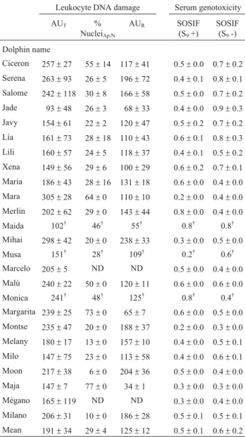
Table 2 shows the total DNA strand breakage in dol-phin leukocytes, expressed as total arbitrary units (AUT) of
the Comet assay. There was significant inter-individual variation (p < 0.05 in ANOVA) in the proportion of DNA strand breakage between the dolphins Jade (93±48) and
Mara (305±28) (mean: 191±34). Table 2 also shows the
percentage of apoptotic/necrotic nuclei (%Napoptotic/necrotic)
for each dolphin; there was significant inter-individual variation (p < 0.05 in ANOVA) between the dolphins Moon (6±0) and Maja (77±0) (mean: 29±4).
The proportion of DNA strand breakage (AUR) not
at-tributable to apoptosis and necrosis was calculated based on the total DNA strand breakage (AUT) estimated with the
Comet assay and the percentages of apoptotic/necrotic nu-clei (%Napoptotic/necrotic). The AURranged from 34 ± 1 for
Maja to 238±33 for Mihai (mean: 125±12). The AUR
esti-mates, but not the AUT, were significantly higher (t
-value = - 2.16, p < 0.03) in males than females (164±30vs.
119±14, respectively), suggesting that AURis a better
in-dicator than AUT for detecting DNA strand breakage in
bottlenose dolphins. This finding contrasts with the results reported for beluga whales using chromosomal aberration and sister chromatid exchange assays (Gauthier et al., 1999), but is not surprising since chromosomal aberrations occur on a much different scale than strand breakage and may be attributed to different factors. Both of the estimates of DNA damage (AUT and AUR) increased significantly
with dolphin age (data not shown), in agreement with previ-ous findings obtained using the micronucleus assay (Za-mora-Perezet al., 2006).
To determine whether the residual (non-apoptotic/ne-crotic) DNA strand breakage (AUR) originated from
expo-sure to genotoxic compounds we examined the genoto-xicity of dolphin serum using the SOS chromotest (Quillardetet al., 1982). This test detects a wide range of genotoxic compounds, including marine contaminants such as polycyclic aromatic hydrocarbons and organochlo-rine compounds (Quillardet and Hofnung, 1993). In experi-ments with metabolic activation, the SOSIF values ranged from 0.2±0.0 to 0.8±0.0 (mean: 0.5±0.2) while in
experi-ments without metabolic activation this indicator ranged from 0.3± 0.0 to 0.9 ±0.6 (mean: 0.6 ±0.4) (Table 2).
These data indicated that there were no genotoxic com-pounds in dolphin blood.
Discussion
In this work, we combined the Comet and DNA diffu-sion assays to measure DNA strand breakage in peripheral blood leukocytes of bottlenose dolphins. Nearly a third (~29%) of the DNA strand breakages arose from apopto-tic/necrotic events and led to overestimation of DNA cleav-age by the Comet assay, as also previously observed for mussels (Steinert, 1996), humans (Ticeet al., 2000) and sea
Table 2- Leukocyte DNA damage and serum genotoxicity in bottlenose dolphins.
Leukocyte DNA damage Serum genotoxicity
AUT %
NucleiAp/N
AUR SOSIF
(S9+)
SOSIF (S9-)
Dolphin name
Ciceron 257±27 55±14 117±41 0.5±0.0 0.7±0.2 Serena 263±93 26±5 196±72 0.4±0.1 0.8±0.1 Salome 242±118 30±8 166±58 0.5±0.0 0.7±0.2 Jade 93±48 26±3 68±33 0.4±0.0 0.9±0.3 Javy 154±61 22±2 120±47 0.5±0.2 0.7±0.2 Lía 161±73 28±18 110±43 0.6±0.1 0.8±0.3 Lili 160±57 24±5 118±37 0.4±0.1 0.5±0.2 Xena 149±56 29±6 100±29 0.6±0.2 0.7±0.1 Maria 186±43 28±16 131±18 0.6±0.0 0.4±0.0 Mara 305±28 64±0 110±10 0.2±0.0 0.4±0.0 Merlin 202±62 29±0 143±44 0.8±0.0 0.4±0.0 Maida 102† 46† 55† 0.8† 0.8†
Mihai 298±42 20±0 238±33 0.3±0.0 0.5±0.0 Musa 151† 28† 109† 0.2† 0.6†
Marcelo 205±5 ND ND 0.5±0.0 0.4±0.0 Malù 240±22 50±0 120±11 0.6±0.0 0.6±0.0
Monica 241† 48† 125† 0.8† 0.4†
Margarita 239±25 73±0 65±7 0.6±0.0 0.5±0.0 Montse 235±47 20±0 188±37 0.2±0.0 0.3±0.0 Melany 180±17 13±0 157±10 0.4±0.0 0.5±0.1 Milo 147±75 23±0 113±58 0.4±0.0 0.6±0.1 Moon 217±38 6±0 204±36 0.5±0.0 0.4±0.0 Maja 147±7 77±0 34±1 0.3±0.0 0.3±0.0 Mègano 165±119 ND ND 0.3±0.0 0.4±0.0 Milano 206±31 10±0 186±28 0.5±0.1 0.5±0.1 Mean 191±34 29±4 125±12 0.5±0.1 0.6±0.2
The values are the mean±S.E.M., where appropriate. AU corresponds to arbitrary units, where AUTis the total DNA damage as measured with the
Comet assay, AURis the remaining non-apoptotic/necrotic DNA damage
and %NAp/Nis the percentage of apoptotic/necrotic nuclei. (†) Only one
lions (El-Zeinet al., 2006). The total DNA strand breakage
(AUT) in dolphin leukocytes was 183±17 AU, indicating a
moderate level of DNA cleavage. However, as indicated above, this value was overestimated because of the influ-ence of apoptotic/necrotic events. We therefore computed the value for residual (non-apoptotic/necrotic) DNA strand breakage (AUR= 125±12) and found this to be negatively
correlated (r = -0.31, p < 0.05) with the percentage of apoptotic/necrotic nuclei. Hence, this parameter may pro-vide a better estimate of non-apoptotic/necrotic DNA strand breakage. The mean AURcorresponded to ~23% of
DNA in the tail and was slightly higher than the limit for non-damaged nuclei (20% of DNA in the tail), as indicated by Collinset al.(1997).
Several studies have shown that marine contaminants such as heavy metals may induce apoptosis (Steinert, 1996; Shenkeret al., 2000; Waalkeset al., 2000). We have
investi-gated the involvement of Fe, Cu and Zn in DNA strand breakage induced by apoptotic/necrotic events and found a weak but significant correlation between serum copper lev-els and apoptotic/necrotic DNA strand breakage (data not shown). Thus, although the copper content of the dolphi-narium water and dolphin serum was consistently low, the apoptosis observed here may have been induced by copper ions; no such relationship was observed for iron or zinc. Other environmental contaminants such as organochlorines and polycyclic aromatic hydrocarbons may also induce apoptosis (Salas and Burchiel, 1998; Shin et al., 2000;
Frenzilliet al., 2004), but we have not detected genotoxic
ac-tivity in dolphin serum using the SOS chromotest. Future studies measuring copper and other genotoxin levels in the environments where the dolphins used in this study normally live should improve our knowledge of the importance of this metal in causing apoptosis-related DNA damage.
The AURvalues clearly indicated that factors other
than apoptosis/necrosis affect the integrity of dolphin leu-kocyte DNA. Based on the negative SOS chromotest re-sults (Table 2) and the classification for DNA strand breakage in which a score of 0-100 indicated no DNA breakage or damage, 101-200 indicated little DNA damage, 201-300 indicated moderate DNA damage, and 301-400 in-dicated severe DNA damage, we expected baseline AUR
values of 0-100 AU,i.e., no DNA damage (Kobayashi et al., 1995; Collinset al., 1997). However, nearly 78% of the
dolphins had higher than expected AURvalues. Using the
Comet assay, Taddei et al. (2001) estimated that DNA
strand breakage in the nuclei of undamaged lymphocytes from bottlenose dolphins resulted in 16%-20% of their total DNA in the tail, which corresponded to 80-100 AU (Collins
et al., 1997). These studies suggest that there may be
differ-ences in the baseline estimates of DNA strand breakage ob-tainedin vitroandin vivo, and that additional factors that
affect the estimates of DNA cleavage must be considered during risk assessment studiesin vivo. Variation in the
ex-tent of DNA strand breakagein vivomay reflect
physiolog-ical conditions, such as a transient increase in oxidative stress caused by the diet or a sub-clinical infection (Collins
et al., 1997). An understanding of the influence of such
fac-tors on DNA cleavage in bottlenose dolphins should im-prove our estimates of baseline values for DNA strand breakage measured with the Comet assay.
Conclusions
To our knowledge, this is the first estimate of DNA strand breakage obtained with the Comet assay in periph-eral blood leukocytes of bottlenose dolphins. Our results in-dicate that this assay is sufficiently sensitive for assessing the influence of genotoxic substances in bottlenose dol-phins. However, the Comet assay overestimates the extent of DNA strand breakage in these cells because of DNA cleavage caused by apoptotic and necrosis. In addition to apoptosis and necrosis, factors other than exposure to geno-toxins may also affect the intactness of bottlenose dolphin DNA. Finally, our results indicate that the DNA diffusion assay is a suitable complementary tool for use alongside the Comet assay during risk assessment studies in bottlenose dolphins.
Acknowledgments
This work was done at CEADEN, Cuba and was sup-ported by a grant (PRMA-2059) from the Cuban Ministry of Science, Technology and Environment.
References
Betti C and Nigro M (1996) The Comet assay for the evaluation of the genetic hazard of pollutants in cetaceans: Preliminary re-sults on the genotoxic effects of methyl-mercury on the bot-tle-nosed dolphin (Tursiops truncatus) lymphocytesin vitro.
Mar Pollut Bull 32:545-548.
Collins AR (2002) The comet assay, principles, applications and limitations. In: Didenko VV (ed)In SituDetection of DNA Damage. Methods and Protocols. V. 203. Humana Press Inc., Totowa, pp 163-177.
Collins A, Dusinska A, Franklin M, Somorovska M, Petrovska H, Duthie S, Fillion L, Panayoitidis M, Raslova K and Vaughan N (1997) Comet assay in human biomonitoring studies: Re-liability, validation and applications. Environ Mol Mutagen 30:139-146.
Cotelle S and Férard JF (1999) Comet assay in genetic ecoto-xicology: A review. Environ Mol Mutagen 34:246-255. Del Barga I, Frenzilli G, Scarcelli V, Nigro M, Malmvärn A,
Asplund L, Förlin L and Sturve J (2006) Effects of algal ex-tracts (Polysiphonia fucoides) on rainbow trout (Oncorhynchus mykiss): A biomarker approach. Mar
Envi-ron Res 62:S283-S286.
El-Zein RA, Hastings-Smith DA, Ammenheuser MM, Trinen-Moslen M, Gulland FM and Ward Jr JB (2006) Evaluation of two different biomarkers for use in the assessment of toxic chemical exposure in California sea lions (Zalophus californianus). Mar Pollut Bull 52:104-120.
Frenzilli G, Scarcelli V, Del Barga I, Nigro M, Förlin L, Bolog-nesi C and Sturve J (2004) DNA damage in eelpout (Zoarces viviparous) from Göteborg harbour. Mutat Res
552:187-195.
Frenzilli G, Nigro M and Lyons BP (2009) The comet assay for the evaluation of genotoxic impact in aquatic environments. Mutat Res 681:80-92.
Fuentes JL, Vernhe M, Cuetara EB, Sánchez-Lamar A, Santana JL and Llagostera M (2006) Tannins from barks ofPinus caribeaeMorelet protectEscherichia colicells against DNA damage induced byg-rays. Fitoterapia 77:116-120. García O, Mandina T, Lamadrid AI, Díaz A, Remigio A,
Gonza-lez Y, Piloto Y, Rodríguez JE and Alvarez A (2004) Sensi-tivity and variability of visual scoring in the comet assay. Result of an inter-laboratory scoring exercise with the use of silver staining. Mutat Res 556:25-34.
Gauthier JM, Dubeau H, Rassart É, Jarman WM and Wells RS (1999) Biomarkers of DNA damage in marine mammals. Mutat Res 444:427-439.
Gichner T, Mukherjee A, Wagner ED and Plewa MJ (2005) Eval-uation of the nuclear DNA diffusion assay to detect apop-tosis and necrosis. Mutat Res 586:38-46.
Godard T, Deslandes E, Lebailly P, Vigreux C, Sichel F, Poul JM and Gauduchon P (1999) Early detection of staurosporine-induced apoptosis by comet and annexin V assays. Histo-chem Cell Biol 112:155-161.
Hartmann A, Agurell E, Beevers C, Brendler-Schawaab S, Bur-linson B, Clay P, Collins A, Smith A, Speit G, Thybaud V,et al.(2003) Recommendations for conducting thein vivo
al-kaline comet assay. Mutagenesis 18:45-51.
Kevekordes S, Mersch-Sunderman SV, Burghaus CM, Spielberg J, Schmeiser HH, Artl VM and Dunkeberg H (1999) SOS in-duction of select naturally occurring substances in Esche-richia coli(SOS chromotest). Mutat Res 445:81-91.
Kobayashi H, Sugiyama C, Morikawa Y, Hayashi M and Sofuny T (1995) A comparison between manual microscopic analy-sis and computerized image analyanaly-sis in the single cell gel electrophoresis assay. MMS Commun 3:103-115.
Lee RF and Steinert S (2003) Use of the single cell gel electropho-resis/comet assay for detecting DNA damage in aquatic (marine and freshwater) animals. Mutat Res 544:43-64. McKelvey-Martin VJ, Green MHL, Schmezer P, Pool-Zobel BL,
Meo MPD and Collins A (1993) The single cell gel electro-phoresis (comet assay): A European review. Mutat Res 288:47-63.
Mitchelmore CL and Chipman JK (1998) DNA strand breakage in aquatic organisms and the potential value of the comet assay in environmental monitoring. Mutat Res 399:135-147. NC:25 (1999) Norma Cubana: Sistema de Normas para la
Pro-tección del Medio Ambiente (Hidrosfera). Especificaciones y Procedimientos para la Evaluación de los Objetos Hídricos de uso Pesquero. Oficina Nacional de Normalización, La Habana, 9 pp.
Nigro M, Frenzilli G, Scarcelli V, Gorbi S and Regoli F (2002) In-duction of DNA strand breakage and apoptosis in the eel
Anguilla anguilla. Mar Environ Res 54:517-520.
Olive PL and Banath JP (1995) Sizing highly fragmented DNA in individual apoptotic cells using the comet assay and a DNA crosslinking agent. Exp Cell Res 221:19-26.
Olive PL, Banath JP and Durand RE (1990) Heterogeneity in radi-ation-induced DNA damage and repair in tumor and normal cells using the comet assay. Radiat Res 122:86-94.
Olive PL, Frazer G and Banath JP (1993) Radiation-induced apoptosis measured in TK6 human B Lymphoblast cells us-ing the comet assay. Radiat Res 136:130-136.
Quillardet P and Hofnung M (1993) The SOS Chromotest. Mutat Res 279:235-279.
Quillardet P, Huisman O, D’Ari R and Hofnung M (1982) SOS Chromotest, a direct assay of induction of an SOS function inEscherichia coliK-12 to measure genotoxicity. Proc Natl Acad Sci USA 79:5971-5975.
Quillardet P, Frelat G, Nguyen VD and Hofnung M (1989). De-tection of ionizing radiations with the SOS Chromotest, a bacterial short-term test for genotoxic agents. Mutat Res 216:251-257.
Salas VM and Burchiel SW (1998) Apoptosis in Daudi human B cell in response to benzo[a]pyrene and benzo[a]pyrene-7,8-dihydrodiol. Toxicol Appl Pharmacol 151:367-376. Shenker BJ, Guo TL and Shapiro IM (2000) Mercury-induced
apoptosis in human lymphoid cells: Evidence that apoptosis pathway is mercurial species dependent. Environ Res 84:89-99.
Shin KJ, Bae SS, Hwang YA, Seo JK, Ryu SH and Suh PG (2000) 2,2’,4,6,6’-pentachlorobiphenyl induces apoptosis in human monocytes cells. Toxicol Appl Pharmacol 169:1-7. Singh NP (2000a) A simple method for accurate estimation of
apoptotic cells. Exp Cell Res 256:328-337.
Singh NP (2000b) Microgels for estimation of DNA strand breaks, DNA protein crosslinks and apoptosis. Mutat Res 455:111-127.
Singh NP (2005) Apoptosis assessment by DNA diffusion assay. Meth Mol Med 111:55-67.
Singh NP, McCoy MC, Tice R and Schnider EL (1988) A simple technique for quantification of low levels of DNA damage in individual cells. Exp Cell Res 175:184-191.
Steinert SA (1996) Contribution of apoptosis to observed DNA damage in mussel cells. Mar Environ 42:253-259.
Taddei F, Scarcelli V, Frenzilli G and Nigro M (2001) Genotoxic hazard of pollutants in cetaceans: DNA damage and repair evaluated in the bottlenose dolphin (Tursiops truncatus) by the Comet assay. Mar Pollut Bull 42:324-328.
Tice RR, Agurell E, Anderson D, Burlinson B, Hartmann A, Kobayashi H, Miyamae Y, Rojas E, Ryu JC and Sasaki YF (2000) Single cell gel/comet assay: Guidelines forin vitro
and in vivo genetic toxicology testing. Environ Mol
Mu-tagen 35:206-221.
Waalkes MP, Fox DA, States JC, Patierno SR and McCabe Jr MJ (2000) Metals and disorders of cell accumulation: Modula-tion of apoptosis and cell proliferaModula-tion. Toxicol Sci 56:255-261.
Wada S, Khoa TV, Kobayashi Y, Funayama T, Yamamoto K, Natsuori M and Ito N (2003) Detection of radiation-induced apoptosis using comet assay. J Vet Med Sci 65:1161-1166. Zamora-Perez A, Camacho-Magaña C, Gómez-Meda B,
Ramos-Ibarra M, Batista-González C and Zuñiga-González G (2006) Importance of spontaneous micronucleated erythro-cytes in bottlenose dolphin (Tusiops truncatus) to marine
toxicology studies. Acta Biol Hung 57:441-448.
Associate Editor: Carlos F.M. Menck