Vol.58, n.5: pp. 789-797, September-October 2015 http://dx.doi.org/10.1590/S1516-89132015050318
ISSN 1516-8913 Printed in Brazil
BRAZILIAN ARCHIVES OF BIOLOGY AND TECHNOLOGY
A N I N T E R N A T I O N A L J O U R N A L
Characterization of Lipase from
Bacillus subtilis
I-4 and Its
Potential Use in Oil Contaminated Wastewater
Syeda Abeer Iqbal and Abdul Rehman
*Department of Microbiology and Molecular Genetics; University of the Punjab; New Campus, Lahore - Pakistan
ABSTRACT
A lipase producing bacterium was isolated from oil contaminated effluents of various industries from Sheikhupura Road, Pakistan, and, on the basis of biochemical and 16S rRNA ribotyping, was identified as Bacillus subtilis. The optimum temperature and pH for the growth of the culture were 37ºC and 7.0, respectively. B. subtilis I-4 had a lag phase of 4 h in LB medium while this phase prolonged to 6 h in oil containing medium. The optimum temperature and pH for the enzyme activity were 50ºC and 7.0, respectively. Maximum lipase activity was found in the presence of Ca ions. Olive oil and Tween 80 induced lipase gene in the bacterium while concentration of oil greater than 2% retarded the growth of the organism. In addition to lipase B. subtilis I-4 also produced alkane hydroxylase and biosurfactant which could make this bacterium potential candidate for lipase production as well as bioremediation of oil-contaminated wastewater.
Key words:Bacillus subtilis I-4, lipase, alkane hydroxylase
*Author for correspondence:[email protected]
INTRODUCTION
Lipases are glycerol ester hydrolases that have the ability to act on acylglycerols to liberate fatty acids and glycerol. They are water-soluble enzymes that act in catalyzing the hydrolysis of ester bonds in water-insoluble lipid substrates (Guo and Xu 2005). Lipases are ubiquitously produced by the plants (Bhardwaj et al. 2001; Belguith et al. 2009); animals (Carriere et al. 1994); and microorganisms (Olempska-Beer et al. 2006; Ramesh et al. 2013). Microbial lipases are the preferred potent source due to several industrial potentials (Hasan et al. 2006; Riaz et al. 2010; Veerapagu et al. 2013). They are present in many bacteria, fungi, plants, and animals (Rabbani et al. 2013; Sugahara et al. 2014). A considerable number of lipase producing microorganisms such as Pseudomonas fragi, Staphylococcus aureus, and
Burkholderia glumae have been described
(Ghanem et al. 2000; Gupta et al. 2004; Sangeetha et al. 2014).
The advent of enzymology represents an important breakthrough in the biotechnology industry, with the worldwide usage of enzymes (Kirk et al. 2002). Recombinant lipase has been developed for use in applications such as baking, laundry detergents and even as biocatalysts (Guo and Xu 2005) in alternative energy strategies to convert the vegetable oil into fuel (Gupta et al. 2004), etc. Biological wastewater treatment could be effective by lipase-producing microorganisms because lipid residues are converted to carbon dioxide, water and biomass (Lefebvre et al. 1998).
The present study was concerned with the isolation
and characterization of B. subtilis I-4 from oil
contaminated environment and lipase production
and bioremediation of oil contaminated
MATERIAL AND METHODS
Samples Collection and Enrichment
Six samples were collected separately from the waste effluents of ICI polyester, Sufi banaspati ghee and Paper mill from Sheikhupura (Pakistan). The temperature and pH of samples at their sites were also noted. The samples were transferred to
the laboratory and were spread on Luria-Bertani
(LB) agar (1% NaCl, 1% tryptone, 0.5% yeast extract and 1.5% agar) plates and incubated at 37°C for 24 h on the same day.
Samples were enriched using the method of Hasanuzzaman et al. (2004) by taking 1.0 mL of sample and inoculated in 100 mL sterile minimal
salt (MS) medium containing FeSO4.7H2O 1.5 mg,
KH2PO4 0.47 g, MgSO4.7H2O 0.1 g, CaCl2.2H2O
0.001 g, Na2HPO4 0.0119 g, NH4NO3 0.4 g,
MnSO4.4H2O 0.001 g in 100 mL distilled water
and containing 1% olive oil as a sole source of carbon. Same samples were inoculated in another
minimal salt medium containing (NH4)2SO4 0.5%,
K2HPO4 0.05%, MgSO4.7H2O 0.03%. All the
samples were incubated at 25°C at 180 rpm for two weeks. Turbidity in the flasks was observed regularly.
Screening of Lipase Producing Bacteria
For selection purpose, 1.0 mL culture broth from the flasks was taken and spread on Peptone Yeast Agar medium (peptone 10 g, yeast extract 10 g, agar 15 g, olive oil 10 mL, Tween80 3.0 mL; final
volume 1000 mL with distilled H20) containing
emulsified olive oil and were incubated at 37°C for 24 h. Colonies showing the zone of lipolysis around them were selected and were further purified on L-agar plates
Selection of Most Efficient Lipase Producing Bacteria
Following screening tests were carried out to select the most efficient bacterial isolates for lipase production.
Olive Oil Hydrolysis Agar
L-agar medium (Trypton 10.0 g, NaCl 5.0 g, yeast extract 5.0 g, agar 15.0 g) containing 1% olive oil was streaked with the isolates and incubated at 37°C for 48 h until the zones of hydrolysis became visible.
Lipase Chromogenic Plate Assay
Bacterial isolates were grown in LB medium containing 1% olive oil for 48 h. A modified
method from Amara et al. (2009) was used and chromogenic substrate plates were prepared by using phenol red (0.01%) along with 2% olive oil
as substrate, 4% Tween 80, 20 mM CaCl2. The pH
was adjusted to 7.3–7.4 by using 0.1 M NaOH,
and mixture was added to the same quantity of 4% melted agar (50°C) to give orange-reddish color. Holes were punched into the medium with the help of Pasteur pipette. Culture supernatants were poured into the holes and incubated at 37°C for 30 min. Zones of yellow color around the wells indicate lipase activity by the isolates.
Lipase Quantification Medium
Bacterial isolates were inoculated in 50 mL LB-broth and incubated at 37°C for 2-3 days on a rotary shaker. Then, 1.0 mL cell supernatant was taken after centrifugation at 6,000 rpm for 5 min. LQ-Agar medium with 1% olive oil were taken. LQ-Agar medium (glucose 10 g, peptone 10 g, yeast extract 3.0 g, agar 15 g, olive oil 10 mL,
Tween80 3.0 mL, NH4Cl, 1.5g, K2HPO4 2.0 g,
MgSO4.7H2O 0.5 g, NaH2PO4 1.0 g; final volume
made to 1000 mL with distilled water) with 1% emulsified olive oil was taken and then a sterile pasture pipette was used to make wells in the LQ-Agar medium. About 60 µL of the culture supernatant was loaded on each well and labeled accordingly. Plates were left at room temperature
for about 2 h and then were incubated at 37oC for
24 h. The formation of clear zones of lipolysis indicated lipase production of by the isolate and measured in millimeters.
Morphological, Biochemical and Molecular Characterization
The isolate was Gram stained. For the biochemical characterization the isolate was tested for catalase activity, oxidase acivity, Voges-Proskauer test, and utilization of different sugars following Benson (1994). For molecular characterization, genomic DNA was extracted as described by Carozzi et al. (1991). The 16S rRNA gene was amplified by the PCR using 16S rRNA primers
(RS-1; 5′-AAACTC-AAATGAATTGACGG-3′,
and RS-3; 5′-ACGGGCGGTGTGTAC-3′)
amplified products were electrophoresed on 1% agarose gel. Sequencing was carried out by
Genetic analysis system model CEQ-800
(Beckman) Coulter Inc. Fullerton, CA, USA. Nucleotide sequence similarities were determined
using BLAST (NCBI database;
http://www.ncbi.nlm.nih.gov/BLAST).
Bacterial Growth Curves
The strain was grown in LB broth and 1% olive oil under similar conditions of temperature and pH. The media (100 mL) were dispensed in 250 mL
flasks and autoclaved at 121oC for 15 min. After
inoculating with 100 μL of log phase growing cells of bacterial isolate, the flasks were incubated at
37oC for 24 h. After each 4 h, optical density of
each flask was taken at 600 nm with the help of a spectrophotometer.
Enzyme Assay
Lipase assay was performed as described by Bussamara et al. (2010) with slight modifications. The reaction was initialized by the addition of 0.1 mL of cell-free culture supernatant to 0.9 mL of substrate solution containing 3.0 mg of pNPP/mL of isopropanol and 9.0 mL of solution B 50 mM Tris-Cl (pH-8.0) 200 mL, Tween80 0.2 mL, Triton X-100 0.8 mL. The mixture was then incubated at 37°C for 30 min after maintaining its pH at 8.0. The increase in absorbance was measured at 410 nm with reference to an enzyme free control.
Effect of Temperatures, pH and Metal Ions on Enzyme Activity
Optimum temperature for enzyme stability was determined by incubating the culture supernatants at different temperatures, i.e., 30, 40, 50, 70 and 90°C for 1 h and then the enzyme assays were performed as described by Bussamara et al. (2010). Optimum pH for enzyme stability was determined by incubating the culture supernatants at different pH, i.e., 5.0, 6.0 7.0, 8.0 and 9.0 for 1 h and then enzyme assays were performed. Lipase activity was also determined in the presence of metal ions by incubating the culture supernatants separately in different flasks with 1mM solution of
each salt, i.e., ZnCl2, MgCl2, CuSO4 and CaCl2 at
37°C for 1 h.
Effect of Tween80, Ethanol, Oil and Glucose on Bacterial Growth
The bacterial isolate was inoculated in phenol red broth (Phenol red 1.0 g, yeast extract 5.0 g,
tryptone 10 g, distilled water 1L) along with 1% olive oil, 0.1% Tween 80, 0.1% ethanol and 0.1% glucose and then incubated at 37°C for 24 h. Effect of different concentrations of olive oil (0.5, 1.0, 1.5, 2.0, 2.5 and 3.0%) was also checked on the bacterial growth after incubated at 37°C for 24 h.
Partial Purification and SDS-PAGE Analysis of Lipase
The bacterial cultures supernatants were harvested by centrifugation at 6000 × g at 4°C for 10 min and concentrated by the addition of solid ammonium sulfate (60%). The precipitate was harvested by centrifuging at 6000 × g for 10 min and dialyzed (Cellu·Sep membrane cat no 5-5050-34; pore size 34 mm for mw of protein 50,000) against 10 mM sodium tartrate buffer (pH-5.5) for overnight. The dialyzed sample was centrifuged at 6000 × g for 1.5 h in centricon Ultracel YM-100 membrane (100,000 NMWL) to remove and concentrate the protein having molecular weight lower than 100 kDa. The protein solution above the centricon was discarded and flow through was taken, having proteins less than 100 kDa. This protein solution was further used in Ultracel YM-20 membrane (YM-20,000 NMWL) to remove the protein having MW 20, or less than 20 kDa. The protein solution in the upper chamber of the centricon was further concentrated in concentrator (Eppendorf concentrator, 5301) at 4°C and was finally analyzed by sodium dodecyl sulfate-polyacrylamide gel electrophoresis (SDS-PAGE) as described by Laemmli (1970). The protein concentration was determined by Bradford assay using bovine serum albumin (BSA) as a standard.
Thin Layer Chromatography (TLC)
The TLC plate was dried after loading the samples, developed with Hexane-Diethyl Ether-Acetic Acid (80:30:1) solvent system and finally
was stained by spraying it with 30% H2SO4 and
0.2% ninhydrin reagents separately. The plates were dried under a hot air drier and were observed for the appearance of color bands. The developed and air dried plated were also visualized using UV light at 366 nm and 254 nm.
Biosurfactant Assay
Drop collapse test
the center of a well and observed after 1 min. The droplet was observed for its shape.
Tube Emulsification Test
In tube emulsification test 2.0 mL petrol and 2.0 mL bacterial culture (16 h grown) were taken and the mixture was vortexed for 10 min. The mixture was observed for layer formation.
Alkane Hydoxylase Assay
B. subtilis I-4 was inoculated in MS media containing 1% kerosene oil and 1% petrol and incubated at 37°C for 48 h at 130 rpm.
Statistical Analysis
All the experiments were repeated two or more times and the results were averaged.
RESULTS AND DISCUSSION
Screening and Identification of Lipolytic Bacteria
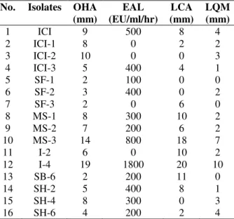
Among the 16 bacterial isolates, only the most potential lipase producer (I-4) was selected for further studies on the basis of different screening tests (Table 1). The temperature and pH of the sample were 30ºC and 7.5, respectively from where the isolate I-4 was isolated.
Table 1 - Screening tests performed for the selection of most efficient lipase producing bacteria.
No. Isolates OHA (mm)
EAL (EU/ml/hr)
LCA (mm)
LQM (mm)
1 ICI 9 500 8 4
2 ICI-1 8 0 2 2
3 ICI-2 10 0 0 3
4 ICI-3 5 400 4 1
5 SF-1 2 100 0 0
6 SF-2 3 400 0 2
7 SF-3 2 0 6 0
8 MS-1 8 300 10 2
9 MS-2 7 200 6 2
10 MS-3 14 800 18 7
11 I-2 6 0 10 2
12 I-4 19 1800 20 10
13 SB-6 2 200 11 0
14 SH-2 5 400 8 1
15 SH-4 8 300 0 3
16 SH-6 4 200 2 4
OHA: Olive oil hydrolysis agar; EAL: Enzymatic assay of lipase; LCA: Lipase chromogenic plate assay; LQM: Lipase quantitation medium.
Olive oil hydrolysis agar was used to analyze the diameter of zones of lipolysis. Zone of 19 mm was produced on OHA medium after 48 h of
incubation at 37°C by B. subtilisI-4. The isolate
showed the zones of 20 and 10 mm on LCA and LQM, respectively (Table 1) and was able to produce lipase (1800 EU/mL). The biochemical tests performed for the isolate are shown in Table 2. The partial 16S rRNA gene was sequenced and the (500 bp) analysis clearly demonstrated that
strain I-4 was a member of the genus Bacillus and
exhibited maximum similarity (Maximum query coverage 100%; identity 92%) with the 16 S rRNA
sequence of Bacillus subtilis. This sequence data
has been submitted to the GenBank databases under accession no. JN546608.
Table 2 - Biochemical tests for the identification of most efficient lipase producing bacterium.
Biochemical tests B. subtilis I-4
Gram staining +
Methyl red -
Voges-Proskauer -
Motility +
Catalase +
Oxidase -
Citrate +
Starch hydrolysis +
Casein hydrolysis +
Indole -
Glucose +
Mannitol +
Inositol +
Sorbitol NA
Rhamnose NA
Arabinose +
Lactose +
Sucrose +
Xylose +
Fructose +
Melibiose NA
Amygdalin NA
+: positive; -: negative; NA: Not applicable
Effect of Olive Oil on the Bacterial Growth
Growth curves of the strain were analyzed in LB and 1% olive oil. LB medium was taken as control for the bacterial growth. Bacterial growth was slightly slowed down in the presence of 1% olive oil as compared to LB medium. The maximum cell density was obtained after 16 h of incubation at
Figure 1 -Growth curves of Bacillus subtilis I-4 in the presence of LB and olive oil over a period of 24 h of incubation at 37ºC.
Effect of pH and temperature on activity and stability of lipase
Bacillus subtilis I-4 showed optimum growth at 37°C and pH of 7.0; however, its growth was not retarded at 45°C. Largely, bacteria prefer pH around 7.0 for best growth and lipase production,
such as in the case of Bacillus sp. (Sugihara et al.
1991), Acinetobacter sp. (Barbaro et al. 2001) and
Burkholderia sp. (Rathi et al. 2001). However, maximum activity at higher pH (>7.0) has been observed in many cases (Wang et al. 1995; Khyami-Horani 1996; Dong et al. 1999; Sharma et
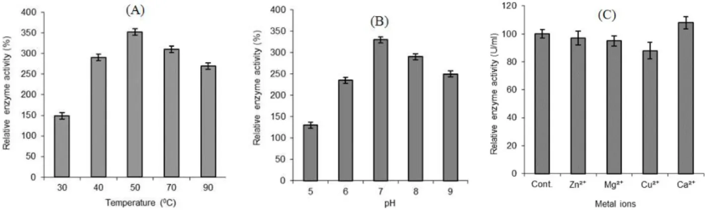
al. 2002). In the present study, lipase from B.
subtilis not only showed stability at higher temperatures but also showed maximum activity at 50°C (Fig. 2A); the optimum pH for enzyme
stability was 7.0 (Fig. 2B). An extracellular lipase
isolated from B. megaterium AKG-1 showed
optimum activity at 55°C and pH 7.0 (Sekhon et al. 2005).
The optimum temperature for lipase production corresponds with the growth temperature of the respective microorganism. For example, the best temperature for the growth and lipase production
in the case of Bacillus sp. RSJ1 was 50°C (Sharma
et al. 2002). Lipase produced by the Bacillus sp.
MPTK 912 had the optimum pH of 8.0 and the optimum temperature was 35°C (Mukeshkumar et al. 2012). Sinchaikul et al. (2001) reported that the purified lipase had an optimum pH of 8.5 and showed maximal activity at 55°C. Shabtai and Daya-Mishne (1992) reported that the enzyme hydrolyzed a variety of fatty acid esters and had an optimum pH of about 7.0. The enzyme retained its full activity at 20 to 55°C.
Divalent cations stimulate, or inhibit enzyme production in microorganisms. The effect of different metal ions on lipase activity was also determined through enzyme assay using 1mM
concentrations of different salts. Lipase from B.
subtilis I-4 was activated by Ca2+, while Mg2+,
Zn2+, and Cu2+ ions inhibited its activity (Fig. 2c).
It has been reported that lipase was inhibited by
Co2+, Ni2+, Fe2+, Fe3+ and EDTA, and activated by
Ca2+, Li+ and SDS (Matsumae et al. 1993). Rathi et
al. (2001) observed stimulation in lipase
production from Burkholderia sp. in the presence
of Ca2+ and Mg2+.
Figure 2 - Effect of temperature (A), pH (B) and metal ions (C) on lipase activity of Bacillus subtilis I-4. .
Sharma et al. (2002) also reported stimulation in
lipase production from Bacillus sp.RSJ1 in the
presence of calcium chloride. However, most other
metal ion salts were inhibitory to lipase
production. The enzyme was inhibited by Al3+,
and Mg2+ ions enhanced the enzyme activity; Na+ ions had no effect on enzyme activity (Kumar et
al. 2005). Mukeshkumar et al. (2012) reported that
Fe2+, Mg2+, Triton X-100, and Tween esters
enhanced lipase production in Bacillus sp. MPTK
912.
Effect of Different Substrates on Lipase Production
Lipase production by the bacterial isolate in the
presence of different substrates and oil
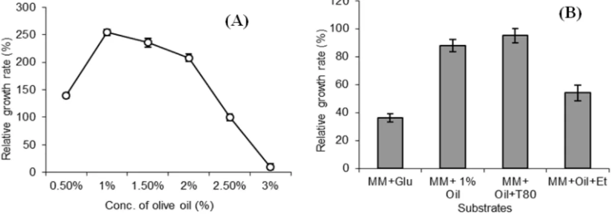
concentrations was analyzed. Olive oil (1%) in broth cultures of isolate induced lipase production significantly as compared to 1% tributyrin. Increasing concentration of olive oil from 0.5 to 3% oil showed an increase in bacterial growth up to 1% and then a gradual decrease in growth with further increase in oil concentration was observed. At 3% oil, growth of the bacterial isolate was completely ceased (Fig. 3a). It could be due to a certain ability of the organism to use it as a carbon source and high concentration of such substrate
could become toxic for the organism. Addition of 0.1% surfactant, i.e., Tween 80 showed significant increase in lipase production. Isolate I-4 showed lipase activity by the yellowing of phenol red medium after 24 h of incubation. The addition of ethanol to the medium showed no positive effect on lipase production; in fact, its production was reduced (Fig. 3b). However, irrespective of inducing compounds added to the medium, both the activity of lipase and bacterial growth were increased when lipids were added to the medium, indicating that they not only acted as lipase inducers, but could also be utilized as energy and carbon sources. Tweens have been shown to
stimulate the production of lipase in Acinetobacter
and Pseudomonas species (Lin et al. 1995; Martinez and Nudel 2002). Olive oil was present in the growth medium as a carbon source. Glucose and peptone acted as the best carbon and nitrogen sources for lipase activity in the production medium (Mukeshkumar et al. 2012).
Figure 3 - Effect of olive oil concentration (A) and different substrates (B) on the growth of B. subtilis I-4.
Presumably, olive oil also functions as an inducer of the lipase operon. This hypothesis was tested by
Boekema et al. (2007), who studied lipase
production after the growth in PG medium supplemented with alternative carbon sources, i.e., glucose and sucrose, which functioned as a good or poor car-bon source. Tween80 dramatically increased the extracellular lipase activity of strain PG1 in the presence of sucrose but much less in the presence of glucose as a carbon source. These results indicated that Tween 80, which, in contrast to Triton X-100, contained a fatty acyl ester bond, functioned as an inducer of the lipase operon and, furthermore, that the expression of this operon in
strain PG1 in the presence of glucose was prone to
catabolite repression. Minakshichouhan and
Dawande (2010) found that the enzymatic hydrolysis was highest with Tween 20, followed by Tween 80.
Thin Layer Chromatography
Bacterial isolate was analyzed for its ability to degrade the lipids through TLC as different components of lipid degradation could be visualized by different staining reagents. Fatty
acids were visualized by 0.2% ninhydrin. B.
indicating the degradation of lipids into its
components. Lipid concentration decreased
significantly in I-4 treated culture as compared to
the control, when sprayed with H2SO4 (results not
shown).
SDS-PAGE
The enzyme molecular mass was determined by the SDS-PAGE and induction of lipase in the presence and absence of olive oil was observed. Isolate I4 showed visible increase in protein production (data not shown), presumptively lipase, visible as band of nearly 53kDa on the gel (Fig. 4). Shabtai and Daya-Mishne (1992) reported that the approximate molecular weight of lipase was 40 kDa. The enzyme hydrolyzed a variety of fatty acid esters and had an optimum pH of about 7.0. Sinchaikul et al. (2001) reported the molecular mass of the lipase determined by the SDS-PAGE was approximately 43 kDa. The apparent molecular masses of LipA and LipB determined by the SDS-PAGE were 50 and 57 kDa, respectively (Salameh and Wiegel 2007). Kumar et al. (2005) reported the molecular weight of
lipase 31 kDa on the SDS–PAGE for B. coagulans
BTS-3. Kumar et al. (2012) reported a single band
of 62.2 kDa lipase from B. pumilus RK31 on
SDS-PAGE. Mukeshkumar et al. (2012) described that the enzyme was purified to homogeneity by Sephadex G-100 column chromatography and revealed molecular weight of 66 kDa.
Figure 4 - SDS-PAGE of partially purified lipase of B. subtilis I-4. M, protein marker. The gel is 12% stained with Coomassie blue.
Biosurfactant Production
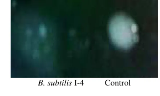
Bacillus subtilis I-4 was also tested for its ability to produce biosurfactants and emulsify immiscible
compounds. The culture showed positive slide drop collapse test as the drop of culture did not remain beaded when applied on a surface coated with oil (Fig. 5).
B. subtilis I-4 Control
Figure 5 - Drop collapse test of biosurfactant
production, isolate I-4 showing
collapse of drop on oil covered surface, and control showing no collapse of culture drop.
Alkane Hydroxylase
The ability of isolate to survive in the presence of hydrocarbons, including kerosene and petrol was analyzed. The culture was able to utilize kerosene oil and petrol when grown in minimal medium having 1% hydrocarbons as sole source of carbon (Fig. 6).
Figure 6 -Growth of B. subtilis I-4 in 1% kerosene oil; turbidity indicates the growth in the medium after 48 h of incubation as compared to control showing no growth.
CONCLUSION
In the present study, lipase producing B. subtilis
optimum temperature and pH for the growth of the
culture were 37ºC and 7.0, respectively. B. subtilis
I-4 had a lag phase of 4 h in LB medium while this phase prolonged to 6 h in oil containing medium. The optimum temperature and pH for the enzyme activity were 50ºC and 7.0, respectively. Maximum lipase activity was in the presence of Ca ions. Olive oil and Tween 80 induced lipase gene in the bacterium while concentration of oil higher than 2% retarded the growth of the
organism. In addition to lipase, B. subtilis I-4 also
produced alkaline hydroxylase and biosurfactant, which could make this bacterium potential candidate for lipase production as well as bioremediation of oil-contaminated wastewater.
ACKNOWLEDGEMENT
This work was supported by the research grant from Research Cell, University of the Punjab, Lahore-54590 (2010-2011).
REFERENCES
Amara AA, Salem SR, Shabeb MSA. The possibility to use bacterial protease and lipase as biodetergent.
Global J Biotechnol Biochem. 2009; 4(2): 104-114. Barbaro SE, Trevors JT, Inniss WE. Effect of low
temperature, cold shock, and various carbon sources on esterase and lipase activities and exopolysaccharide production by a psychrotrophic
Acinetobacter sp. Can J Microbiol. 2001; 47: 194-205.
Belguith H, Fattouch S, Jridi T, Ben Hamida J. Immunopurification and characterization of a rape (Brassica napus L.) seedling lipase. Afr J Biotechnol. 2009; 12(21): 3224-3234.
Benson HJ. Microbiological applications.Laboratory manual in general microbiology. Wan C. Brown Publishers, Dubuque, 1994.
Bhardwaj K, Raju A, Rajasekharan R. Identification, purification and characterization of a thermally stable lipase from rice bran. A new member of the phospholipase family. Plant Physiol. 2001; 127: 1728-1738.
Boekema BKHL, Beselin A, Breuer M, Hauer B, Koster M, Rosenau F, et al. Hexadecane and Tween80 stimulate lipase production in
Burkholderiaglumaeby different mechanisms. Appl Environ Microbiol. 2007; 73(12): 3838-3844.
Bussamara R, Fuentefria AM, Oliveira ES, Broetto L, Simcikova M, Valente P, et al. Isolation of a lipase-secreting yeast for enzyme production in a pilot-plant scale batch fermentation. Bioresource Technol. 2010; 101(1): 268-275.
Carozzi NB, Kramer VC, Warren GW, Evola S, Koziel MG. Prediction of insecticidal activity of Bacillus thuringiensis strains by polymerase chain reaction product profiles. Appl Environ Microbiol. 1991; 57: 3057-306.
Carriere F, Thirstrup K, Hjorth S. Cloning of the classical guinea pig pancreatic lipase and comparison with the lipase related protein2. FEBS Lett. 1994; 388: 63-68.
Dong H, Gao S, Han S, Cao S. Purification and characterization of a Pseudomonas sp. lipase and its properties in non-aqueous media. Appl Microbiol Biotechnol. 1999; 30: 251-256.
Ghanem EH, Al-Sayeed HA, Saleh KM. Analkalophilic thermostable lipase produced by a new isolate of
Bacillus alcalophilus. World J Microbiol Biotechnol. 2000; 16: 459-464.
Guo Z, Xu X. New opportunity for enzymatic modification of fats and oils with industrial potentials. Org Biomol Chem. 2005; 3(14): 2615-2619.
Gupta R, Gupta N, Rathi P. Bacterial lipases: an overview of production, purification and biochemical properties. Appl Microbiol Biotechnol. 2004; 64(6): 763-781.
Hasan F, Shah AA, Hameed A. Industrial applications of microbial lipases. Enzyme Microb Technol. 2006; 39: 235-251.
Hasanuzzaman M, Kathryn M, Umadhay-Briones SM, Morita N, Nodasaka Y, Yumoto I, et al. Isolation, identification, and characterization of a novel, oil-degrading bacterium, Pseudomonas aeruginosa T1.
Curr Microbiol. 2004;49: 108-114.
Khyami-Horani H. Thermotolerant strain of Bacillus licheniformis producing lipase. World J Microbiol Biotechnol. 1996; 12: 399-401.
Kirk O, Borchert TV, Fuglsang CC. Industrial enzyme applications. Curr Opin Biotechnol. 2002; 13: 345-351.
Kumar R, Sharma A, Kumar A, Singh D. Lipase from
Bacillus pumilus RK31: Production, purification and some properties. World Appl Sci J. 2012; 16(7): 940-948.
Laemmli UK. Cleavage of structural proteins during the assembly of the head of bacteriophage T4. Nature.
1970; 227: 680-685.
Lefebvre X, Paul E, Mauret M, Baptiste P, Capdeville B. Kinetic characterization of saponified domestic lipid residues aerobic biodegradation. Water Res.
1998; 32(10): 3031- 3038.
Lin S-F, Chiou C-M, Tsai Y-C. Effect of triton X-100 on alkaline lipase production by Pseudomonas pseudoalcaligenes F-111. Biotechnol Lett. 1995; 17: 959-962.
Martinez DA, Nudel BC. The improvement of lipase secretion and stability by addition of inert compounds into Acinetobacter calcoaceticus cultures. Can J Microbiol. 2002; 48: 1056-1061.
Matsumae H, Furui M, Shibatani T. Lipase-catalyzed asymmetric hydrolysis of 3-phenylglycidic acid ester, the key intermediate in the synthesis of diltiazem hydrochloride. J Ferment Bioeng. 1993; 75: 93-98. Minakshichouhan M, Dawande AY. Partial
purification, characterization of lipase produced from
P. aeruginosa. Asiatic J Biotech Res. 2010; 1: 29-34. Mukeshkumar DJ, Rejitha R, Devika S, Balakumaran
MD, Immaculate NRA, Kalaichelvan PT. Production, optimization and purification of lipase from Bacillus
sp. MPTK 912 isolated from oil mill effluent. Adv Appl Sci Res. 2012; 3(2): 930-938.
Olempska-Beer ZS, Merker RI, Ditto MD. Food processing enzymes from recombinant microorganisms-a review. Reg Toxicol Pharmacol. 2006; 45: 144-158.
Rabbani, M, Bagherinejad MR, Sadeghi HM, Shariat ZS, Etemadifar Z, Moazen F, et al. Isolation and characterization of novel thermophilic lipase-secretingnbacteria. Braz J Microbiol. 2013; 44: 1113-1119.
Ramesh S, Kumar R, Devi A, Balakrishnan, K. Isolation of a lipase producing bacteria for enzyme synthesis in shake flask cultivation. Int J Curr Microbiol App Sci. 2014; 3(3): 712-719.
Riaz M, Shah AA, Hameed A, Hasan F. characterization of lipase produced by Bacillus sp. FH5 in immobilized and free state. Ann Microbiol. 2010; 60: 169-175.
Rathi P, Saxena RK, Gupta R. A novel alkaline lipase from Burkholderia cepacia for detergent formulation.
Process Biochem. 2001; 37: 187-192.
Rehman A, Ali A, Muneer B, Shakoori AR. Resistance and biosorption of mercury by bacteria isolated from industrial effluents. Pakistan J Zool. 2007; 39: 137-146.
Salameh MA, Wiegel J. Purification and characterization of two highly thermophilic alkaline lipases from Thermosyntropha lipolytica. Appl Environ Microbiol. 2007; 73 7725-7731.
Sangeetha R; Arulpandi I, Geetha, A. Molecular characterization of a proteolysis-resistant lipase from Bacillus pumilus SG2. Braz J Microbiol.
2014; 45: 389-393.
Sekhon A, Dahiya N, Tiwari RP, Hoondal GS. Properties of a thermostable extracellular lipase from
Bacillus megaterium AKG-1. J Basic Microbiol. 2005; 45(2): 147-154
Shabtai Y, Daya-Mishne N. Production, purification, and properties of a lipase from a bacterium (Pseudomonas aeruginosa YS-7) capable of growing in water-restricted environments. Appl Environ Microbiol. 1992; 58(1): 174-180.
Sharma R, Soni SK, Vohra RM, Jolly RS, Gupta LK, Gupta JK. Production of extracellular alkaline lipase from a Bacillus sp. RSJ1 and its application in ester hydrolysis. Ind J Microbiol. 2002; 42: 49-54.
Sinchaikul S, Sookkheo B, Phutrakul S, Pan FM, Chen ST. Optimization of a thermostable lipase from
Bacillus stearothermophilus P1: overexpression, purification, and characterization. Protein Expr Purif. 2001; 22(3): 388-398.
Sugihara A, Tani T, Tominaga Y. Purification and characterization of a novel thermostable lipase from
Bacillus sp.J Biochem. 1991; 109: 211-216.
Sugahara VH, Varea GS. Immobilization of Beauveria bassiana lipase on silica gel by physical adsorption. Braz Arch Biol Technol. 2014; 57: 842-850.
Veerapagu M, Sankara DR, Narayanan A, Ponmurugan K, Jeya KR. Screening selection identification production and optimization of bacterial lipase from oil spilled soil. Asian J Pharm Clin Res. 2013; 6: 62-67.
Wang Y, Srivastava KC, Shen G-J, Wang HY. Thermostable alkaline lipase from a newly isolated thermophilic Bacillus strain A30-1 (ATCC 53841). J Ferment Bioeng.1995; 79: 433-438.