Thermostability in Variants of Lipase A from
Bacillus subtilis
Prakash Chandra Rathi1, Karl-Erich Jaeger2,3, Holger Gohlke1*
1Institute of Pharmaceutical and Medical Chemistry, Heinrich-Heine-University, Düsseldorf, Germany,
2Institute of Molecular Enzyme Technology, Heinrich-Heine-University, Düsseldorf, Germany,3Institute of Bio- and Geosciences IBG-1: Biotechnology, Research Centre Jülich, Jülich, Germany
Abstract
Understanding the origin of thermostability is of fundamental importance in protein biochem-istry. Opposing views on increased or decreased structural rigidity of the folded state have been put forward in this context. They have been related to differences in the temporal reso-lution of experiments and computations that probe atomic mobility. Here, we find a signifi-cant (p= 0.004) and fair (R2= 0.46) correlation between the structural rigidity of a well-characterized set of 16 mutants of lipase A fromBacillus subtilis(BsLipA) and their thermo-dynamic thermostability. We apply the rigidity theory-based Constraint Network Analysis (CNA) approach, analyzing directly and in a time-independent manner the statics of the BsLipA mutants. We carefully validate the CNA results on macroscopic and microscopic experimental observables and probe for their sensitivity with respect to input structures. Fur-thermore, we introduce a robust, local stability measure for predicting thermodynamic ther-mostability. Our results complement work that showed for pairs of homologous proteins that raising the structural stability is the most common way to obtain a higher thermostability. Furthermore, they demonstrate that related series of mutants with only a small number of mutations can be successfully analyzed by CNA, which suggests that CNA can be applied prospectively in rational protein design aimed at higher thermodynamic thermostability.
Introduction
Sufficiently high thermostability of proteins is important for both organisms living in high temperature environments and for biotechnological applications where enzymes are used as biocatalysts under often harsh reaction conditions [1,2]. From a mechanistic point of view,
“protein thermostability”embraces at least two different meanings [3,4]: (1)thermodynamic
thermostability describes the folded-unfolded equilibrium of a protein, and (2)kinetic thermo-stability refers to the length of time a protein remains active before undergoing irreversible denaturation at an elevated temperature. Several factors have been frequently attributed to ele-vated protein thermostability including improved hydrogen bonding [5], ion pair and salt
OPEN ACCESS
Citation:Rathi PC, Jaeger K-E, Gohlke H (2015) Structural Rigidity and Protein Thermostability in Variants of Lipase A fromBacillus subtilis. PLoS ONE 10(7): e0130289. doi:10.1371/journal.pone.0130289
Editor:Annalisa Pastore, National Institute for Medical Research, Medical Research Council, London, UNITED KINGDOM
Received:October 1, 2014
Accepted:May 18, 2015
Published:July 6, 2015
Copyright:© 2015 Rathi et al. This is an open access article distributed under the terms of the
Creative Commons Attribution License, which permits unrestricted use, distribution, and reproduction in any medium, provided the original author and source are credited.
Data Availability Statement:All relevant data are within the paper and its Supporting Information files.
Funding:The study was supported by the Ministry of Innovation, Science, and Research of North Rhine-Westphalia and Heinrich-Heine-University Düsseldorf (HHU) by a scholarship to PCR within the CLIB2021 Graduate Cluster Industrial Biotechnology. The funders had no role in study design, data collection and analysis, decision to publish, or preparation of the manuscript.
bridge networks [6], better hydrophobic packing [7], shortened loops [8], and higher secondary structure content [9], in all favoring an increased structural rigidity of the folded state [10–13].
As an opposing view, proteins from thermophilic organisms have been reported to be as flexi-ble as or even more flexiflexi-ble than homologs from mesophilic organisms [14–17].
These different views on the relation between protein thermostability and structural rigidity have been a matter of ongoing discussion [10,18–23]. In particular, it has been argued that
atomic movements, which are the primary mobility data from which information on protein statics (rigidity and flexibility) is derived, cover a wide range of timescales within a protein [15, 24,25]. Hence, depending on the temporal resolution of the experimental technique or compu-tational analysis used to detect such movements, (parts of) a protein can come out as rigid or flexible [26–32]. Here, we address the question of the relation between protein thermostability
and structural rigidity by analyzingdirectlythe static properties of a well-characterized set of 16 mutants of lipase A fromBacillus subtilis(BsLipA). We do so by applying the rigidity the-ory-based Constraint Network Analysis (CNA) approach developed by us [33–35], thereby
considering theBsLipA variants to be in static equilibrium, hence avoiding that the results depend on the temporal resolution of the approach.
BsLipA is an important member of the lipase class of enzymes and used in diverse biotech-nological applications [36,37]. Owing to its importance,BsLipA has been extensively studied with respect to structure [38–41] and thermostability [42–48]. As to the latter, Reetzet al.
applied iterative saturation mutagenesis on the most flexible amino acids as identified by crys-tallographic B-factors, which resulted inBsLipA mutants that were more thermostable than the wild type showing an increase inT5060(the temperature required to reduce the initial enzy-matic activity by 50% within 60 min) of45 K [42]. Subsequent biophysical characterization of the three most thermostable mutants revealed that the improved activity retention resulted from a reduced rate of protein unfolding and a reduced precipitation of the unfolding interme-diates, i.e., due to kinetic reasons [49]. In contrast, Raoet al. sequentially developed several thermostableBsLipA mutants using directed evolution assisted by structural information. These mutants were shown to be more thermostable than the wild type due to predominantly thermodynamic reasons [44–48,50]; the most thermostable mutant displayed an increase in
the melting temperatureTmof ~22 K.
In the CNA approach, a protein is modeled as a constraint network where bodies (repre-senting atoms) are connected by sets of bars (constraints, repre(repre-senting covalent and noncova-lent interactions) [51]. A rigidity analysis performed on the network [52,53] results in a decomposition into rigid parts and flexible links in between. By analyzing a series of“
per-turbed”networks in which noncovalent interactions are included in a temperature-dependent
manner [11,13,54], the loss of rigidity of a protein is simulated, which can be related to ther-mal unfolding [12,13,54]. Results of these analyses can be linked to biologically relevant char-acteristics of a biomolecular structure by a set of global and local indices [55]. In particular, a phase transition pointTpcan be identified during the thermal unfolding simulation at which a largely rigid network becomes almost flexible; this phase transition point has been related to the thermodynamic thermostability of a protein [11–13]. For improving the robustness of the
analyses, the rigidity analyses are performed on ensembles of network topologies (ENTFNC) [56]. That way, thermal fluctuations of a protein are considered without actually sampling conformations.
thermodynamic thermostability, which complements the detection of the (global) phase transi-tion pointTp. As theBsLipA variants are sequentially closely related, these results have impor-tant implications for applying CNA in a prospective manner in rational protein design aimed at higher thermodynamic thermostability. Finally, we discuss our results in terms of potentially different mechanisms underlying the increased protein thermostabilities of mutants isolated by Reetzet al.and Raoet al.
Materials and Methods
Data set
The wild type structure ofBsLipA with the highest resolution (PDB ID: 1ISP; resolution = 1.3 Å) was obtained from the Protein Data Bank (PDB;www.pdb.org) [57]. For probing the sensi-tivity of the CNA results on the conformation of the input structures, five additional crystal structures of wild typeBsLipA were analyzed (PDB IDs: 1I6W, 1R4Z, 1R50, 2QXT, 2QXU). We included in our study all mutants from Raoet al. for whichTmvalues were determined [44–48]. In addition, we included the three most thermostable mutants developed in the last
rounds of iterative saturation mutagenesis by Reetzet al. [42]. Models of mutant structures for which crystal structures were not available in the PDB were generated with the SCWRL pro-gram [58], using the respectiveBsLipA structure as a template that is closest in sequence to the mutant. SCWRL constructs mutant models by predicting backbone-dependent side chain con-formations with the help of a rotamer library; coordinates of backbone atoms remain
unchanged. Conformations of side chains of all residues within 8 Å of a mutated residue were re-predicted in order to allow for a local structural relaxation. For all structures, hydrogen atoms were added using REDUCE [59]; side chains of Asn, Gln, and His were flipped in this stage if necessary to optimize the hydrogen bond network. All water molecules, buffer ions, and crystal solvents were removed from the structures. Finally, all structures were minimized by 5000 steps of conjugate gradient minimization (including an initial steepest descent minimi-zation for 100 steps) or until the root mean-square gradient of the energy was<1.010−4kcal mol-1Å-1. The energy minimization was carried out with Amber11 [60] using the Cornellet al. force field [61] with modifications for proteins (ff99SB) [62] and the GBOBCgeneralized Born
model [63]. All variants ofBsLipA used in this study are summarized inTable 1.
Construction of the constraint network and rigidity analysis
As described in the previous section, only the protein part was considered for network con-struction, i.e., all non-protein molecules including water molecules were discarded. This was done based on previous findings that including water molecules does not significantly change the rigidity analysis results [64,65]. Proteins were modeled as constraint networks in a body-and-barrepresentation (see section“Body-and-bar networks”inS1 File) [66,67] using the
CNA software [35] that acts as a front- and back-end to the Floppy Inclusion and Rigid Sub-structure Topography (FIRST) program [51,68]. Once the constraint network is built, rigidity analysis is carried out, which identifies (rigid) clusters of atoms with no internal motion and flexible links in between, using the pebble game algorithm [52,53] as implemented in the FIRST software [51].
Thermal unfolding simulation
bonds break at higher temperatures than weaker ones [69]. As such, only hydrogen bonds with an energyEHBEcut(σ) were included in the network of stateσ. A thermal unfolding trajectory
of 60 network states was generated for each input network by decreasingEcutfrom−0.1 kcal
mol−1to−6.0 kcal mol−1with a step size of 0.1 kcal mol−1. According to the linear relationship
betweenEcutand the temperatureTintroduced by Radestock and Gohlke on 20 pairs of ortho-logs from mesophilic and thermophilic organisms, respectively (Eq 1) [12,13], the range of
Ecutused in this study is equivalent to increasing the temperature of the system from 302 K to 420 K with a step size of 2 K. Because hydrophobic interactions remain constant or become even stronger as the temperature increases [70,71], the number of hydrophobic tethers were kept unchanged throughout the thermal unfolding simulation. Rigidity analysis was performed on all such generated network states, and then local and global rigidity characteristics were cal-culated (see section“Local and global rigidity indices”inS1 File). The setup of the thermal
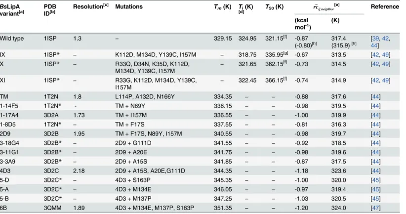
Table 1. Summary ofBsLipA variants used in the study. BsLipA
variant[a]
PDB ID[b]
Resolution[c] Mutations Tm(K) Ti(K)
[d] T50(K) rceij;neighbor
[e] Reference
(kcal mol-1)
(K)
Wild type 1ISP 1.3 − 329.15 324.95 321.15[f] -0.87
(-0.80)[h] 317.4(315.9)[h] [4439],42,
IX 1ISP* − K112D, M134D, Y139C, I157M − 318.75 335.95[g] -0.67 313.5 [42,49]
X 1ISP* − R33Q, D34N, K35D, K112D,
M134D, Y139C, I157M − 321.65 362.15
[f] -0.73 314.5 [42,49]
XI 1ISP* − R33G, K112D, M134D, Y139C,
I157M −
322.45 366.15[f] -0.74 314.9 [42,49]
TM 1T2N 1.8 L114P, A132D, N166Y 334.35 − − -0.88 317.6 [44]
1-14F5 1T2N* - TM + N89Y 336.15 − − -0.98 319.5 [44]
1-17A4 3D2A 1.73 TM + I157M 336.55 − − -1.00 319.9 [44]
1-8D5 1T2N* − TM + F17S 337.55 − − -0.81 316.3 [44]
2D9 3D2B 1.95 TM + F17S, N89Y, I157M 340.55 − − -0.98 319.7 [44]
3-18G4 3D2B* − 2D9 + G111D 341.55 − − -0.92 318.5 [44]
3-11G1 3D2B* − 2D9 + A20E 341.75 − − -0.98 319.6 [44]
3-3A9 3D2B* − 2D9 + A15S 341.85 − − -0.87 317.5 [44]
4D3 3D2C 2.18 2D9 + A15S, A20E,G111D 344.35 − − -1.18 323.6 [44]
5-D 3D2C* − 4D3 + S163P 345.35 − − -1.00 320.0 [45]
5-A 3D2C* − 4D3 + M134E 346.05 − − -0.97 319.4 [45]
5-B 3D2C* − 4D3 + M137P 347.25 − − -1.03 320.5 [45]
6B 3QMM 1.89 4D3 + M134E, M137P, S163P 351.35 − − -1.20 324.0 [47]
[a]Names ofBsLipA structures are taken from the respective references.
[b]A PDB ID marked with an asterisk indicates that the model of the corresponding variant was built using the structure with that PDB ID as a template. [c]InÅ.
[d]The temperature at which the unfolding transition begins.
[e]Median stability of rigid contacts between residue neighbors computed by applying the ENTFNCapproach (see section
“Median stability of rigid contacts
between residue neighbors as a new measure for predicting thermodynamic thermostability”) (left column). Values in the right column were obtained by
converting the median stabilities to a temperature scale according toEq 1.
[f]
T5060values, i.e., the temperature required to reduce the initial enzymatic activity by 50% within 60 min. [g]T
5015values, i.e., the temperature required to reduce the initial enzymatic activity by 50% within 15 min. [h]Averagerce
ij;neighborover six wild type structures (see the main text for details).
unfolding simulation and the subsequent rigidity analysis were performed using the CNA soft-ware [35], which is available fromhttp://cpclab.uni-duesseldorf.de/software. A web service for performing CNA analysis can be accessed viahttp://cpclab.uni-duesseldorf.de/cna[34].
T¼ 20K
kcalmol 1Ecutþ300K ð1Þ
Ensemble of networks generated by using fuzzy noncovalent constraints
For improving the robustness of rigidity analyses, CNA is generally carried out on an ensemble of structures (e.g., generated by molecular dynamics (MD) simulations), and then results are averaged [11,64]. The preceding MD simulation compromises the efficiency of the rigidity analysis, however. To overcome this drawback, Pflegeret al. [56] recently introduced an approach that performs rigidity analyses on an ensemble of network topologies (ENTFNC)gen-erated from a single input structure by using fuzzy noncovalent constraints. Here, the number and distribution of non-covalent constraints (hydrogen bonds and hydrophobic tethers) are modulated by random components within certain ranges as specified in ref. [56], thus simulat-ing thermal fluctuations of a biomacromolecule without actually movsimulat-ing atoms. An ensemble of 2000 network configurations was generated using these definitions of fuzzy noncovalent constraints for allBsLipA variants, respectively. Finally, average local indices were calculated, as were average phase transition temperatures identified by the global index cluster configura-tion entropyHtype2. The indexHtype2monitors the degree of disorder in the realization of a given network stateσ: As long as a network is dominated by a very large rigid cluster,Htype2
tends to be low because there are only a few configurations of a system with a large rigid cluster possible;Htype2increases when larger rigid clusters break down in smaller clusters (see section
“Local and global rigidity indices”inS1 Fileand ref. [55] for details).
Clustering of unfolding pathways
Recently, we showed that curves of the rigidity order parameter, which characterizes the gen-eral percolation behavior of a constraint network during thermal unfolding, for mesophilic proteins and their thermophilic counterparts are almost identical except for a shift of the curve of the thermophilic protein to higher temperatures [12]. This finding supported the hypothesis of corresponding states according to which mesophilic and thermophilic enzymes are in corre-sponding states of similar rigidity and flexibility at their respective optimal temperature [12]. The percolation indexpiis a local analog to the rigidity order parameter. It monitors for each bond when it segregates from the largest rigid cluster present at the beginning of a thermal unfolding simulation (see section“Local and global rigidity indices”inS1 Fileand ref. [55] for
details). That way, a residue-wisepiprofile of a protein, generated by taking the lower of thepi
values of the two backbone bonds for each residue, expresses the hierarchical break-down of the largest rigid cluster during a thermal unfolding simulation.
medoids of the 10 clusters are shown inFig 1). A clustering in more than 10 clusters essentially created additional clusters that were very similar to other clusters. From this, the cluster distri-bution (frequencies of network topologies in each of the 10 clusters out of in total 2000 network topologies) for eachBsLipA variant was calculated by counting the number of networks that
belongs to each of the 10 clusters. A high (low) correlation between cluster distributions for twoBsLipA variants then indicates that both variants unfold in a similar (different) manner. Finally, a matrix of all pairwise correlations of cluster distributions ofBsLipA variants was generated.
Fig 1. Residue-wisepiplots for medoids of the 10 clusters.Secondary structure elements as computed by the DSSP program [88,89] are indicated on
the top of the plots and are labeled:α-helix (red rectangle),β-strands (green rectangle), loop (black line).
Results
Data set
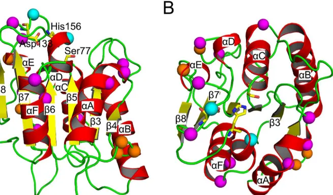
BsLipA is a protein of 181 amino acids with a minimalα/βhydrolase fold; in this fold, a central parallelβ-sheet of sixβ-strands is surrounded by sixα-helices. Ser77, Asp133, and His156
con-stitute the catalytic triad (Fig 2). Unlike other lipases, the catalytic site inBsLipA is not covered with a lid. Hence,BsLipA does not show interfacial activation [40]. The data set used in this study contains structures of the wild typeBsLipA, thirteen mutants from Raoet al. [44–48],
and three mutants from Reetzet al. [42,49] (Table 1). The mutants differ from the wild type by three to twelve mutations, i.e., the sequence identity is>93%. Models for the mutants for which X-ray structures were not available were built using the SCWRL program. As the num-ber of mutations in the modeled variants is7 with respect to the template structures (<4% with respect to the sequence length) (Table 1), an overall similar backbone confirmation can be expected as can be an overall reliable modeling of side chain conformations by SCWRL. This was also evident from a very good structural alignment and low root-mean-square deviations (RMSD) between the wild type and those mutants for which crystal structures were available (Cαatom-based RMSD values between the wild type and the mutants<0.38 Å). The high
structural similarity allows a direct comparison of results from rigidity analyses for these struc-tures [11–13].
The melting temperatureTmof the wild type is 329.15 K. TheTmvalues of the mutants of Raoet al. range from 334.35 to 351.35 K (Table 1). For the mutants of Reetzet al. noTmvalues are available. Rather, unfolding initiation temperaturesTiwere reported, which are lower by
Fig 2. Cartoon representation of wild typeBsLipA with mutated residues indicated by spheres of their Cαatoms (mutations from Raoet al. [44–48]:
magenta; Reetzet al. [42,49]: orange; mutations common in both data sets: cyan).The catalytic triad (Ser77-Asp133-His156) is shown in stick representation with yellow carbons. The protein is colored according to secondary structure (α-helices: red;β-sheets: yellow; loops: green). The right view (B)
differs from the left (A) by an anti-clockwise rotation of ~90° about a horizontal axis. All figures ofBsLipA structures were generated with PyMOL (http://www. pymol.org).
2.5 to 6.2 K than that of the wild type. This suggests that mutants of Reetzet al. are thermody-namically less thermostable than the wild type [49], in contrast to mutants from Raoet al. [44–
48]. However, we note that, whileTmreports on the temperature at which 50% of the protein is unfolded and, hence, properly describes the folded-unfolded equilibrium of a protein,Tionly reports on the temperature at which the unfolding transition begins. Therefore, we will only consider relations within mutants of Raoet al. and to the wild type and distinguish those from relations within mutants of Reetzet al. and to the wild type. Finally, theT50tvalues of the mutants of Reetzet al. are higher than that of the wild type (Table 1), showing that these mutants more efficiently refold upon cooling after incubation at high temperatures than does the wild type. The location of mutations in all of the mutants investigated in this study is shown inFig 2; all mutations are located on the protein surface.
Thermal unfolding pathway of
Bs
LipA
From monitoring the loss in rigidity percolation during thermal unfolding simulations, major phase transitions in the protein can be identified that relate to the unfolding pathway [11–13,
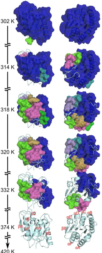
54,73]. Here, we describe the loss of rigidity percolation of the wild typeBsLipA (PDB ID 1ISP) as an example. Similarity or dissimilarity, respectively, of the unfolding pathways across all variants is described below. During the thermal unfolding, a giant rigid cluster that exists at low temperature (equivalent to a highEcut) breaks down in smaller sub-clusters until, finally, the whole protein becomes flexible at a high temperature (Fig 3; see alsoS1 Videoshowing the loss of rigidity percolation during the thermal unfolding of the wild type). As such, nearly the entire protein structure constitutes a single giant rigid cluster initially (at 302 K;Fig 3). As the temperature increases, loops segregate first from the giant rigid cluster. Then, at 314 K,α-helix D (αD) andαE segregate to form individual small rigid clusters (Fig 3), as doαA andαF at 318
K. The giant rigid cluster at this temperature is formed by the centralβ-sheet region and the two helicesαB andαC (Fig 3). Next, theβ-sheet region becomes sequentially flexible,
begin-ning withβ4 andβ8 at 320 K (Fig 3). Then, the remainingβ-strands become flexible in the orderβ3,β7, andβ5−β6, leading to a completely flexibleβ-sheet region at 332 K (Fig 3The
immediate next step at whichαB andαC become two separate rigid clusters is identified as a phase transition point: Now most of the structure has become flexible. This transition is most prominent with respect to going from a structurally stable wild typeBsLipA to an unfolded one (Figure B inS1 File). After this phase transition point, the remaining rigidity is sequentially lost, and the structure finally becomes completely flexible at 374 K (Fig 3).
During the thermal unfolding ofBsLipA, helices segregate from the giant rigid cluster as independent small rigid clusters. This is due to two reasons: First, in thebody-and-barnetwork representation, a helix with a minimum of seven amino acids is already rigid by itself due to constraints arising from covalent and backbone hydrogen bonds [66]. Second, with the current energy functionEHB[69], all backbone hydrogen bonds are assigned a very similar strength, irrespective of their location along a helix. Thus, a helix will persist as an independent rigid cluster during the thermal unfolding simulation until all backbone hydrogen bonds break almost simultaneously at a high temperature, which most likely represents an overstabilization of a helix [74]. Considering this behavior, the unfolding pathway identified for the wild type
BsLipA is in good agreement with respect to the early segregation ofα-helices with experimen-tal findings on the unfolding of proteins with anα/βhydrolase fold [75,76]. This indicates that
Prediction of thermodynamic thermostability of
Bs
LipA variants based
on the global index
H
type2From the thermal unfolding simulations, the temperature of the phase transition pointTpwas identified as described in the section“Local and global rigidity indices”inS1 File. Note thatTp
values determined that way should be considered relative values only, as stated in previous studies [12,34,35]. Initially, we calculated phase transition points using single network topolo-gies generated from the input structures of wild typeBsLipA and mutants of Raoet al.; how-ever, this resulted in a very poor prediction of thermodynamic thermostability with a coefficient of determination (R2) for a linear fit between experimentalTmand predictedTpof 0.22 (Figure C inS1 File). We anticipated that this result reflects the high sensitivity of CNA on the conformation of the input structures as also found previously [11,56,64,65]. We thus resorted to averagingTpvalues over an ensemble ofBsLipA, applying the recently developed ENTFNCapproach. This approach generates an ensemble of network topologies from a single input structure and has been shown to yield results of rigidity analyses both at the local and global level that agree almost perfectly with those obtained from MD simulations-generated ensembles of structures [56]. However, this yielded a significant (p= 0.002) correlation betweenTpandTmwithR2= 0.58 only if the two structures with the lowest (wild type) and highest (mutant 6B)Tmwere considered outliers (Fig 4A; see below for an explanation regard-ing the outliers; note that removregard-ing the two outliers in the case of usregard-ing sregard-ingle network topolo-gies only marginally improvedR2from 0.22 to 0.29). The mutants IX, X and XI of Reetzet al. were predicted to be slightly less thermostable than the wild type (Fig 4A). This is in line with experimental findings by Reetzet al. that suggest that these mutants are thermodynamically less stable than the wild type [49]. In summary, these results suggest that CNA coupled with the ENTFNCapproach can sense effects on the thermodynamic thermostability that arise from
only a few sequence variations (pairwise sequence identity>93%; pairwise RMSD<0.38 Å). However, the false predictions for wild typeBsLipA and mutant 6B are dissatisfying.
Difference in unfolding pathways explains outliers
Next, we investigated why the thermostabilities of the wild type and the mutant 6B were pre-dicted falsely. Since the precision of the computations shown inFig 4Ais high (the standard error in the mean is<0.38 K in all cases), we reasoned that the false prediction must arise from a systematic difference between the wild type and 6Bversusall other mutants of Raoet al. Thus, we mutually compared all unfolding pathways of the systems as described in“Materials
and Methods”. After partitioning unfolding pathways ofBsLipA variants characterized on a
residue basis by the percolation indexpiinto 10 clusters (seeFig 1for thepiprofiles of the 10 cluster medoids), we calculated correlation coefficients from the resulting cluster distributions for all pairs of variants (Fig 5; Tables A and B inS1 File).
thermal unfolding simulation (302 K), almost the complete structure is part of the giant rigid cluster; in contrast, the structure becomes completely flexible at temperatures374 K. The right views differ from the left ones by an anti-clockwise rotation of ~90° about a horizontal axis. Important secondary structure elements are labeled. Note that the unfolding pathway shown here represents anaverage loss of rigidity percolationcalculated from a stability map (see section“Local and global rigidity indices”inS1 File) averaged over all unfolding trajectories obtained for the ensemble of 2000 network topologies. Hence, the temperature at the phase transition point identified that way (Figure B inS1 File) cannot be compared to theaverage phase transition temperature, which is obtained from 2000 individualTpvalues and used for predicting the
thermodynamic thermostability ofBsLipA variants (see section“Prediction of thermodynamic thermostability ofBsLipAvariants”)
These results revealed that the wild type enzyme shows an unfolding pathway distribution very distinct from otherBsLipA variants from Raoet al. with correlation coefficientsrranging from−0.69 to 0.54 (Fig 5, Table A inS1 File). The averagervalue for the wild type against all
Fig 4. Correlation between predicted and experimental thermostabilities (Tmvalues) ofBsLipA
variants; for the predictions, the ENTFNCapproach was used. A: Correlation betweenT
pderived from the
global indexHtype2andTmvalues for thermodynamically thermostabilized mutants from Raoet al. Data
points colored red were considered outliers (see main text for explanation) and excluded when calculatingR2
values and the correlation lines.B: Correlation betweenrceij;neighborandTmvalues for thermodynamically
thermostabilized mutants from Raoet al. Data points shown as empty squares representrceij;neighborvalues for
five additional wild type crystal structures (see main text for details; two of the squares closely overlap; mean
e
rcij;neighborover all six data points for wild type structures is shown as a small horizontal line: 315.9±0.6 K). A
and B: Error bars represent the standard error in the mean.Tpandrceij;neighborvalues for kinetically
thermostabilized mutants from Reetzet al. are marked by arrows on the corresponding ordinates.
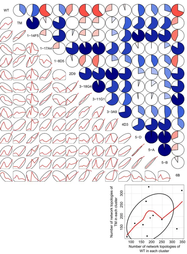
Fig 5. Pairwise correlations of cluster distributions (using 10 clusters) of unfolding pathways of wild typeBsLipA and mutants from Raoet al.The
upper triangle shows pairwise correlation coefficients as dial plots where a filled portion of a pie indicates the magnitude of the correlation (r) and blue (red)
color indicates a positive (negative) correlation. The lower triangle shows 68% data ellipses (depicting the bivariate mean±1 standard deviation) [90] and scatterplots of the respective cluster distributions (frequencies of network topologies in each of the 10 clusters) of the twoBsLipA variants as red lines
smoothed by locally-weighted polynomial regression [91]. Data points and axes for the plots in the lower triangle are omitted for clarity. The blow-up in the right bottom corner depicts exemplarily for the TMversusWT case axes, axes labels, data points, the smoothing line obtained by locally-weighted polynomial regression, and the 68% data ellipsis. The figure was generated using the“corrgram”package [92] of the R program (http://www.r-project.org).
other variants from Raoet al. is−0.06 ± 0.14 (mean ± SEM), which is lower than that of the
other variants (°0.16 except for the outlier 6B) (Table A inS1 File). The second outlier, mutant 6B, has an averagervalue of 0.12 ± 0.16 when comparing its unfolding pathway distri-bution to those of other variants from Raoet al. This averagervalue is lower than the corre-sponding averagervalues of all other mutants from Raoet al. (Table A inS1 File). The thermal unfolding pathway of 6B is shown in Figure D inS1 File. While the overall unfolding pathway of 6B is comparable with that of the wild typeBsLipA in that the helices segregate from the giant rigid cluster as individual rigid clusters in the early phase of unfolding, they do so in a dif-ferent order (αD,αA−αF,αE,αB−αC; Figure D inS1 File). A probability density function
(PDF) ofrvalues of unfolding pathway distributions of the two outliers wild type and mutant 6B with all other variants shows a bimodal distribution and is shifted towards lowerrvalues compared to the PDF of thervalues of other mutants from Raoet al. Furthermore, about half of this distribution is related to negativervalues (Figure E inS1 File). In all, this suggests that the two outliers have unfolding pathways different from all other mutants from Raoet al. for which the prediction of thermodynamic thermostability was successful. Finally, we note that the unfolding pathway distributions of the wild type and the three mutants from Reetzet al. are highly similar to each other (r>0.79;p0.001; Table B inS1 File).
These findings have important implications: First, the results strongly suggest that the mis-prediction of the thermostabilities of the wild type and mutant 6B arises from them showing different unfolding pathways from all of the remaining mutants from Raoet al.. Apparently, the present approach of identifying phase transition points by monitoring theglobalindex
Htype2(see section“Local and global rigidity indices”inS1 File) is too sensitive with respect to
the details of such pathways. Consequently, alternative methods should be explored (see sec-tion“Median stability of rigid contacts between residue neighbors as a new measure for
pre-dicting thermodynamic thermostability”). Second, the results suggest that the history of the
generation of theBsLipA structures may play a role for the observed differences in the unfold-ing pathways: generally, the most similar unfoldunfold-ing pathways (Tables A and B inS1 File) (and then the most coherentTppredictions) are found for those variants that originate from a com-mon structural“ancestor”(Table 1). Third, the results propose to apply the
similarity/dissimi-larity of unfolding pathway distributions as a measure to judge the reliability of thermostability predictions in future studies: the lower the similarity for two variants, the less confident should one be that relative thermostability predictions are correct. Finally, we cannot exclude at the present stage that thermostabilizing mutations lead to an unfolding pathway that is different from the one of the wild type. Considering that intrinsic and extrinsic modifications in other systems that led to thermostabilization have been shown to influence not just the folded state but the entire (un)folding free energy landscape [77,78], this possibility also exists forBsLipA mutants [45,47].
Median stability of rigid contacts between residue neighbors as a new
measure for predicting thermodynamic thermostability
The above findings called for predicting the thermodynamic thermostability in a way that is less sensitive to the details of the unfolding pathway than the present approach relying on the
composed of modules. As a consequence, often more than one pronounced jump inHtype2is observed, which then makes it difficult to assign a phase transition point (Figure B inS1 File).
As an alternative, we set out to characterize thermodynamic thermostability at thelocal
level [55], i.e., by monitoring residue pair-wise descriptors of local stability within a protein structure as a function of the temperature. The most comprehensive information in that direc-tion is provided by stability mapsrcij[12], which depict when a rigid contactrcbetween two residuesiandjceases to exist along a thermal unfolding trajectory. As such,rcijcontains infor-mation cumulated over all statesσof a network along the trajectory as to which parts of the network are (locally) mechanically stable at a given stateσ, and which are not [12,55]. Of note, this stability information is not only available in a qualitative manner (i.e., in terms of local rigidity and flexibility) but also quantitatively in that eachrcijhas associated with it the energy
Ecutat which this rigid contact is lost. Thus,∑i,j>ircijrepresents the chemical potential energy due to non-covalent bonding, obtained from the coarse-grained, residue-wise network repre-sentation of the underlying protein structure. With respect to a reference state where no non-covalent interactions are present anymore (i.e., an unfolded state),∑i,j>ircijcan be considered
an unfolding energy then. Three modifications were applied to∑i,j>ircijhere for technical rea-sons. I) In order to stress the locality of interactions within a protein, which will later aid in understanding how structural differences relate to thermostability differences (see section
“Influence of mutations on local structural rigidity”), we focused on the stability of rigid
con-tactsrcij,neighborbetween structurally close residues only (i.e., those residues where at least one pair of respective atoms is within 5 Å distance). II) To suppress the influence of extreme values in the double summation on the outcome of the unfolding energy, we used the median stability of rigid contactsrceij;neighborinstead. Such extreme values can occur in regions that are highly sta-bilized by interactions to hydrophobic atoms [56]. III) Applying the ENTFNCapproach, e
rcij;neighborwere averaged over ensembles of 2000 constraint networks, which has been shown to significantly improve the robustness of rigidity analyses [56]. Therceij;neighborvalues are given in Table 1. In addition,Table 1andFig 4Bshow these values after converting them to a tempera-ture scale viaEq 1
A significant and fair linear correlation ofrceij;neighborwithTmvalues of the thermodynami-cally stable mutants from Raoet al. is obtained (R2= 0.46,p= 0.004;Fig 4B). No outlier is observed now, indicating that our definition of an average local stability correctly reflects dif-ferences in the thermodynamic thermostability. Thisfinding substantiates our above interpre-tation ofrceij;neighboras an approximation to the unfolding energy, because under the condition of a temperature-independent heat capacity the unfolding energy is linearly correlated to the melting temperature, with the heat capacity as the scaling factor [80]. The slope of the correla-tion line (0.26) inFig 4Bdeviates from unity. This indicates that the linear relationship inEq 1 used for convertingrceij;neighborto a temperature scale, which was derived forHtype2-based ther-mostability prediction [12,13], may need to be reparameterized for application withrceij;neighbor. In this case, as the heat capacity has been shown to scale linearly with the number of residues for small globular proteins [80,81], a normalization with respect to protein size needs to be applied. When only considering the six X-ray structures in the dataset of Raoet al., a good cor-relation ofrceij;neighborwithTmvalues ofR
2= 0.87 (p= 0.007) is found (Table 1). In contrast, a
Therceij;neighbor-based measure is apparently less sensitive to differences in the unfolding path-way because the wild type and mutant 6B are now much better ranked. However, comparing the prediction of thermostabilities byrceij;neighborandHtype2, the latter yields a better correlation withTmfor mutants with similar unfolding pathways. From an application point of view, we thus recommend usingHtype2-derivedTpvalues for comparing thermostabilities of variants of a protein unless the underlying unfolding pathways are dissimilar; in that case, we recommend usingrceij;neighbor.
When applied to hen egg white lysozyme the ENTFNCapproach has been shown to
signifi-cantly improve the robustness of rigidity analyses with respect to the conformation of the input structures [56]. To probe if this also holds forBsLipA investigated here, we computedrceij;neighbor using the ENTFNCapproach for
five additional crystal structures of wild typeBsLipA (see sec-tion“Materials and methods”). The standard error of the mean inrceij;neighborover all six wild
typeBsLipA structures is 0.57 K (Fig 4B) including PDB ID 1ISP discussed so far. This error is likely within the experimental uncertainty, confirming our previous results of robust rigidity analyses with ENTFNC[56]. Still, if the average
e
rcij;neighborover all six crystal structures (315.9 K; see horizontal line inFig 4B;Table 1) is considered for therceij;neighborversus Tmcorrelation, the quality of the correlation improves considerably toR2= 0.55 (p= 0.001) compared to if only e
rcij;neighborof PDB ID 1ISP is used (see above). This indicates that the use of multiple input struc-tures in connection with the ENTFNCapproach further increases the accuracy of
thermostabil-ity predictions.
Influence of mutations on local structural rigidity
Considering that the average local stability defined above correctly reflects differences in the (macroscopic) thermodynamic thermostability, we analyzed on a residue basis how changes in thermostability relate to changes in local structural stability (rigidity). First, we compared sta-bility maps of variants from Raoet al. with distinct thermostabilities to analyze the effect of mutations on the local rigidity. In particular, we compared the wild type to a more thermosta-ble variant 1-14F5 and the most thermostathermosta-ble variant 6B. We averaged stability maps of the six wild type structures (see above andFig 4B) and used this average for comparison against the thermostable variants ofBsLipA. Difference stability maps for 1-14F5/wild type (Fig 6A) and 6B/wild type (Fig 6B) pairs demonstrate that mutations in general improve the strength of rigid contacts to and in between neighboring residues of the mutations (lower triangles inFig 6A and 6B) but also in between residue pairs not in contact distance (upper triangles inFig 6A and 6B). This effect is more pronounced for 6B/wild type than 1-14F5/wild type.
In more detail, the four mutations (indicated by arrows inFig 6Aand shown inFig 6D) on 1-14F5 stabilize contacts ofαD with its neighboring helixαC and contacts ofαA withαF (Fig
6A and 6D). More importantly, the contacts of helicesαA andαF with their neighboringβ -strands in the centralβ-sheet region are stabilized, which delays the early loss of these helices
observed during the thermal unfolding of the wild type (Fig 3). Similarly, the contacts between
αB and the centralβ-sheet region also become stronger, which delays the decay of structural
stability of theβ-sheet during thermal unfolding. On average, contacts between all residue neighbors are ~−0.1 kcal mol−1or ~2 K more stable in 1-14F5 than in the wild type.
Residues mutated in 6B (indicated by arrows inFig 6Band shown inFig 6E) include the mutations already found in 1-14F5. This explains a strengthening of inter-helical contacts and of the contacts betweenαhelices and the centralβ-sheet region as discussed already for 1-14F5 (Fig 6D and 6E). However, the additional mutations in 6B stabilize contacts between otherα
αF and theβ-sheet. On average, contacts between all residue neighbors are ~−0.4 kcal mol−1or
~8 K more stable in 6B than in the wild type (Fig 6E).
Taken together, contacts between peripheral helices and the centralβ-sheet region are
stron-ger in 6B than in 1-14F5. This delays the loss ofα-helices during thermal unfolding (Fig 3) to a larger extent in 6B than in 14F5, explaining at a structural level why 6B is more stable than 1-14F5. Remarkably, many of these stabilizations must arise from the long-range aspect of rigid-ity percolation [52,64,82,83], because almost all mutations in 6B are on the surface, i.e., far from the centralβ-sheet region. In contrast, inter-helical contacts of theαB/αC helix pair become weaker in the mutants than in wild type (Fig 6D and 6E) indicating that the strength-ened stability between these helices and the centralβ-sheet region is sufficient to keep the struc-ture folded. At last, for all other thermodynamically more thermostable mutants, a similar profile of changes in contact stability between various secondary structure elements was observed (Figure F inS1 File). Not unexpected, the increase in contact stability compared to wild type (Figure F inS1 File) was generally the more pronounced the higher the thermody-namic thermostability is of the mutant (Table 1).
Second, we compared the mutants from Reetzet al. to the wild type. Regarding mutant X, seven residues have been mutated (indicated by arrows inFig 6Cand shown inFig 6G). In strict contrast to what was observed for the thermodynamically thermostabilized mutants, this mutant showed a destabilization of rigid contacts both locally and globally (Fig 6C and 6F; see also Figure G inS1 File, where a similar finding is depicted for mutants IX and XI). For mutant X, the average decrease in stability over all residue neighbors is ~0.06 kcal mol-1or ~1.2 K. The
destabilization found on the local scale agrees with results of a lowerTpfound when analyzing the mutants globally. Furthermore, the results are in line with experimental findings which suggest that the mutants are thermodynamically less stable than the wild type (Table 1) [49]. Our findings are also in good agreement with results obtained by comparative crystal structure analysis of wild type and variant X [49]: Loop region 14–21, for which lower B-factors in X
than in the wild type structure were observed, shows increased contact stabilities with its neigh-boring residues in X (Fig 6C and 6F; Figure H in theS1 File). Likewise, regions 129–153 and
177–181, for which higher B-factors in X than in the wild type structure were observed, show
decreased contact stabilities with their neighboring residues in X (Fig 6C and 6F; Figure H in theS1 File). However, region 60–70 shows increased contact stabilities in X (Fig 6C and 6Fand
Figure H in theS1 File) despite higher B-factors observed in the comparative crystal structure analysis. The latter may reflect increased motions of a stabilized region as a whole, taking into consideration that B-factors can report on rigid body motions of a structurally stable part [84].
Finally, it would be very satisfying from both the biochemical and structural biology point of view, if the effects of the three to twelve mutations on increased or decreased local rigidity could be immediately related to the changes in specific interactions with neighboring residues. Our above observation for the mutants from Raoet al. that many of the stability changes must Fig 6. Differences in the stability of rigid contacts between wild type and mutants ofBsLipA.Maps depict differences between stability maps of the respective mutants and an average stability map of the six wild type structures (see the main text for explanation) forA: mutant 1-14F5,B: mutant 6B, andC:
mutant X. A red (blue) color indicates that a rigid contact in the mutant is more (less) stable than in the wild type (see color scale at the bottom). The upper triangles show differences in the stability values for all residue pairs; the lower triangles show differences in the stability values only for residue pairs that are within 5Åof each other, with values for all other residue pairs colored gray. Secondary structure elements as computed by the DSSP program [88,89] are indicated on both abscissa and ordinate and are labeled:α-helix (red rectangle),β-strands (green rectangle), loop (black line). Arrows represent the mutation
positions with respect to the wild type sequence: Common mutations in 1-14F5 (A) and 6B (B) are shown in magenta, unique mutations in 6B (B) are shown in green, and mutations in X (C) are shown in orange. The differences in the stability of rigid contacts for residue neighbors is also displayed on the structures of the mutants by sticks connecting Cαatoms of residue pairs colored according to the color scale of the maps forD: 1-14F5,E: 6B, andF: X. Only those contacts that are stabilized by4 K or destabilized by3 K are shown for clarity; for the same reason, contacts between two residues of the same secondary structure element are not shown. Mutated residues are shown as sticks and a sphere at their Cαatoms (D, E, and F) in the same color used for arrows (A, B, and C).
arise from the long-range aspect of rigidity percolation [52,64,82,83] speaks against such an endeavor. Nevertheless, we analyzed differences in the per-residue number of hydrogen bonds and hydrophobic tethers of the variants 1_14F5, 6B, and X with respect to the wild type (Figure I inS1 File). While these analyses reveal differences in the number of interactions>1 indeed only for a small set of residues, between ~10 and ~35% of all residues show differences of 1. This finding is remarkable given the small number of mutations in the variants and the high structural similarity between crystal structures of the wild type and mutants found for backbone atoms above; apparently, these differences arise from subtle changes in the confor-mations of the side chains due to the mutations. This finding also suggests that an interpreta-tion of the relainterpreta-tionship between increased or decreased local rigidity due to changes in specific interactions of a mutated residue and a change in thermostability of a mutant may fall short of the actual complexity underlying this relationship.
Discussion
Understanding the origin of thermostability is of fundamental importance in protein biochem-istry. Here, we have probed the relation between protein thermostability and structural rigidity by directly analyzing static properties of a well-characterized set of 16BsLipA mutants. The main outcome of this work is the finding of a good correlation between the structural rigidity of allBsLipA variants and their thermodynamic thermostability. This finding of a quantitative relation between structural rigidity and thermodynamic thermostability within a series of closely related protein variants complements a previous study that showed for pairs of homolo-gous proteins from thermophilic and mesophilc organisms that raising the structural stability is the most common way (~77% of all cases) to obtain a higher thermostability [85].
Intense discussions are ongoing regarding the question if elevated protein thermostability is related to increased or decreased structural rigidity of the folded state [10,18–23]. Part of this
discussion is related to how information on structural rigidity is derived from information on mobility, in particular with respect to the temporal resolution of the experimental techniques and computational analysis [26–32]. In this context, the finding we describe here is highly
rele-vant. As the rigidity theory-based CNA approach applied characterizes rigidity and flexibility of proteins directly, i.e., without the requirement of information on atomic movements, it does not suffer from such time dependence. Another part of the discussion is related to the fact that changes in the enthalpy, entropy and/or heat capacity can lead to thermodynamic stabilization; these changes can be linked to distinct effects on the structural stability of the folded state [19]. It was thus instructive to observe that the general increase in rigidity in the mutants of Rao
et al. is accompanied by certain inter-helical contacts becoming weaker than in the wild type; these weakened contacts between the“modular”helices may increase the entropy of the folded
state and so may further contribute to the overall stability of the systems [17,86,87]. This find-ing again calls attention to analyzfind-ing the origin of thermostability with methods that cover a wide range of temporal and spatial resolution because otherwise one effect may be hidden beneath another.
Our results are backed up with a careful validation of the accuracy and robustness of the CNA approach on the data set both from a macroscopic and microscopic point of view. As to the former, good and statistically significant correlations between experimental melting tem-peratures (Tm) of mutants of Raoet al. and predicted thermodynamic thermostabilities have been found based on two independent measures (Htype2andrceij;neighbor), as was correctly pre-dicted that the thermodynamic thermostability of the mutants of Reetzet al. is lower than that
within experimental error, confirming previous results of robust rigidity analyses when apply-ing the ENTFNCapproach [56]. As to the latter, the detailed analysis of the unfolding pathway of wild typeBsLipA revealed a good agreement with respect to the early segregation ofα-helices
with experimental observations on other proteins with anα/βhydrolase fold. Thesefindings are in line with previous successful applications of CNA in predicting melting temperatures and identifying structural weak spots [11–13].
From a methodological point of view, some additional comments are in order. First, in the present study we successfully predicted the thermodynamic thermostability for mutants that differ by as few as three to twelve mutations from the wild type. Compared to previous applica-tions of CNA on either pairs of mesophilic and thermophilic homologues [12,13] or a series of homologous proteins from different organisms living at varying temperatures [11], this finding considerably broadens the application domain of CNA towards data-driven protein engineer-ing: There, related series of mutants with only a small number of respective mutations will be the major focus of investigations. Second, we introduced a measure for the similarity/dissimi-larity of unfolding pathways of mutants and used it for explaining false thermostability predic-tions. We suggest to use the measure in future studies as a significance criterion to judge the reliability of thermostability predictions from CNA. Third, we introduced the median stability of rigid contacts as a new local measure for predicting thermodynamic thermostability and showed that this measure is less sensitive to details of the unfolding pathway. The measure is thus recommended for comparing thermostabilities of mutants the underlying unfolding path-ways of which are dissimilar.
Finally, regarding the subset of mutants of Reetzet al., we find a decreased local rigidity compared to wild type, in line with findings of lower unfolding initiation temperatures, yet the mutants are more“thermostable”than the wild type in that they preserve enzymatic activity
better after subjecting them to higher temperatures [42]. It would have been tempting to inves-tigate how this relates to a potential kinetic stabilization of the mutants. However, we refrained from doing so due to the lack of direct experimental evidence for such a kinetic stabilization [49]. In turn, this finding draws attention to the fact that the term“protein thermostability”is
often used in a non-discriminating sense, i.e., data reported in the literature does not allow to establish whether a protein is thermodynamically or kinetically stable [49]. This adds another layer of complexity to the question of the relation between protein thermostability and struc-tural rigidity as it may be required to decouple observations on“increasedvs. decreased
struc-tural rigidity”from the general description of“protein thermostability”in future studies.
Supporting Information
S1 File. The file contains additional information to the manuscript: Pairwise Pearson cor-relation coefficientsrand correspondingpvalues between cluster distributions ofBsLipA variants from Raoet al.(Table A) and Reetzet al. (Table B), objective function of the cluster-ing (Figure A), cluster configuration entropyHtype2vs. temperature obtained from theaverage loss of rigidity percolationof wild typeBsLipA (Figure B), correlation between predictedTp
derived from the global indexHtype2and experimental thermostabilities (Tmvalues) ofBsLipA variants using single input structures. (Figure C), average loss of structural rigidity of mutant 6B during a thermal unfolding simulation (Figure D), probability density functions (PDFs) of all pairwise Pearson correlation coefficients between cluster distributions ofBsLipA variants (Figure E), differences in the stability of rigid contacts between wild type and variants of
wild type (Figure I). (PDF)
S1 Video. The video file shows the average loss of structural rigidity during thermal unfold-ing of wild typeBsLipA and the corresponding global rigidity indexHtype2vs. temperature plot.See captions ofFig 3in the main text and Figure B inS1 Filefor details.
(AVI)
Acknowledgments
We are grateful to the Ministry of Innovation, Science, and Research of North Rhine-Westpha-lia and Heinrich-Heine-University Düsseldorf (HHU) for a scholarship to PCR within the CLIB2021Graduate Cluster Industrial Biotechnology. We acknowledge the„Zentrum fuer
Informations- und Medientechnologie“(ZIM) at HHU for computational support. We thank
Anuseema Bhadauriya, HHU, for fruitful discussions on the experimental data of the investi-gated mutants.
Author Contributions
Conceived and designed the experiments: HG. Performed the experiments: PCR. Analyzed the data: PCR KEJ HG. Wrote the paper: PCR KEJ HG.
References
1. Demirjian DC, Moris-Varas F, Cassidy CS. Enzymes from extremophiles. Current Opinion in Chemical Biology. 2001; 5(2):144–51. Epub 2001/04/03. S1367-5931(00)00183-6 [pii]. PMID:11282340.
2. Van den Burg B. Extremophiles as a source for novel enzymes. Current Opinion in Microbiology. 2003;
6(3):213–8. Epub 2003/07/02. S1369527403000602 [pii]. PMID:12831896.
3. Ó’Fágáin C. Engineering protein stability. In: Walls D, Loughran ST, editors. Protein Chromatography:
Springer; 2011. p. 103–36.
4. Polizzi KM, Bommarius AS, Broering JM, Chaparro-Riggers JF. Stability of biocatalysts. Current Opin-ion in Chemical Biology. 2007; 11(2):220–5. Epub 2007/02/20. doi:10.1016/j.cbpa.2007.01.685PMID:
17307381.
5. Vogt G, Woell S, Argos P. Protein thermal stability, hydrogen bonds, and ion pairs. Journal of Molecular Biology. 1997; 269(4):631–43. Epub 1997/06/20. S0022-2836(97)91042-1 [pii] doi:10.1006/jmbi.1997.
1042PMID:9217266.
6. Kumar S, Tsai CJ, Nussinov R. Factors enhancing protein thermostability. Protein Engineering. 2000; 13(3):179–91. Epub 2000/04/25. PMID:10775659.
7. Gromiha MM, Pathak MC, Saraboji K, Ortlund EA, Gaucher EA. Hydrophobic environment is a key fac-tor for the stability of thermophilic proteins. Proteins. 2013; 81(4):715–21. doi:10.1002/Prot.24232 PMID:ISI:000316003500015.
8. Russell RJ, Hough DW, Danson MJ, Taylor GL. The crystal structure of citrate synthase from the ther-mophilic archaeon, Thermoplasma acidophilum. Structure. 1994; 2(12):1157–67. Epub 1994/12/15. PMID:7704526.
9. Querol E, PerezPons JA, MozoVillarias A. Analysis of protein conformational characteristics related to thermostability. Protein Engineering. 1996; 9(3):265–71. doi:10.1093/protein/9.3.265PMID:ISI: A1996UN04900003.
10. Vihinen M. Relationship of protein flexibility to thermostability. Protein Engineering. 1987; 1(6):477–80. Epub 1987/12/01. doi:10.1093/protein/1.6.477PMID:3508295.
11. Rathi PC, Radestock S, Gohlke H. Thermostabilizing mutations preferentially occur at structural weak
spots with a high mutation ratio. Journal of biotechnology. 2012; 159(3):135–44. Epub 2012/02/14. doi: 10.1016/j.jbiotec.2012.01.027PMID:22326626.
12. Radestock S, Gohlke H. Protein rigidity and thermophilic adaptation. Proteins. 2011; 79(4):1089–108.
Epub 2011/01/20. doi:10.1002/prot.22946PMID:21246632.
14. Hernandez G, LeMaster DM. Reduced temperature dependence of collective conformational opening in a hyperthermophile rubredoxin. Biochemistry-Us. 2001; 40(48):14384–91. doi:10.1021/Bi0112560
PMID:ISI:000172465100009.
15. Hernandez G, Jenney FE, Adams MWW, LeMaster DM. Millisecond time scale conformational flexibility in a hyperthermophile protein at ambient temperature. P Natl Acad Sci USA. 2000; 97(7):3166–70. doi: 10.1073/pnas.040569697PMID:ISI:000086195200039.
16. Fitter J, Heberle J. Structural equilibrium fluctuations in mesophilic and thermophilic alpha-amylase. Biophys J. 2000; 79(3):1629–36. PMID:ISI:000089060900043.
17. Danciulescu C, Ladenstein R, Nilsson L. Dynamic arrangement of ion pairs and individual contributions to the thermal stability of the cofactor-binding domain of glutamate dehydrogenase from Thermotoga maritima. Biochemistry-Us. 2007; 46(29):8537–49. doi:10.1021/Bi7004398PMID:
ISI:000248073200005.
18. Jaenicke R. Do ultrastable proteins from hyperthermophiles have high or low conformational rigidity? Proceedings of the National Academy of Sciences. 2000; 97(7):2962–4. doi:10.1073/pnas.97.7.2962
19. Jaenicke R, Böhm G. The stability of proteins in extreme environments. Current Opinion in Structural Biology. 1998; 8(6):738–48. PMID:ISI:000077614300011.
20. Kalimeri M, Rahaman O, Melchionna S, Sterpone F. How Conformational Flexibility Stabilizes the
Hyperthermophilic Elongation Factor G-Domain. J Phys Chem B. 2013; 117(44):13775–85. doi:10. 1021/jp407078zPMID:24087838
21. Basu S, Sen S. Do Homologous Thermophilic-Mesophilic Proteins Exhibit Similar Structures and
Dynamics at Optimal Growth Temperatures? A Molecular Dynamics Simulation Study. J Chem Inf Model. 2013; 53(2):423–34. doi:10.1021/Ci300474hPMID:ISI:000315478900012.
22. Oyeyemi OA, Sours KM, Lee T, Kohen A, Resing KA, Ahn NG, et al. Comparative Hydrogen-Deuterium
Exchange for a Mesophilic vs Thermophilic Dihydrofolate Reductase at 25 degrees C: Identification of a Single Active Site Region with Enhanced Flexibility in the Mesophilic Protein. Biochemistry-Us. 2011; 50(38):8251–60. doi:10.1021/Bi200640sPMID:ISI:000295058700016.
23. Marcos E, Jimenez A, Crehuet R. Dynamic Fingerprints of Protein Thermostability Revealed by Long
Molecular Dynamics. J Chem Theory Comput. 2012; 8(3):1129–42. doi:10.1021/Ct200877zPMID: ISI:000301396300036.
24. Henzler-Wildman K, Kern D. Dynamic personalities of proteins. Nature. 2007; 450(7172):964–72. Epub 2007/12/14. doi:10.1038/nature06522PMID:18075575.
25. Henzler-Wildman KA, Lei M, Thai V, Kerns SJ, Karplus M, Kern D. A hierarchy of timescales in protein
dynamics is linked to enzyme catalysis. Nature. 2007; 450(7171):913–6. Epub 2007/11/21. doi:10. 1038/nature06407PMID:18026087.
26. Ishima R, Torchia DA. Protein dynamics from NMR. Nature structural biology. 2000; 7(9):740–3. Epub
2000/08/31. doi:10.1038/78963PMID:10966641.
27. Englander SW, Kallenbach NR. Hydrogen exchange and structural dynamics of proteins and nucleic acids. Quarterly reviews of biophysics. 1983; 16(4):521–655. Epub 1983/11/01. PMID:6204354.
28. Weiss S. Fluorescence spectroscopy of single biomolecules. Science (New York, NY. 1999; 283 (5408):1676–83. doi:10.1126/science.283.5408.1676PMID:10073925.
29. Zhang XJ, Wozniak JA, Matthews BW. Protein flexibility and adaptability seen in 25 crystal forms of T4 lysozymes. Journal of Molecular Biology. 1995; 250(4):527–52. doi:10.1006/jmbi.1995.0396PMID: 7616572.
30. Frank J, Agrawal RK. A ratchet-like inter-subunit reorganization of the ribosome during translocation. Nature. 2000; 406(6793):318–22. Epub 2000/08/05. doi:10.1038/35018597PMID:10917535.
31. Karplus M, McCammon JA. Molecular dynamics simulations of biomolecules. Nature structural biology.
2002; 9(9):646–52. doi:10.1038/nsb0902-646PMID:12198485
32. Case DA. Normal mode analysis of protein dynamics. Current Opinion in Structural Biology. 1994; 4 (2):285–90. doi:10.1016/S0959-440X(94)90321-2
33. Rathi PC, Pfleger C, Fulle S, Klein DL, Gohlke H. Statics of biomacromolecules. In: Comba P, editor. Molecular Modeling. Weinheim: Wiley-VCH; 2011. p. 281–99.
34. Kruger DM, Rathi PC, Pfleger C, Gohlke H. CNA web server: rigidity theory-based thermal unfolding simulations of proteins for linking structure, (thermo-)stability, and function. Nucleic acids research. 2013; 41(Web Server issue):W340–W8. Epub 2013/04/24. doi:10.1093/nar/gkt292PMID:23609541; PubMed Central PMCID: PMC3692064.
35. Pfleger C, Rathi PC, Klein DL, Radestock S, Gohlke H. Constraint Network Analysis (CNA): A Python software package for efficiently linking biomacromolecular structure, flexibility, (thermo-)stability, and function. J Chem Inf Model. 2013; 53(4):1007–15. Epub 2013/03/23. doi:10.1021/ci400044mPMID:
36. Jaeger KE, Eggert T. Lipases for biotechnology. Curr Opin Biotech. 2002; 13(4):390–7. doi:10.1016/ S0958-1669(02)00341-5PMID:ISI:000177248000017.
37. Jaeger KE, Ransac S, Dijkstra BW, Colson C, Vanheuvel M, Misset O. Bacterial Lipases. Fems Micro-biol Rev. 1994; 15(1):29–63. doi:10.1111/j.1574-6976.1994.tb00121.xPMID:ISI:A1994PH57500003.
38. Droge MJ, Boersma YL, van Pouderoyen G, Vrenken TE, Ruggeberg CJ, Reetz MT, et al. Directed
evolution of Bacillus subtilis lipase A by use of enantiomeric phosphonate inhibitors: crystal structures and phage display selection. Chembiochem: a European journal of chemical biology. 2006; 7(1):149–
57. Epub 2005/12/13. doi:10.1002/cbic.200500308PMID:16342303.
39. Kawasaki K, Kondo H, Suzuki M, Ohgiya S, Tsuda S. Alternate conformations observed in catalytic ser-ine of Bacillus subtilis lipase determser-ined at 1.3 A resolution. Acta crystallographica Section D, Biological crystallography. 2002; 58(Pt 7):1168–74. Epub 2002/06/22. PMID:12077437.
40. van Pouderoyen G, Eggert T, Jaeger KE, Dijkstra BW. The crystal structure of Bacillus subtilis lipase: a minimal alpha/beta hydrolase fold enzyme. Journal of Molecular Biology. 2001; 309(1):215–26. Epub 2001/08/09. PMID:11491291.
41. Rajakumara E, Acharya P, Ahmad S, Sankaranaryanan R, Rao NM. Structural basis for the remarkable stability of Bacillus subtilis lipase (Lip A) at low pH. Bba-Proteins Proteom. 2008; 1784(2):302–11. doi: 10.1016/j.bbapap.2007.10.012PMID:ISI:000253353600006.
42. Reetz MT, Carballeira JD, Vogel A. Iterative saturation mutagenesis on the basis of B factors as a strat-egy for increasing protein thermostability. Angewandte Chemie International Edition. 2006; 45 (46):7745–51.
43. Abraham T, Pack SP, Yoo YJ. Stabilization of Bacillus subtilis Lipase A by increasing the residual pack-ing. Biocatal Biotransfor. 2005; 23(3–4):217–24. doi:10.1080/10242420500193013PMID:
ISI:000233591500012.
44. Ahmad S, Kamal MZ, Sankaranarayanan R, Rao NM. Thermostable Bacillus subtilis lipases: In vitro evolution and structural insight. Journal of Molecular Biology. 2008; 381(2):324–40. doi:10.1016/j.jmb. 2008.05.063PMID:ISI:000258483600008.
45. Ahmad S, Rao NM. Thermally denatured state determines refolding in lipase: Mutational analysis.
Pro-tein Sci. 2009; 18(6):1183–96. doi:10.1002/Pro.126PMID:ISI:000267882100007.
46. Acharya P, Rajakumara E, Sankaranarayanan R, Rao NM. Structural basis of selection and
thermosta-bility of laboratory evolved Bacillus subtilis lipase. Journal of Molecular Biology. 2004; 341(5):1271–81. doi:10.1016/j.jmb.2004.06.059PMID:ISI:000223480000011.
47. Kamal MZ, Ahmad S, Molugu TR, Vijayalakshmi A, Deshmukh MV, Sankaranarayanan R, et al. In Vitro
Evolved Non-Aggregating and Thermostable Lipase: Structural and Thermodynamic Investigation. Journal of Molecular Biology. 2011; 413(3):726–41. doi:10.1016/j.jmb.2011.09.002PMID: ISI:000296950200018.
48. Kamal MZ, Ahmad S, Yedavalli P, Rao NM. Stability curves of laboratory evolved thermostable mutants of a Bacillus subtilis lipase. Bba-Proteins Proteom. 2010; 1804(9):1850–6. doi:10.1016/j.bbapap.2010. 06.014PMID:ISI:000280976800018.
49. Augustyniak W, Brzezinska AA, Pijning T, Wienk H, Boelens R, Dijkstra BW, et al. Biophysical charac-terization of mutants of Bacillus subtilis lipase evolved for thermostability: factors contributing to increased activity retention. Protein Sci. 2012; 21(4):487–97. Epub 2012/01/24. doi:10.1002/pro.2031 PMID:22267088; PubMed Central PMCID: PMC3375749.
50. Kamal MZ, Mohammad TAS, Krishnamoorthy G, Rao NM. Role of Active Site Rigidity in Activity: MD Simulation and Fluorescence Study on a Lipase Mutant. Plos One. 2012; 7(4):e35188. ARTN e35188 doi:10.1371/journal.pone.0035188PMID:ISI:000305341600126.
51. Jacobs DJ, Rader AJ, Kuhn LA, Thorpe MF. Protein flexibility predictions using graph theory. Proteins. 2001; 44(2):150–65. doi:10.1002/prot.1081PMID:11391777.
52. Jacobs DJ, Thorpe MF. Generic rigidity percolation: the pebble game. Physical review letters. 1995; 75 (22):4051–4. Epub 1995/11/27. doi:10.1103/PhysRevLett.75.4051PMID:10059802.
53. Jacobs DJ, Hendrickson B. An algorithm for two-dimensional rigidity percolation: the pebble game.
Journal of Computational Physics. 1997; 137(2):346–65. doi:10.1006/jcph.1997.5809
54. Rader AJ, Hespenheide BM, Kuhn LA, Thorpe MF. Protein unfolding: rigidity lost. P Natl Acad Sci USA. 2002; 99(6):3540–5. Epub 2002/03/14. doi:10.1073/pnas.062492699PMID:11891336; PubMed
Cen-tral PMCID: PMC122559.
55. Pfleger C, Radestock S, Schmidt E, Gohlke H. Global and local indices for characterizing biomolecular flexibility and rigidity. Journal of computational chemistry. 2013; 34(3):220–33. Epub 2012/09/26. doi:
![Fig 1. Residue-wise p i plots for medoids of the 10 clusters. Secondary structure elements as computed by the DSSP program [88, 89] are indicated on the top of the plots and are labeled: α-helix (red rectangle), β-strands (green rectangle), loop (black lin](https://thumb-eu.123doks.com/thumbv2/123dok_br/16426681.195680/6.918.56.748.110.786/residue-secondary-structure-elements-computed-indicated-rectangle-rectangle.webp)