Ciência Rural, v.46, n.2, fev, 2016.
Ehrlichia canis
detection in dogs from Várzea Grande: a
comparative analysis of blood and bone marrow samples
Detecção de Ehrlichia canis em cães domiciliados em Várzea Grande: análise comparativa entre amostras de sangue e medula óssea
Herica MakinoI Valéria Régia Franco SousaII Mahyumi FujimoriIII Juliana Yuki RodriguesIII Alvaro Felipe Lima Ruy DiasI Valéria DutraII
Luciano NakazatoII Arleana do Bom Parto Ferreira de AlmeidaII*
ISSN 0103-8478
Received 01.26.15 Approved 07.23.15 Returned by the author 09.24.15 ABSTRACT
The objective of this study was to compare the DNA detection of Ehrlichia canis in blood and bone marrow to determine the prevalence of the agent in Várzea Grande, Mato Grosso. Blood samples and bone marrow from 80 dogs of both sexes, different breeds and age, were collected and processed for a cross-sectional study performed using nested PCR. Of the 80 dogs, 61 (76.3%) had E. canis DNA in one of the samples. The buffy coat was positive in 42 dogs (52.5%) and the bone marrow was positive
in 33 (41.3%). There was no significant association between the
positive biological samples of either the buffy coat or bone marrow and the presence or absence of clinical signs (P=0.49). No risk factor was associated with infection in the studied area. The bone
marrow samples were efficient for the molecular diagnosis of
canine ehrlichiosis, particularly when there was a negative blood sample, although infection was present.
Key words: canine ehrlichiosis, diagnosis, nested PCR, biological samples.
RESUMO
Este trabalho teve por objetivo comparar a detecção de DNA de Ehrlichia canis em amostras de sangue e medula óssea, além de determinar a ocorrência do agente em Várzea Grande, Mato Grosso. Amostras de sangue e medula óssea de 80 cães, de ambos os sexos, diferentes raças e idade, foram coletados em estudo seccional e processados para realização de nested PCR. Dos 80 cães, 61 (76,3%) apresentaram DNA de E. canis em uma das amostras pesquisadas. A capa leucocitária foi positiva em 42 (52,5%) e a medula óssea em 33 (41,3%). Não foi observada associação
significativa com a positividade das amostras biológicas, sangue ou
medula óssea, e a presença ou ausência de sinais clínicos (P=0,49). Nenhum fator de risco foi associado à infecção na área pesquisada. A amostra de medula óssea mostrou-se bom sítio para o diagnóstico
molecular da erliquiose canina, principalmente quando da infecção com negatividade da amostra sanguínea.
Palavras-chave: erliquiose canina, diagnóstico, nested PCR, amostra biológica.
INTRODUCTION
Ehrlichia canis, the etiological agent of canine monocytic ehrlichiosis (CME), is a bacteria distributed worldwide that may cause lethal disease in dogs (AGUIAR et al., 2015). The pathogenesis of the disease involves an incubation period of 8 to 20 days, followed by an acute, subclinical and sometimes chronic phase (HARRUS & WNER, 2011). Normally, during the acute phase, infected dogs recover spontaneously. However, when they enter the subclinical stage, the dogs remain infected for longer periods. At this stage, dogs do not eliminate the agent from the body, and they develop the chronic phase of the disease, characterised by bone marrow suppression and bleeding, followed by death (WANER & HARRUS, 2013).
Because the clinical signs associated
with the disease are nonspecific, clinical diagnosis is difficult. Therefore, a laboratory diagnosis of infections
caused by E. canis morulae is performed using the visualisation of mononuclear cells, the detection of
IPrograma de Pós-graduação em Ciências Veterinárias, Universidade Federal de Mato Grosso (UFMT), Cuiabá, MT, Brasil.
IIDepartamento de Clínica Médica Veterinária, Faculdade de Agronomia, Medicina Veterinária e Zootecnia, UFMT, Av. Fernando Correa
da Costa, 2367, Boa Esperança, 78060-900, Cuiabá, MT, Brasil. E-mail: arleferreira@gmail.com. *Corresponding author. IIIMédica Veterinária Autônoma, Cuiabá, MT, Brasil.
antibodies using serological techniques and molecular analysis using PCR (TANIKAWA et al., 2013).
The introduction of molecular techniques for the detection of E. canis has allowed for the rapid,
sensitive and specific diagnosis of acute and chronic
phases of the disease. Various PCR techniques can be used, such as nested PCR (OLIVEIRA et al., 2009), RFLP-PCR (restriction fragment length polymorphism) and real-time PCR (BANETH et al., 2009). Several target genes, including p28, p30, dsb, and VirB9, and PCR for the 16S rRNA gene and p30 are the most commonly used targets (HARRUS & WANER, 2011). Different biological sites can also be used (WANER & HARRUS, 2013). Animal health and CME is of global importance. The objective of this study was to compare the presence of E. canis DNA in the blood and bone marrow of dogs, as well as to determine the occurrence of E. canis in Várzea Grande, Mato Grosso.
MATERIALS AND METHODS
Animals and study area
Dogs in this study were obtained from a cross-sectional study for canine visceral leishmaniasis in the municipality of Várzea Grande, Mato Grosso, the neighbourhoods of São Matheus, Jardim Eldorado and Parque Sabia, coordinates 15º57’55” S 54º58’06” W. The survey was conducted by home visits, considering
one residence for every five, totalling 521 dogs. The
bone marrow was obtained from approximately 10% of the population studied. Dogs of all ages, both sexes and different breeds were included in this study with prior permission of the owner. The dogs were clinically evaluated for the presence of clinical signs of infection with E. canis,such as apathy, anorexia, weight loss, lymphadenopathy, hepatomegaly, splenomegaly and ophthalmopathy and the clinical and epidemiological characteristics of the registered disease.
With the consent of the owners, the dogs were mechanically restrained and underwent sedation with ketamine hydrochloride (10mg kg-1) and
acepromazine (0.2mg kg1). Approximately 5ml blood
was collected by cephalic or jugular puncture into tubes containing anticoagulant for the recovery of the buffy coat by centrifugation. Bone marrow samples (0.5mL) were obtained via aspiration of the sternum manubrium and were stored in microtubes containing anticoagulant after prior asepsis and local anaesthesia. The biological specimens were stored at -20° until use.
DNA extraction and PCR
Extraction of DNA samples was performed using phenol/chloroform/isoamyl alcohol according to GOMES et al. (2007). E. canis DNA detection
was performed using nested PCR. The primers used
for the first amplification step were as follows: ECC
(5’-AGAACGAACGCTGGCGGCAAGC-3’) and ECB (5’-3’CGTATTACCGCGGCTGCTGGCA-3’). Primers for the second stage were ECAN (5’-CAATTATTTATAGCCTCTGGCTATAGGA-3’) and HE3 (5’-TATAGGTACCGTCATTATCTTCCCTAT-3’). These primers amplify fragments of 458 and 398 bp, respectively, of the 16S rRNA gene (MURPHY et al., 1998). Positive control was (dog 3577) an animal positive for E. canis, and a negative control (DNA-free reaction) was also included in all PCR
experiments. Amplified products were fractionated
by agarose gel electrophoresis, stained with 1.5% gel Red and visualised transilluminator (UV-300 nm). To verify that no Leishmania DNA was amplified, the primers used were tested using the DNA reference strain L. (L.) infantum (MHOM/BR/1974/PP75),
and no nonspecific amplification was observed. To confirm the species, eight (10%) samples were
sequenced at the Sanger Sequencing facility (Applied Biosystems® Genetic Analysis, Foster City, CA)
according to the manufacturer’s recommendations.
Statistical analysis
Data were transferred to a database and analysed with the software Epi Info 3.3.2 (CDC, Atlanta, GA, USA) using Fisher’s exact or chi-square tests to assess the association between independent variables, the presence of E. canis DNA positive results, the differences in the frequency for each clinical sample and the comparison of the clinical
status of dogs at the 5% significance level.
RESULTS
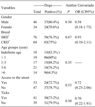
Of the 80 dogs surveyed, 61 (76.3%) had E. canis DNA in one of the surveyed samples. No association was observed between gender, age, race, origin of dogs, free access to the street, or the presence of ticks (Table 1). In the analysis of biological samples, the buffy coat were positive in 42 (52.5%) and the bone marrow was positive in 33 (41.3%) of 80 dogs. Of the
positive dogs, 14 (17.5%) showed DNA amplification
of E. canis in both samples. The PCR products of eight dogs (10%) were sequenced, resulting in DNA sequences 100% identical to the sequences of E. canis in GenBank (accession numbers KP844663.1, KJ995842.1, KF972452.1, and JX118827.1).
Of all the dogs, 29 (36.3%) were asymptomatic, and 51 (63.8%) showed some clinical signs consistent with infection by E. canis, of which, 21 (72.4%) and 40 (78.4%), respectively, were
positive by PCR, with no statistically significant
observed were apathy, weight loss, lymphadenopathy, splenomegaly, hepatomegaly and ophthalmopathies.
For clinical the presence or absence of
clinical signs, there was no significant association
between the positivity of the biological samples, buffy coat and bone marrow (Table 2). There was
no significant association between the two types of
biological samples within the asymptomatic dog group (P=0.24) and symptomatic group (P=0.49).
DISCUSSION
The high incidence of infection with E. canis found in this study is consistent with data obtained in different regions of Brazil (DAGNONE et
al., 2009; TANIKAWA et al., 2013) and Mato Grosso (MELO et al., 2011) based on serological analysis. In this study, nested PCR of buffy coat and bone marrow samples showed an occurrence of infection in 76%
of dogs. This finding differs from that of WITTER
et al. (2013), who analysed buffy coat samples and observed E. canis infection in 23.3% of dogs. The high occurrence can be related using two sites where bacteraemia is observed during different stages of the disease (MILONAKIS et al., 2003).
Direct exposure to the vector tick, R. sanguineus (CARLOS et al., 2011) and age (COSTA JUNIOR et al., 2007) were considered risk factors for infection with E. canis in other regions; however, these factors were not associated with infection in the dogs surveyed, as described by SILVA et al. (2012).
In epidemiological analyses, the blood sample is easily obtainable and provides good results (SILVA et al., 2012; SANTOS et al., 2013). However, the detection of the agent can decrease at this site with the progression of infection or the treatment of bacteraemia compared to other sites (HARRUS et al., 2004; BANETH et al., 2009). In this study, the
highest percentage of DNA amplification in buffy coat
indicates acute infection as demonstrated by BANETH et al. (2009), who analysed an experimental infection with E. canis in dogs in blood and spleen. However, determining the stage of infection in dogs naturally
infected with canine ehrlichiosis is difficult because
of the possible presence of similar acute and chronic infection clinical signs (HARRUS & WANER, 2011).
The absence of apparent clinical signs and the long duration of the subclinical stage may hinder the detection of infection (HARRUS & WANER, 2011; WANER & HARRUS, 2013), likely represented in this study by the asymptomatic group. HARRUS et al. (1998) observed the increased detection of E. canis in spleen samples in the subclinical stage, and in blood and bone marrow in the acute phase, whereas the number of positive animals detected by PCR using blood and spleen samples was similar (HARRUS et al., 2004). However, spleen samples are not routinely
Table 1 - Epidemiological factors associated with positive PCR for Ehrlichia canis in a population of pet dogs in Várzea Grande, Mato Grosso.
---Dogs--- Análise Univariada Variables
Total Positive (%) P OR (CI95%) Gender
Male 46 37(80.4%) 0.30
Female 34 24(70.6%)
0.58 (0.18-1.75) Breed
SRD* 76 58(76.3%) 0.67
CRD* 04 03(75%)
0.93 (0.10-2.11) Age groups (year)
Indefinite age 18 15(83.3% )
< 1 10 06(60%)
1-3 17 15(88.2%)
3-6 21 16(76.2%)
>6 14 9(64.3%)
0.35
---Access to the street
Yes 33 24(72.7%)
No 47 37(78.7%) 0.53
0.72 (0.23-2.06) Ticks
Yes 41 30(73.2%)
No 39 31(79.5%) 0.50
0.70 (0.22-1.91)
*SRD - Without defined Race; *CRD - With defined Race.
Table 2 - DNA detection of E. canis using nested PCR in the buffy coat and bone marrow compared to clinical signs observed in dogs in Várzea Grande, Mato Grosso.
---Biological Samples---Clinic Analysis
Negative + / (%)
Buffy Coat + / (%)
Bone Marrow + / (%)
Both + / (%)
χ2 P
Asymptomatic 8 (10) 12 (15) 6 (7.5) 3 (3.8) 2.42 0.49
Symptomatic 11 (13.8) 16 (20) 13 (16.3) 11 (13.8)
used in clinical practice because obtaining the samples is invasive (BANETH et al., 2009).
MYLONAKIS et al. (2004) and SIARKOU et al. (2007) detected E. canis DNA in bone marrow samples of 68.42% and 75% of surveyed dogs, respectively, during the chronic phase of infection. Despite no association between clinical signs and samples, there was a higher rate of observation of E. canis DNA in blood samples, independent of the analysed group. However, MOREIRA et al.
(2005) identified a greater number of developmental
forms of E. canis in the bone marrow compared to dog blood during the acute phase of the disease. MYLONAKIS et al. (2003) reported the occurrence of E. canis sequestration in the spleen during the subclinical stage and chronic disease as well as in the bone marrow during the chronic phase.
CONCLUSION
Infection with E. canis is highly prevalent in dogs in the city of Várzea Grande, Mato Grosso, and bone marrow is a good site for the molecular diagnosis of canine ehrlichiosis, particularly when infection is suspected despite of negative blood sample.
ACKNOWLEDGEMENTS
The Coordenação de Aperfeiçoamento de Pessoal de Nível
Superior (CAPES) Brasil, AUXPE nº 3531/2014, for financial support..
BIOETHICS AND BIOSSECURITY COMMITTEE APPROVAL
The Ethics Committee on Animal Use approved this study (CEUA - UFMT) Protocol 23108.018081/12-0.
REFERENCES
AGUIAR, D.M.; MELO, A.L. Divergence of the TRP36 protein (gp36) in Ehrlichia canis strains found in Brazil. Ticks and Tick-borne Diseases, n.6, p.103-105, 2015. Available from: <http:// www.sciencedirect.com/science/article/pii/S1877959X14001952>. Accessed: Dec. 20, 2014. doi: 10.1016/j.ttbdis.2014.10.003.
BANETH, G. et al. Longitudinal quantification ofEhrlichia canis in experimental infection with comparison to natural infection. Veterinary Microbiology, n.136, p.321-325, 2009. Available from: <http://www. sciencedirect.com/science/article/pii/S0378113508005488>. Accessed: Nov. 17, 2014. doi: 10.1016/j.vetmic.2008.11.022.
CARLOS, R.S.A. et al. Risk factors and clinical disorders of canine ehrlichiosis in the South of Bahia, Brazil. Revista Brasileira de Parasitologia Veterinária, v.16, p.210-214, 2011. Available from: <http://www.scielo.br/scielo.php?pid=S1984-29612011000300006&script=sci_arttext>. Accessed: Nov. 17, 2014. doi: 10.1590/S1984-29612011000300006.
COSTA JR, L.M. et al. Sero-prevalence and risk indicators for canine ehrlichiosis in three rural areas of Brazil. Veterinary Journal, v.174, n.3, p.673-676, 2007. Available from: <http:// www.sciencedirect.com/science/article/pii/S1090023306002413>. Accessed: Nov. 17, 2014. doi: 10.1016/j.tvjl.2006.11.002. DAGNONE A.S. et al. Molecular diagnosis of Anaplasmataceae organisms in dogs with clinical and microscopical signs of ehrlichiosis. Revista Brasileira de Parasitologia Veterinária, v.18, n.4, p.20-25, 2009. Available from: <http://www.scielo.br/ scielo.php?script=sci_arttext&pid=S1984-29612009000400004>. Accessed: Nov. 17, 2014. doi: 10.4322/rbpv.01804004.
GOMES, A.H.S. et al. PCR identification of Leishmania in diagnosis and control of canine leishmaniasis. Veterinary Parasitology, n.144, p.234-241, 2007. Available from: <http:// www.sciencedirect.com/science/article/pii/S0304401706006030>. Accessed: Nov. 17, 2014. doi: 10.1016/j.vetpar.2006.10.008. HARRUS, S. et al. Investigation of splenic functions in canine monocytic ehrlichiosis. Veterinary Immunology and Immunopathology, v.62, p.15-27, 1998. Available from: <http://www.sciencedirect.com/ science/article/pii/S016524279700127X?np=y>. Accessed: Nov. 17, 2014. doi: 10.1016/S0165-2427(97)00127-X.
HARRUS, S. et al. Comparison of simultaneous splenic sample PCR with blood sample PCR for diagnosis and treatment of experimental Ehrlichia canis infection. Antimicrobial agents and chemoteraphy, v.48, n.11, p. 4488-4490, 2004. Available from: <http://aac.asm.org/content/48/11/4488.short>. Accessed: Nov. 17, 2014. doi: 10.1128/AAC.48.11.4488-4490.2004. HARRUS, S.; WANER, T. Diagnosis of canine monocytotropic ehrlichiosis (Ehrlichia -canis): An overview. Veterinary Journal, n.187, p.292-296, 2011. Available from: <http://www.sciencedirect. com/science/article/pii/S1090023310000353>. Accessed: Nov. 17, 2014. doi: 10.1016/j.tvjl.2010.02.001.
MELO, A.L.T. et al. Seroprevalence and risk factors to Ehrlichia
spp. and Rickettsia spp. in dogs from the Pantanal Region of Mato Grosso State, Brazil. Ticks and Tick-borne Diseases n.2, p.213-218, 2011. Available from: <http://www.sciencedirect.com/ science/article/pii/S1877959X11000689>. Accessed: Nov. 17, 2014. doi: 10.1016/j.ttbdis.2011.09.007.
MOREIRA, S.M. et al. Detection of Ehrlichia canisin bone marrow aspirates of experimentally infected dogs. Ciência Rural, v.35, n.4, p.958-960, 2005. Available from: <http://www.scielo. br/scielo.php?pid=S0103-84782005000400038&script=sci_ arttext&tlng=es>. Accessed: Nov. 17, 2014. doi: 10.1590/S0103-84782005000400038.
MYLONAKIS, M.E. et al. Evaluation of cytology in the diagnosis
of acute canine monocytic ehrlichiosis: a comparison between five
methods. Veterinary Microbiology, v.91, n.2, p.197-204, 2003. Available from: <http://www.sciencedirect.com/science/article/pii/ S0378113502002985>. Accessed: Nov. 17, 2014. doi: 10.1016/ S0378-1135(02)00298-5.
SANTOS, L.G.F.D. et al. Molecular detection of Ehrlichia canis
in dogs from the Pantanal of Mato Grosso State, Brazil. Revista Brasileira de Parasitologia Veterinária, v.22, n.1, p.114-118, 2013. Available from: <http://www.scielo.br/scielo.php?pid=S1984-29612013000100114&script=sci_arttext>. Accessed: Nov. 17, 2014. doi: 10.1590/S1984-29612013005000013.
MURPHY, G.L. et al. Molecular and serologic survey of Ehrlichia canis, Ehrlichia chaffeensis, and E. ewingii in dogs and ticks from Oklahoma. Veterinary Parasitology, v.79, p.325-339, 1998. Available from: <http://www.sciencedirect.com/science/article/pii/ S0304401798001794>. Accessed: Nov. 17, 2014. doi: 10.1016/ S0304-4017(98)00179-4.
OLIVEIRA L. et al. First report of Ehrlichia ewingii detected by molecular investigation in dogs from Brazil. Clinical Microbiology Infection, n.15, p.55-56, 2009. Available from: <http://onlinelibrary. wiley.com/doi/10.1111/j.1469-0691.2008.02635.x/epdf>. Accessed: Nov. 17, 2014. doi: 10.1111/j.1469-0691.2008.02635.x.
SIARKOU, V.I. et al. Sequence and phylogenetic analysis of the 16S rRNA gene of Ehrlichia canis strains in dogs with clinical monocytic ehrlichiosis. Veterinary Microbiology, n.125, p.304– 312, 2007. Available from: <http://www.sciencedirect.com/ science/article/pii/S0378113507002684>. Accessed: Nov. 17, 2014. doi: 10.1016/j.vetmic.2007.05.021.
SILVA, G.C.F. et al. Occurrence of Ehrlichia canis and
Anaplasma platys in household dogs from northern Parana. Revista Brasileira de Parasitologia Veterinária, v.21, n.4, p.379-385, 2012. Available from: <http://www.scielo.br/scielo. php?script=sci_arttext&pid=S1984-29612012005000009&lng= en&nrm=iso&tlng=en>. Accessed: Nov. 17, 2014. doi: 10.1590/ S1984-29612012005000009.
TANIKAWA, A. et al. Ehrlichia canis in dogs in a semiarid region of Northeastern Brazil: Serology, molecular detection and associated factors. Research in Veterinary Science, n.94, p.474-477, 2013. Available from: <http://www.ncbi.nlm.nih.gov/pubmed/23141416>. Accessed: Nov. 17, 2014. doi: 10.1016/j.rvsc.2012.10.007. WANER, T.; HARRUS, S. Canine monocytic ehrlichiosis–from pathology to clinical manifestations. Israel Journal of Veterinary Medicine, v.68, p.1, 2013. Available from: <http://www.ijvm. org.il/sites/default/files/canine_mononocytic_ehrlichiosis.pdf>. Accessed: Jan. 22, 2015.
