Vol.57, n.6: pp. 851-859, November-December 2014 http://dx.doi.org/10.1590/S1516-8913201402555
ISSN 1516-8913 Printed in Brazil
BRAZILIAN ARCHIVES OF BIOLOGY AND TECHNOLOGY
A N I N T E R N A T I O N A L J O U R N A L
Induction
and
Morpho-Ultrastructural
Analysis
of
Organogenic Calli of a Wild Passionfruit
Milene Alves de Figueiredo Carvalho
1*, Renato Paiva
2, Vanessa Cristina Stein
3, Raírys
Cravo Herrera
4, Jorge Marcelo Padovani Porto
5, Daiane Peixoto Vargas
6and Eduardo
Alves
71Embrapa Café; Brasília - DF - Brasil. 2Departamento de Biologia; Setor de Fisiologia Vegetal; Universidade
Federal de Lavras, Lavras - MG - Brasil. 3Universidade Federal de Goiás; Jataí - GO - Brasil. 4Universidade Federal do Pará; Altamira - PA - Brasil. 5Instituto Federal do Espírito Santo; Nova Venécia - ES - Brasil.
6Embrapa Clima Temperado; Pelotas - RS - Brasil. 7Departamento de Fitopatologia; Laboratório de Microscopia
Eletrônica e Análise Ultraestrutural; Universidade Federal de Lavras; Lavras - MG - Brasil
ABSTRACT
This work studied a new protocol for organogenic calli induction and characterization of the morphology and ultrastructure of callogenesis in leaf explants of Passiflora gibertii N. E. Brown, a native passion fruit species from Brazil. Calli induction was performed in different growth conditions (light and dark), different MS medium salt concentrations (MS and MS half strength) and the presence or absence of coconut water. The leaf explants maintained in the dark were more responsive to bud formation. In order to reduce spending on in vitro culture, the most suitable induction medium for P. gibertii organogenesis could, therefore be the MS half strength salt concentration medium maintained in the dark. The addition of coconut water to the culture medium was essential for both calli induction and bud formation. The morphological and ultrastructural features of the organogenic calli were isodiametric cells, characterized by an organized cellular system, nucleus with prominent nucleoli, presence of starch grains and dense cytoplasm rich in endoplasmic reticulum. The scanning electron microscopy demonstrated that buds were present on these calli.
Key words: Passifloraceae, callogenesis, electromicroscopy, optic microscope, tissue culture
*Author for correspondence:[email protected]
INTRODUCTION
Brazil is the center of diversity for the Passifloraceae family. It hosts more than 150
native species, including some wild Passiflora
species that are not commercially cultivated (Bernacci et al. 2008). Breeding programs may contribute to improving disease resistance in
existing commercial cultivars. One native
Brazilian species with considerable potential in
this context is Passiflora gibertii N. E. Brown, a
fast growing vine with attractive mauve/purple
flowers, which is apparently resistant to several important passion fruit diseases such as premature death and other diseases caused by soil-borne pathogens (Junqueira et al. 2005).
In vitro culture of native Passiflora spp. is a key
tool for the production of clones with enhanced resistance and other desirable traits. Numerous
tissue culture studies in Passiflora have been
al. 2012). However, this is not the case for P.
gibertii, where there have been limited studies on
in vitro cultivation (Faria et al. 2006; Faria et al.
2007; Carvalho et al. 2013).
The cytokinin 6-benzylaminopurine
(benzyladenine) (BA) is very effective at promoting multiplication in several species (Lima-Brito et al. 2011; Sá et al. 2012), including
Passiflora spp. such as P. cincinnata Mast.
(Lombardi et al. 2007; Silva et al. 2011), P.
suberosa L. (Garcia et al. 2011), P. edulis Sims
(Silva et al. 2011) and P. alata Curtis (Pacheco et
al. 2012). Coconut water contains substances that are essential for the growth and for regeneration of
many species such as P. edulis x P. edulis var
flavicarpa (Hall et al. 2000), P. edulis (Fernando
et al. 2007) and P. alata Curtis (Pacheco et al.
2012). It is used as a growth supplement in plant tissue culture and its chemical composition is rich in sugars, vitamins, minerals, amino acids and phytohormones such as cytokinins that stimulate the division of mature plant cells (Yong et al. 2009).
Histological, morphological and ultrastructural analyses can be performed to monitor the regeneration and to differentiate organogenesis (Fernando et al. 2007; Silva et al. 2011; Rocha et al. 2012) from somatic embryogenesis (Pinto et al. 2011). By characterizing the regeneration path, the best growth conditions can be established allowing the development of efficient protocols for the induction and production of plants. There are no
reports of organogenic calli induction in P. gibertii
in the literature. It would, therefore, be useful to provide a detailed account of the morphological
and ultrastructural changes during in vitro
organogenic callus induction to form the seedlings from this native passion fruit. In this context, the objective of this study was to establish a protocol for organogenic calli induction on leaf explants of
P. gibertii and to characterize the morphology and
ultrastructure of these calli.
MATERIAL AND METHODS
Seeds of P. gibertii (access CPAC MJ-22-01) were
obtained from the Embrapa Cerrados (CPAC) germplasm collection, Planaltina, DF. They were
germinated in Plantmax HT® and the parent plants
were maintained in a growth room at 25±2°C,
photon irradiance of 43 µmol m-2 s-1 and subjected
to a 16 h photoperiod. The plants were 52 and 109
days old for the first and second experiment, respectively. The leaves collected from mother plants were used as explants for the experiments. In a laminar growth chamber, the leaves were first
decontaminated by immersion in sodium
hypochlorite solution (NaOCl) containing 0.5% active chlorine and Tween 20 (one drop per 100 mL NaOCl) for 10 min and then washed three times with sterilized distilled water. After
decontamination, leaves were excised (± 1 cm2)
and the surfaces of the explants were given small cuts with a scalpel to induce callogenesis. The explants were then inoculated through placing the abaxial surface in contact with the medium. In the first experiment, different growth conditions (light and dark) and different MS (Murashige and Skoog 1962) medium salt concentrations (full and half MS strengths) were tested. The media were supplemented with 8.88 µm BA, 3% sucrose, 5%
coconut water and 0.5% agar (Vetec®). The second
experiment assessed the influence of presence or absence of 5% coconut water in the MS half strength medium salt concentration supplemented with 8.88 µm BA, 3% sucrose and 0.5% agar. To obtain the organogenic calli, explants with calli from both experiments, were transferred to MS
half strength salt concentration medium,
supplemented with 2.22 µm BA, 3% sucrose and 0.5% agar. The organogenic calli were transferred to MSM medium (Monteiro et al. 2000a) with 3%
sucrose and 2.89 µm gibberellic acid (GA3). The
pH for all the media was adjusted to 5.8 ± 0.1 before autoclaving at 120°C for 20 min. After inoculation, the explants from the first experiment were maintained under dark or in growth room
under 36 µmol m-2 s-1 irradiance with 16 h
photoperiod (light), both at 25 ± 2°C for 30 days. For the second experiment, the explants were maintained under dark and for calli from both the experiments and for the organogenic calli, the explants were cultivated in the light, all of them for a period of 30 days each.
correlations and Kruskal-Wallis tests, performed in SAS (SAS Institute, 2004).
Morphological and ultrastructural analyses were performed on calli resulting from the first experiment, cultivated in the MS half strength medium salt concentration, supplemented with 8.88 µm BA, 3% sucrose, 5% coconut water and 0.5% agar, maintained 30 days on dark or light and calli from dark sub-cultured to a new medium (MS
half strength salt concentration medium
supplemented with 2.22 µm BA, 3% sucrose and 0.5% agar) and maintained for seven days in a
growth room under 36 µmol m-2 s-1 irradiance, at
25 ± 2°C and 16 h photoperiod. In order to analyze the calli under scanning and transmission electron microscopy, they were fixed in modified
Karnovisky [glutaraldehyde (2.5%) and
paraformaldehyde (2.5%) in cacodylate buffer, pH 7.2] at room temperature for at least 24 h. The calli were then washed in 0.05 M cacodylate buffer (three times every 10 min) and subsequently fixed in a solution containing 1% osmium tetroxide and 0.05 M cacodylate buffer for 4 h. The calli were then dehydrated in an ascending acetone gradient (25, 50, 75 and 90%) for 10 min and three times (10 min each) in 100% acetone.
Transmission Electron Microscopy (TEM)
analysis was performed immediately after
dehydration The calli were put in an ascending gradient acetone/Spurr resin 30% for 8 h, 70% for 12 h and finally twice at 100% with a 24 h interval. To polymerize, the tissues were molded in pure silicon resin and dried in a forced-air oven at 70°C for 48 h. The blocks obtained were subjected to thinning, using a razor blade to section the excessive resin. Subsequently, the blocks were cut into semi-thin (1 µm) and ultrathin (<100 nm) sections by using a Reichert-Jung (ultra-cut) ultra-microtome, with a diamond blade.
The semi-thin sections were collected with a gold ring and put on glass slides and were later stained with toluidine blue (1.0 g toluidine blue, 1.0 g sodium borate and 100 mL water purified in Millipore 0.2 µm filter) and permanently mounted in Permoult medium. The ultra-thin sections were collected on formar-coated slot grids (Rowley and Moran 1975). These sections were post-stained with uranyl acetate, followed by lead acetate for 3 min and later examined in a Zeiss transmission electron microscope (EM 902 to 80Kv model). For Scanning Electron Microscopy (SEM) analysis, after dehydration, the calli were dried in
a critical point dryer CPD 030 using liquid CO2.
The samples were then sputtered with gold prior to SEM analysis. Observations were made by using an electron microscope (LEO Evo 040) operating between 10 and 20 kV.
RESULTS AND DISCUSSION
There was no statistical difference in calli induction between the different concentrations of MS medium under different environmental conditions (light and dark), with average score of 0.5. When calli explants were transferred to the MS half strength salt concentration medium, supplemented with BA (2.22 µm) and maintained in a growth room under light, only a few explants formed in the dark showed buds after 30 days. After this period, the calli were transferred to
MSM culture medium with 2.89 µm GA3 and
maintained in a growth room under light. Several replicates from the calli originated from dark and sub-cultured to this new medium presented buds (Fig. 1A), while the calli cultured in the light showed brown color and only one replicate from this calli produced the buds (Fig. 1B).
Figure 1 - Organogenic calli obtained from Passiflora gibertii leaf segments from dark (A) and light
Monteiro et al. (2000b) also observed bud
formation when they cultured leaves of P.
suberosa in vitro in MS medium in the dark at 26
± 2°C for four weeks and then transferred calli with organogenic aspects to MSM medium with
2.89 µm GA3 under light. However, when tissues
of P. suberosa were incubated in the dark without
a subsequent period in the light, no significant shoot formation occurred (Garcia et al. 2011). The browning in calli occurs mainly due to the oxidation of phenolic compounds by phenol oxidase (Erland and Mahmoud 2014) and it is likely that the products of this oxidation process are formed in the light (Afshari et al. 2011; Ndakidemi et al. 2014). Phenolic secretions and other exudates in plant tissue culture systems lessen explant initiation, growth and development (Ndakidemi et al. 2014; Yildirim and Turker 2014). The light source also causes an increase in lipid peroxidation leading to lower rates of physiological processes (Tariq et al. 2014; Yildirim and Turker 2014) as observed in the
present work with P. gibertii calli cultured in the
light.
The use of coconut water to the culture medium
significantly influenced calli induction. Higher levels of calli induction were observed using coconut water (score of 0.72) compared with the absence of coconut water (score 0.28) (Fig. 2). The calli from the medium with coconut water presented clear bud formation (Fig. 3A), while calli from the medium without coconut water did not produce buds (Fig. 3B) after 30 days transferred to a new medium and maintained in the light.
Figure 2 - Callus induction in Passiflora gibertii leaf
segments in the presence and absence of coconut water in a MS half strength salt concentration medium with BA (8.88 µ m) at 30 days of cultivation.
Figure 3 - Organogenic calli obtained from Passiflora gibertii leaf segments grown in culture medium
with (A) and without (B) coconut water. Bar = 3 mm (A, B).
These results were in general agreement with several studies (Hall et al. 2000; Pacheco et al. 2012), indicating that coconut water and BA added
to the culture medium of Passiflora spp. promoted
the organogenesis. Kantharajah and Dodd (1990) reported that the addition of 20% coconut water to MS medium with BA 8.88 µm significantly
increased bud production of P. edulis var. Norfolk
Island. Similarly, Fernando et al. (2007) reported
that the direct organogenesis from leaf discs of P.
edulis occurred only when leaf explants were
cultured on MS medium supplemented with 4.44
µm BA and 5% of coconut water. Coconut water
may also be beneficial for the in vitro cultivation
of other species. For example, Al-Khayri et al. (1992) observed that the addition of 15% coconut water to the culture medium increased callus growth, regenerative potential and bud growth in
spinach (Spinacia oleracea L.).
The composition of coconut water, the liquid
endosperm of coconut (Cocos nucifera L.),
chemical composition of sugars, vitamins,
minerals, proteins, amino acids and
phytohormones and it is extensively used as a growth-promoting supplement in plant tissue culture (Ma et al. 2008; Yong et al. 2009). Coconut water, contains zeatin cytokinin, normally used to induce plantlet regeneration from the callus in plant tissue culture by stimulating the division of mature plant cells (Yong et al. 2009). It also contains indole-3-acetic acid auxin (Ma et al. 2008) that has the function of patterned differentiation of cells in meristems and immature organs (Uphade et al. 2008) and the vitamin C, which prevents the oxidation and protects the calli and shoot tissues and vitamins that are
water-soluble and are required as coenzymes for enzymatic reactions essential for cellular function (Ma et al. 2008; Prades et al. 2012). The above could explain the organogenic calli obtained in the present study.
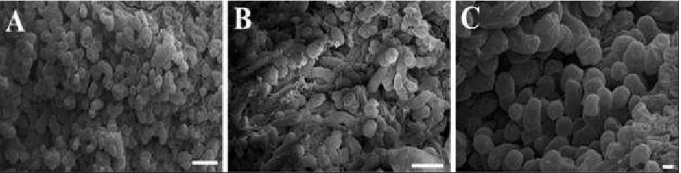
Morphological and ultrastructural differences were observed among the cells from different calli. Calli grown on the medium with BA and maintained in the light were characterized by the cells with both isodiametric (Fig. 4A) and elongated shape (Fig. 4B). In contrast, calli cultured on the medium with BA and maintained in the dark showed well-organized cell proliferation with an isodiametric structure (Fig. 4C).
Figure 4 - Scanning electron micrographs of Passiflora gibertii calli grown in MS half strength salt
concentrations medium with BA, and maintained in the light (A, B) and in the dark (C). Bars = 100 µm (A, B), 20 µm (C).
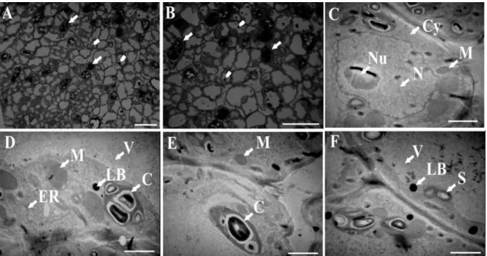
Studies using light microscopy (LM) and transmission electron microscopy (TEM) indicated that calli cells were dividing in an organized system in the calli cultured in the light (Fig. 5A and B) and in the dark (Fig. 6A and B). These calli also showed some common characteristics: a nucleus with prominent nucleoli (Figs. 5C and 6C), granular vacuole (Figs. 5D and F and 6C and D), dense cytoplasm rich in endoplasmic reticulum (Figs. 5C and D and 6C and F), starch grains (Figs. 5B and 5F and 6B and D), lipid bodies (Figs. 5D and F and 6F) and phenolic compounds (Figs. 5A and B and 6A, B and E). However, calli cultured in the light also had a cytoplasm rich in mitochondria and immature chloroplasts (Figs. 5C and E).
Deposition of phenolic compounds, such as tannins on calli cultivated in the light and on the dark is mainly found in the plant vacuoles (Parham and Kaustinen 1977) where they do not interfere with cellular metabolism unless an injury or
cellular death happen. Knowledge of the
biochemical makeup of the Passiflora genus
comes from mainly studies on P. incarnata, P.
edulis and P. alata and phytochemical studies are
generally scarce for this genus (Costa and Tupinambá 2005). The chemical groups most
frequently reported are indole alkaloids,
flavonoids, sterols, lignans and cyanoglucosides. Since there has been reports on the presence of
phenols for the Passiflora genus and tannins are
polyphenols, there could be a possibility that this
metabolite was also present in P. gibertii.
The ultrastructural cell characteristics observed in the present study have also been reported by other
authors. Meristemoids cells in P. edulis
(Rocha et al. 2012) and Bauhinia forficata
and Glycine max (Appezzato-da-Glória and
Machado 2004) presented dense cytoplasm,
numerous mitochondria, prominent nuclei,
concentrically arranged rough endoplasmic
Figure 5 - Photomicrographs (A-B) and transmission electron micrographs (C-F) of Passiflora gibertii callus cells grown in a MS half strength salt concentrations medium with BA and maintained in the light. M = mitochondria, ER = endoplasmic reticulum, Cy = cytoplasm, V = vacuole, LB = lipid bodies, S = starch, N = nucleus, Nu = nucleolus, C = chloroplast. In A and B, cells with phenolic compound accumulation = arrows, starch grains = arrowheads. Bars = 50 µm (A, B), 2 µm (C, D, E, F).
Figure 6 - Photomicrographs (A-B) and transmission electron micrographs (C-F) of Passiflora
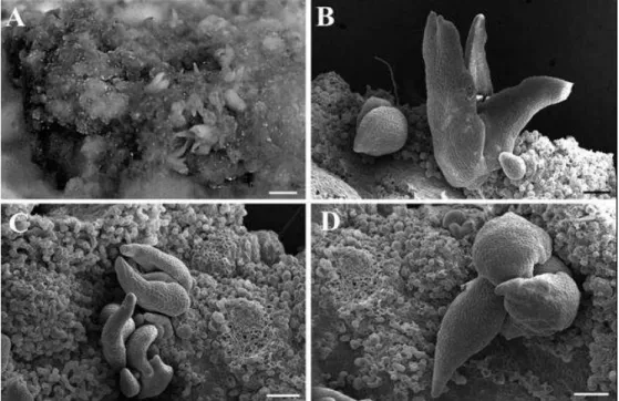
The meristemoids of B. forficata and G. max did not originate from typical meristemoids cells, but rather from highly vacuolated cells (Appezzato-da-Glória and Machado 2004). Similar results were observed in the present study where cells with organogenic potential (Fig. 7A) were highly vacuolated with cytoplasm rich in organelles and starch grains (Figs. 5 and 6). These calli cells, formed in the dark and subsequently transferred to a MS half strength salt concentrations medium
with 2.22 µm BA maintained in the light, became organogenic (Fig. 7A), forming buds and foliar primordia after seven days (Fig. 7) in a new
medium. The SEM results demonstrated
morphological characterization of shoot buds based on emergence of the foliar primordia from a common axis (Figs. 7B, C and D) and not direct insertion of the base into the explants (isolated leaf structures).
Figure 7 - Organogenic calli obtained from leaves of Passiflora gibertii grown in MS half strength
salt concentrations medium with BA and maintained in the dark and seven days after transferring to new medium in the light. General aspect (A), scanning electron micrographs (B-D). Bars = 2 mm (A), 200 µ m (B, C, D).
Appezzato-da-Glória et al. (2005) described SEM
observations of in vitro structures that were
essential for quick morphological characterization, allowing the distinction between bud and leaf primordia. According to these authors, in
Passiflora, the formation of leaf structures was
frequently misinterpreted as buds that did not
elongate. Structural analysis of the in vitro
organogenesis in P. edulis f. flavicarpa Deg. and
P. cincinnata Mast. demonstrated the strong
tendency of explants to form leaf primordial that was not observed in the present work. Apparently this organogenic path compromises the success of
in vitro culture, because the presence of the shoot
apical meristem is essential for shoot production.
CONCLUSIONS
Passiflora gibertii calli produced in the dark were
more responsive to bud formation. In order to
reduce the spending on in vitro culture, the most
suitable induction medium for P. gibertii
organogenesis could, therefore, be the MS half strength salt concentration medium maintained in the dark. However, the addition of coconut water to the culture medium was essential for both callus induction and bud formation.
starch grains and dense cytoplasm rich in endoplasmic reticulum. The scanning electron microscopy demonstrated that the buds were present on these calli.
ACKNOWLEDGEMENTS
This work was financially supported by the following Brazilian agencies: Fundação de Amparo à Pesquisa do Estado de Minas Gerais (FAPEMIG), Coordenação de Aperfeiçoamento de Pessoal de Nível Superior (CAPES) and Conselho Nacional de Desenvolvimento Científico e Tecnológico (CNPq).
REFERENCES
Afshari RT, Angoshtari R, Kalantari S. Effects of light and different plant growth regulators on induction of callus growth in rapeseed (Brassica napus L.) genotypes. Plant Omics J. 2011; 4(2): 60-67.
Al-Khayri JM, Huang FH, Morelock TE, Busharar TA. Spinach tissue culture improved with coconut water. Hort Science. 1992; 27(4): 357-358.
Anand SP, Jayakumar E, Jeyachandran R, Nandagobalan V, Doss A. Direct organogenesis of Passiflora foetida L. through nodal explants. Plant Tissue Cult & Biotech. 2012; 22(1): 87-91.
Appezzato-da-Glória B, Fernando JA, Machado SR, Vieira MLC. Estudos morfológicos, anatômicos, histoquímicos e ultra-estruturais da organogênese in vitro do maracujazeiro. In: Faleiro FG, Junqueira NTV, Braga MF, editors. Maracujá: germoplasma e melhoramento genético. Planaltina: Embrapa Cerrados; 2005. p. 382-407.
Appezzato-da-Glória B, Machado SR. Ultrastructural analysis of in vitro direct and indirect organogenesis. Rev Bras Bot. 2004; 27(3): 429-437.
Bernacci LC, Soares-Scott MD, Junqueira NTV, Passos IRS, Meletti LMM. Passiflora edulis Sims: the correct taxonomic way to cite the yellow passion fruit (and of others colors). Rev Bras Frutic. 2008; 30(2): 566-576.
Carvalho MAF, Paiva R, Alves E, Nogueira RC, Stein VC, Castro EM, et al. Morphogenetic potential of native passion fruit (Passiflora gibertii N. E. Brown.) calli. Braz J Bot. 2013; 36(2): 141-151.
Costa AM, Tupinambá DD. O maracujá e suas propriedades medicinais: estado da arte. In: Faleiro FG, Junqueira NTV, Braga MF, editors. Maracujá: germoplasma e melhoramento genético. Planaltina: Embrapa Cerrados; 2005. p. 474-506.
Erland LAE, Mahmoud SS. An efficient method for regeneration of lavandin (Lavandula x intermedia cv. 'Grosso'). In vitro Cell Dev Biol Plant. In Press. 2014. DOI: 10.1007/s11627-014-9614-4.
Faria GA, Costa MAPC, Junghans TG, Ledo CAS, Souza AS. Efeito da sacarose e sorbitol na conservação in vitro de Passiflora giberti N. E. Brown. Rev Bras Frutic. 2006; 28(2): 267-270. Faria GA, Costa MAPC, Ledo CAS, Junghans TG,
Souza AS, Cunha MAP. Meio de cultura e tipo de explante no estabelecimento in vitro de espécies de maracujazeiro. Bragantia. 2007; 66(4): 535-543. Fernando JA, Vieira MLC, Machado SR,
Appezzato-da-Glória B. New insights into the in vitro organogenesis process: the case of Passiflora. Plant Cell Tiss Org. 2007; 91(1): 37-44.
Garcia R, Pacheco G, Falcão E, Borges G, Mansur E. Influence of type of explant, plant growth regulators, salt composition of basal medium, and light on callogenesis and regeneration in Passiflora suberosa L. (Passifloraceae). Plant Cell Tiss Org. 2011; 106(1): 47-54.
Hall RM, Drew RA, Higgins CM, Dietzgen RG. Efficient organogenesis of an Australian passionfruit hybrid (Passiflora edulis x Passiflora edulis var flavicarpa) suitable for gene delivery. Aust J Bot. 2000; 48(5): 673-680.
Junqueira NTV, Braga MF, Faleiro FG, Peixoto JR, Bernacci LC. Potential of wild species of passionfruit as sources of resistance to diseases. In: Faleiro FG, Junqueira NTV, Braga MF, editors. Passionfruit: germplasm and breeding. Planaltina: Embrapa Cerrados; 2005. p. 143-148.
Kantharajah AS, Dodd WA. In vitro micropropagation of Passiflora edulis (purple passionfruit). Ann Bot. 1990; 65(3): 337-339.
Lima-Brito A, Resende SV, Lima COC, Alvim BM, Carneiro CE, Santana JRF. In vitro morphogenesis of
Syngonanthus mucugensis Giul. subsp. mucugensis.
Ciênc Agrotec. 2011; 35(3): 502-510.
Lombardi SP, Passos IRS, Nogueira MCS, Appezzato-da-Glória B. In vitro shoot regeneration from roots and leaf discs of Passiflora cincinnata Mast. Braz Arch Biol Technol. 2007; 50(2): 239-247.
Ma Z, Ge L, Lee ASY, Yong JWH, Tan SN, Ong ES. Simultaneous analysis of different classes of phytohormones in coconut (Cocos nucifera L.) water using high-performance liquid chromatography and liquid chromatography-tandem mass spectrometry after solid-phase extraction. Anal Chim Acta. 2008; 610(2): 274-281.
Monteiro ACBA, Nakazawa GT, Mendes BMJ, Rodriguez APM. Regeneração in vitro de Passiflora suberosa a partir de discos foliares. Sci Agric. 2000b; 57(3): 571-573.
Murashige T, Skoog F. A revised medium for rapid growth and bioassays with tobacco tissue cultures. Physiol Plantarum. 1962; 15(3): 473-497.
Ndakidemi CF, Mneney E, Ndakidemi PA. Effects of ascorbic acid in controlling lethal browning in in vitro culture of Brahylaena huillensis using nodal segments. Am J Plant Sci. 2014; 5(1): 187-191. Pacheco G, Garcia R, Lugato D, Vianna M, Mansur E.
Plant regeneration, callus induction and establishment of cell suspension cultures of Passiflora alata Curtis. Sci Hortic. 2012; 144: 42-47.
Parham RA, Kaustinen HM. On the site of tannin synthesis in plant cells. Bot Gaz. 1977; 138(4): 465-467.
Pinto APC, Monteiro-Hara ACBA, Stipp LCL, Mendes BMJ. In vitro organogenesis of Passiflora alata. In vitro Cell Dev Biol Plant. 2010; 46(1): 28-33. Pinto DLP, Almeida AMR, Rêgo MM, Silva ML,
Oliveira EJ, Otoni WC. Somatic embryogenesis from mature zygotic embryos of commercial passionfruit (Passiflora edulis Sims) genotypes. Plant Cell Tiss Org. 2011; 107(3): 521-530.
Prades A, Dornier M, Diop N, Pain J-P. Coconut water uses, composition and properties: a review. Fruits. 2012; 67(2): 87-107.
Rocha DI, Vieira LM, Tanaka FAO, Silva LCS, Otoni WC. Anatomical and ultrastructural analyses of in vitro organogenesis from root explants of commercial passion fruit (Passiflora edulis Sims). Plant Cell Tiss Org. 2012; 111(1): 69-78.
Rowley CR, Moran DT. A simple procedure for mounting wring wrinkle - free sections on formvar - coated slot grids. Ultramicrotomy. 1975; 1(1): 151-155.
Sá AJ, Lédo AS, Lédo CAS, Pasqual M, Silva AVC, Silva Junior JF. Sealing and explant types on the mangaba micropropagation. Ciênc Agrotec. 2012; 36(4): 406-414.
SAS Institute. SAS user’s guide: statistic: version 9.1.3. Cary: SAS Institute; 2004.
Silva CV, Oliveira LS, Loriato VAP, Silva LC, Campos JMS, Viccini LF, et al. Organogenesis from root explants of commercial populations of Passiflora edulis Sims and a wild passionfruit species, P.
cincinnata Masters. Plant Cell Tiss Org. 2011;
107(3): 407-416.
Tariq U, Ali M, Abbasi BH. Morphogenic and biochemical variations under different spectral lights in callus cultures of Artemisia absinthium L. J Photoch Photobio B. 2014; 130(5): 264-271.
Uphade BK, Shelke SS, Thorat DG. Studies on some physico-chemical characteristics of coconut water near sugar and chemical factory, Kopergaon (M. S.). Int J Chem Sci. 2008; 6(4): 2052-2054.
Yildirim AB, Turker AU. Effects of regeneration enhancers on micropropagation of Fragaria vesca L. and phenolic content comparison of field-grown and
in vitro-grown plant materials by liquid
chromatography-electrospray tandem mass spectrometry (LC–ESI-MS/MS). Sci Hortic. 2014; 169: 169-178.
Yong JWH, Ge L, Ng YF, Tan SN. The chemical composition and biological properties of coconut (Cocos nucifera L.) water. Molecules. 2009; 14(12): 5144-5164.