Article
J. Braz. Chem. Soc., Vol. 29, No. 12, 2636-2645, 2018 Printed in Brazil - ©2018 Sociedade Brasileira de Química
*e-mail: [email protected]
Preparation of Magnetoliposomes with a Green, Low-Cost, Fast and Scalable
Methodology and Activity Study against
S.
aureus
and
C.
freundii
Bacterial Strains
Rosangela M. F. da Costa e Silva,*,a Luciano R. S. Lara,a Jorge L. López,b
Ângela L. Andrade,c Junnia A. C. Oliveira,d Jacqueline A. Takahashi,a
Henriete S. Vieira,a Tulio Matencio,a Humberto O. Stumpfa and Rosana Z. Dominguesa
aDepartamento de Química, Universidade Federal de Minas Gerais (UFMG),
Av. Antonio Carlos 6627, Pampulha, 31270-901 Belo Horizonte-MG, Brazil
bCentro de Ciências Biológicas e da Natureza (CCBN), Universidade Federal do Acre (UFAC),
Rodovia BR 364, km 04, s/n, Distrito Industrial, 69915-900 Rio Branco-AC, Brazil
cDepartamento de Química, Universidade Federal de Ouro Preto (UFOP), Morro do Cruzeiro,
35400-000 Ouro Preto-MG, Brazil
dInstituto de Ciências Biológicas, Universidade Federal de Minas Gerais (UFMG),
Av. Antonio Carlos 6627, Pampulha, 31270-901 Belo Horizonte-MG, Brazil
A novel, fast, low-cost and scalable methodology to prepare stable magnetoliposomes (MGLs), without the use of organic solvents, is described. The concept of the work is based on the dual use of soy lecithin associated to a new liposome preparation methodology. Soy lecithin was used to coat the nanoparticles of magnetite (Fe3O4@lecithin) and for encapsulation of Fe3O4@lecithin
(Lip-Fe3O4@lecithin). Liposomes with size less than 160 nm, polydispersity index of 0.25 and
zeta potential of –41 mV, were prepared with the use of autoclave and sonication. The liposomal formulations containing magnetite and stigmasterol (Lip-Fe3O4@lecithin, Lip-Stigma and
Lip-Stigma-Fe3O4@lecithin) were shown to be promising for the application as antibacterial. The
liposomal formulation and magnetite were characterized by the following techniques: conventional and high-resolution transmission electron microscopy (TEM/HRTEM), energy-filtered transmission electron microscopy (EFTEM), proton nuclear magnetic resonance (1H NMR), Fourier transform
infrared spectroscopy (FTIR), X-ray powder diffraction (XRPD), dynamic light scattering (DLS) and zeta potential. The Lip-Fe3O4@lecithin had a minimum inhibitory concentration (MIC) of
8.4 µg mL-1 in the presence of 200 Oe magnetic field against S. aureus.
Keywords: soy lecithin, magnetite, liposomes, S. aureus, C. freundii
Introduction
The disordered use of antibiotics has increased the inefficiency of several classes of antibacterial agents in combating microorganisms called multiresistant. Such microorganisms are present in hospitals of large metropolises or even diverse environments of small communities.1-5 The Gram-positive bacterium
Staphylococcus aureus is the leading cause of human bacterial infections worldwide, being endemic and readily resistant to antimicrobials. S. aureus is listed in the World Health Organization (WHO)6 as one of the six pathogens
with priority II (high) in the development of new antibiotics. Regarding Gram-negative bacteria, the WHO warns of resistance to multiple antibiotics and the ability to transmit genetic material to other bacteria. An example of Gram-negative bacteria, Citrobacter freundii is a fecal coliform that resides in water, soils, and intestines of humans.7
Inorganic pharmaceuticals containing metal oxides may utilize different action mechanisms and can, therefore, be considered a viable alternative to traditional antibiotics.8
One option is the use of iron oxide magnetic nanoparticles (IOMNPs). IOMNPs have been extensively explored in several areas, such as nanocatalysis,9 hyperthermia,10
magnetic resonance imaging11 and cancer treatment.12 The
applications, such as high surface area and heat generation capacity by the application of an external magnetic field.9-13
Several studies on the antibacterial activity of IOMNPs, in particular the magnetite nanoparticles (NPs) are described in the literature.14-17 Limitations due to agglomeration and
interactions with biological media have been overcome by coating the NPs with several classes of stabilizing agents.18-21 Another strategy to increase the interaction of
NPs with biological media is encapsulation in liposomes, preparing so-called magnetoliposomes (MGLs). The encapsulation in liposomal vesicles permits reduced contact between drugs and healthy cells; as well as increased efficiency in traversing the diseased cell membrane to reach the target.18,22
In this paper, soy lecithin, a combination of phospholipids from renewable sources, was used to coat NPs (Fe3O4@lecithin)
and then as liposome for encapsulating the Fe3O4@lecithin.
Two previous studies23,24 using soy lecithin as the coat of
NPs have been described. However, none of the studies used lecithin as a liposome. The addition of the second step for encapsulation of Fe3O4@lecithin was determinant
to obtain more stable MGLs than Fe3O4@lecithin in
the aqueous medium. Another factor that distinguishes our paper from the previous publications is the order of addition of reagents. In our work, soy lecithin was added after the co-precipitation, reducing the possibility of encapsulation residuals of initial reagents in the soy lecithin clusters. Therefore, dual use of the soy lecithin biomimetic agent25 increased the interaction between
Fe3O4 and liposome vesicles in the Lip-Fe3O4@lecithin,
creating new possibilities for the preparation of MGLs. Another enhancement was enabling the MGLs to be highly water-dispersive using a novel methodology that is fast, low-cost and scalable without the use of organic solvents, which were not previously described in the literature. Traditional methodologies such as film hydration26 and
sonication27 require a lot of preparing time and the use of
organic solvents to achieve small quantities of product.28
Subsequently, they are still subjected to a sterilization step.29
Therefore, a new methodology was proposed to prepare sterile liposomes. This methodology involves hydrating the soy lecithin in an autoclave at 120 °C for 15 min, cooling to 4 °C, liposome size reduction, encapsulating stigmasterol and/or Fe3O4@lecithin and using ultrasound tip. The use
of an initial autoclave stage allows preparation of large amounts of the liposomes. The production is only limited by to the capacity of the autoclave equipment that can reach industrial scales. In addition, the influence of use of stigmasterol (Stigma), a molecule similar to cholesterol, was also investigated.28,29 The liposomes prepared were
tested against S. aureus and C. freundii microorganisms in
the presence of a magnetic field (MF41) of 200 Oe, 315 kHz and also in its absence at room temperature (WMF25) and at temperature of 41 °C (WMF41).
Experimental
Materials
Stigmasterol (Sigma-Aldrich Chemical Company, USA), soy lecithin (CRQ, Brazil), iron sulfate II ammoniacal hexahydrate (Vetec, Brazil), iron chloride III hexahydrate (Vetec, Brazil), CDCl3 (Sigma-Aldrich
Chemical Company, USA), ammonium hydroxide PA (Synth, Brazil), sodium hydroxide PA (Synth, Brazil), citric acid (Synth, Brazil), brain heart infusion (BHI) agar (Kasvi, Brazil), BHI broth (Kasvi, Brazil) were used without any purification. The Gram-positive and Gram-negative bacteria used were Staphylococcus aureus (ATCC 29212), and Citrobacter freundii (ATCC 8090), respectively. The ultra-purified water was obtained by Milli-Q system (Millipore, Brazil).
Preparation of the bare liposome (Lip)
Bare liposomes were prepared by hydration of soy lecithin (100 mg mL-1) in ultra-purified water. The
autoclavable flask containing the components were autoclaved (CS Prismatec Autoclave, Brazil) for 15 min at 120 °C. Bare liposomes were maintained at 4 °C for 24 h. Then, the bare liposomes were sonicated on tip ultrasound (Disruptor Unique Model DES 500, Brazil) for reducing sizes.26,30 Standards of 4 cycles × 4 min were performed
with intervals of 1 min for each sequence, at 95% power (500 W). The temperature of sample was maintained below 75 °C. The sample was noted Lip.
Synthesis of Fe3O4@lecithin
Magnetite coated with soy lecithin (Fe3O4@lecithin)
was prepared in a one-pot reaction using the modified co-precipitation methodology of the literature.31,32 Briefly,
in a two-neck round bottom flask, under nitrogen flow, 500 mL of ultra-purified and degassed water were mixed to Fe(NH4)2(SO4)2.6H2O (0.0459 mol), and FeCl3.6H2O
(0.08879 mol). The solution was stirred for 5 min at room temperature. After that, 200 mL of NH4OH was added
under stirring for synthesis of the nanoparticles magnetite (Fe3O4) and stirred for additional 20 min. Sequentially,
100 mL of Lip (100 mg mL-1) was added under constant
stirring for 10 min in the bottom flask. The Fe3O4 coated
water.31,33 The sample was denominated Fe
3O4@lecithin and
was conditioned in Falcon tubes, lyophilized and stored at 0 °C. The yield of this operation was 92%.
Preparation of liposome formulations
Aliquots of 10 mL of the Lip were separated for stigmasterol and/or magnetite components incorporation. The liposomal preparations were named according to their composition: stigmasterol (Lip-Stigma), stigmasterol and coated magnetite (Lip-Stigma-Fe3O4@lecithin), and the
coated magnetite (Lip-Fe3O4@lecithin). A bare liposomal
formulation containing the phospholipid (Lip) was also prepared as standard sample. Lip, Lip-Stigma, Lip-Stigma-Fe3O4@lecithin, Lip-Fe3O4@lecithin were submitted to
the same ultrasound cycle defined previously. Then, the formulations were ultracentrifuged34 for 20 min at 10000 × g
in Eppendorf model 5810R centrifuge (Germany). The supernatant was separated and the formulation resuspended in ultra-purified water. The procedure was performed until all unencapsulated components of Lip, Lip-Stigma, Lip-Stigma-Fe3O4@lecithin and Lip-Fe3O4@lecithin were
removed from the formulations. The formulations were suspended in phosphate buffered saline (PBS buffer) at pH 7.3 ± 0.1, leading to the final concentrations (Table 1) determined by atomic absorption spectroscopy (AAS) and proton nuclear magnetic resonance (1H NMR).
Characterization of Fe3O4@lecithin and liposomes
Zeta potential (ζ) was performed for Fe3O4@lecithin,
L i p , L i p - S t i g m a , L i p - F e3O4@ l e c i t h i n a n d
Lip-Stigma-Fe3O4@lecithin (Zetasizer Nano ZS equipment,
United Kingdom) in triplicates at 25.0 °C for 14 days. The same apparatus was used for analyses of liposome size using dynamic light scattering (DLS). The samples were lyophilized in a Terroni Lyophilizer model LS 3000 (Brazil) for 1H NMR, infrared spectroscopy technique
(FTIR), X-ray powder diffraction (XRPD) and for magnetics measurements. The initial and final mass of all lyophilized samples were measured on analytical balance (Shimadzu, model AUW220D, Brazil). Crystallographic
phases of Fe3O4@lecithin, Lip-Fe3O4@lecithin,
Lip-Stigma-Fe3O4@lecithin were characterized by
XRPD, using Rigaku-Geigerflex equipment (Japan) and Siemens-D5000 diffractometer (Germany), with CuKα
radiation in the range of 5 to 80° and 1° min-1. The iron
oxide phase was determined by fitting (311) peak by Voigt function.35,36 FTIR (PerkinElmer FT-IR GX model, USA)
were performed to evaluate the coating of Fe3O4@lecithin,
iron oxide phase and to verify the absence of hydrolysis of the esters or oxidation in Lip, Lip-Stigma, Lip-Fe3O4@lecithin
and Lip-Stigma-Fe3O4@lecithin. The solids were
homogenized in KBr (1% m/m) and disc-pressed. The FTIR spectra were recorded in transmittance mode in the 4000 to 400 cm-1 range. Morphologies, particle size
distribution, and phases by micrographs were recorded on transmission electron microscope Tecnai G2-20 SuperTwin FEI, 200 kV (USA). The samples were diluted in water and one drop was deposited on holey carbon-Cu, 300 mesh, 50 micron screens.37 The micrographs were obtained
in the conventional and high-resolution transmission electron microscopy (TEM/HRTEM) and energy-filtered transmission electron microscopy (EFTEM) modes. The mean diameter of Fe3O4 in the Fe3O4@lecithin was
calculated by the ImageJ software package. The HRTEM images were processed in the Digital Micrograph program in the fast Fourier transform mode (FFT) for determination of the interplanar spacing.38 Spectroscopic analyses of 1H NMR were recorded on DPR 400 Bruker Avance II
DRX 400 equipment (USA) operating at 400 MHz. This technique was used to determine phospholipids/steroids molar ratio of soy lecithin, stigmasterol in the Lip and Lip-Stigma and to verify the absence of phospholipid hydrolysis. The samples Lip and Lip-Stigma were lyophilized and solubilized in CDCl3. The hysteresis
curves of Fe3O4@lecithin and Lip-Fe3O4@lecithin at 310 K
were obtained on a SQUID (superconducting quantum interference device) Magnetometer Cryogenic S700X (Argentina) with magnetic field capacity up to 7 T. FAAS was used to determine iron content in Fe3O4@lecithin,
Lip-Fe3O4@lecithin and Lip-Stigma-Fe3O4@lecithin on a
Hitachi-Z8200 spectrometer (Japan) coupled to a graphite furnace.
Biological activity without magnetic field at room temperature (WMF25), without magnetic field at 41 ± 1 °C (WMF41) and with magnetic field at 41 ± 1 °C (MF41)
Antibacterial activity of the liposomes was assayed by the agar dilution method.39 Bacteria S. aureus (ATCC 29213)
and C. freundii (ATCC 8090) were individually inoculated in vials containing BHI broth and incubated in an oven at 37 °C
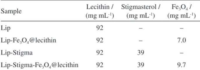
Table1. Content of soy lecithin, stigmasterol and magnetite
Sample Lecithin /
(mg mL-1)
Stigmasterol / (mg mL-1)
Fe3O4 /
(mg mL-1)
Lip 92 – –
Lip-Fe3O4@lecithin 92 – 7.0
Lip-Stigma 92 39 –
for 24 h. Inoculum containing approximately 1 × 10⁶ colony
forming units (CFU) of each microorganism per mL of sterile water were prepared in sterile vials (InocSol). Liposomes samples described in Table 1 and InocSol were added to Eppendorfs containing BHI broth. The final concentrations of liposomes were presented in the Table 2 with total volume of the 1 mL per Eppendorf.
Tests for S. aureus and C. freundii inhibition were done against the liposomes samples prepared under three different conditions: (i) 25 °C for 30 min (WMF25); (ii) oven heating at 41 ± 1 °C for 30 min (WMF41); and (iii) magnetic field for 30 min at 41 ± 1 °C (MF41). The sample with number 2 was tested against S. aureus and C. freundii in the condition: (iii) magnetic field for 30 min at 41 ± 1 °C (MF41). The liposome samples were diluted in BHI agar plates and maintained at 37 °C for 24 h. Then the CFUs were quantified. The procedure was performed in triplicate. Quantification of inhibition of bacterial growth was done according to the literature.39,40 Samples
submitted to MF41 treatment were accompanied by a reference sample (no inoculum) of the same concentration and volume for temperature control. Samples (1 mL) were placed in the center of the three-turn coil with internal diameter of 3.2 cm and a magnetic field of 200 Oe, 300 A and frequency of 315 kHz (Easyheat Ambrell, models 0224, USA) was applied for 30 min. When the temperature reached about 41.8 °C, the equipment was switched off and restarted only when the temperature was below 40.5 °C. The total time of 30 min in the magnetic field did not consider the time required for the sample to be cooled so that the temperature did not reach values higher than 42 °C. The samples were kept inside the coil throughout the treatment. The temperature variation in the reference sample was measured through a fiber optic thermometer (New Star 5kW RF Power Supply, Ameritherm, Inc., USA) located in the center of the Eppendorf (reference sample). The heat rate measurements were recorded by a Photon Control software TPMeter 10FTC-DIN-GT-HT.
Results and Discussion
The first step in preparing the MGLs involved the synthesis of magnetite through a modified co-precipitation method previously described31,32 and then the modification
of its surface in a one-pot reaction is shown in Figure 1 (steps 1a, 1b and 2) to prepare Fe3O4@lecithin.
Characterization of Fe3O4@lecithin
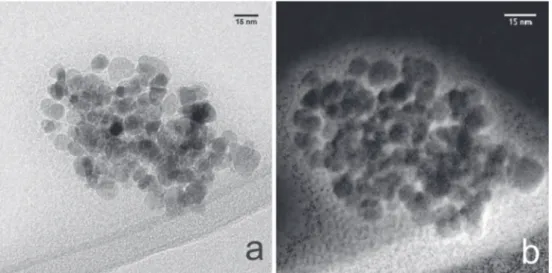
The TEM image (Figure S1a, Supplementary Information (SI) section) of the Fe3O4@lecithin shows the nanoclusters
formation.9,23 NPs of the Fe
3O4 in the Fe3O4@lecithin
(Figure S1b, SI section) have a mean size of 13 nm. The magnetite phase of Fe3O4@lecithin (Figure 2) was
determined by X-ray powder diffraction (XRPD) according to JCPDS card 19-629. The mean diameter of 15 nm of the crystallite was determined by the Debye-Scherer equation.41,42
The structure determined was of cubic face centered (fcc) Fd-3m without deformation. The calculated net parameter was a = 8.41 Å and the density was 5.18 g cm-3.9,10
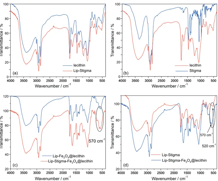
The FTIR spectra for Fe3O4@lecithin (Figure 3)
show the vibration frequency related to the Fe–O bond (561 cm-1) of magnetite and the spectra confirm the surface
functionalization by soy lecithin of Fe3O4@lecithin.
Magnetite (88 ± 1% m/m) contents in Fe3O4@lecithin were
determined by AAS.
The saturation magnetization (Ms) of the Fe3O4@lecithin
was 62 emu g-1 (Figure 4), magnetic moment of
µ = 1.86 × 10⁻¹⁹ J T-1 and magnetic susceptibility at high
fields was 0.96 × 10⁻⁴ emu g-1 at 310 K. The hysteresis
curves were adjusted using the Fortran 90 program based on the Langevin model in the super paramagnetic region (equation S1, SI section).43 The average magnetic core Table2. Content of lecithin, stigmasterol and magnetite
Sample (mg mLLecithin / -1) Stigmasterol / (mg mL-1) Fe3O4 /
(mg mL-1)
Lip-Fe3O4@lecithin1 0.80 – 0.059
Lip-Fe3O4@lecithin2 0.11 – 0.0084
Lip-Stigma1 0.80 0.32 –
Lip-Stigma2 0.11 0.045 –
Lip-Stigma-Fe3O4@lecithin1 0.80 0.32 0.081
Lip-Stigma-Fe3O4@lecithin2 0.11 0.045 0.012
diameter calculated using the Fortran 90 software program was 12.7 ± 0.2 nm confirming the values determined by TEM and XRPD. This value is consistent with nanoparticle super paramagnetic behavior.40 The zeta potential (ζ)
values found for Fe3O4@lecithin were –19 ± 1 mV. The
zeta potential (ζ) confirms the stability of NPs with high water-dispersion in an aqueous medium.
The second step in MGLs preparation involves the encapsulation of the stigmasterol (Lip-Stigma), of F e3O4@ l e c i t h i n ( L i p - F e3O4@ l e c i t h i n ) a n d
encapsulation of the stigmasterol and Fe3O4@lecithin
(Lip-Stigma-Fe3O4@lecithin) as shown in Figure 1 (step 3).
The samples were compared to the bare liposome (Lip).
Characterization of liposomes
The liposome size, polydispersity index (PI), zeta potential (ζ), pH and the FTIR of the liposomes (Table 1)
were evaluated for 14 days (Table 3). Liposomes exhibited temporal stability in aqueous medium and PI compatible with pharmaceutical applications. The zeta potential of the liposomes shows high values without the tendency toward agglomeration corroborating with the observed stability.44,45 The pH of the liposome samples remained
stable at pH 7.3 ± 0.1.
Figures 5a and 5b show the overlap of lecithin spectra with Lip-Stigma (Figure 5a) and lecithin with stigmasterol (Figure 5b). The overlap of the FTIR spectrum of lecithin and Lip-Stigma (Figure 5a) shows characteristic bands of soy lecithin components: 1741 cm-1 (ester carbonyl); 1261
and 1044 cm-1 (P–O–R bonds); 1630 cm-1 (–C=C–, linoleic
acid). It was possible to observe that there were no changes in the spectra pattern due to autoclaving and ultrasound of the sample in the Lip-Stigma. The absence of new bands in the 1600-1800 cm-1 in relation to soy lecithin corroborates Figure2. XRPD of soy lecithin, Fe3O4@lecithin and Lip-Stigma-Fe3O4@lecithin.
Figure3. FTIR spectra of Fe3O4@lecithin and Lip-Fe3O4@lecithin. Figure4. Hysteresis curve of Fe3O4@lecithin and Lip-Fe3O4@lecithin
with the observation of absence of free fatty acids, due to the process of the autoclaving and sonication in the Lip and Lip-Stigma.46-48 Comparison of the soy lecithin sample
with the stigmasterol pattern (Figure 5b) shows an overlap of the stigmasterol bands with the soy lecithin bands. This
can be explained by the presence of steroids in the initial lecithin sample hindering the detection of stigmasterol. The band at 570 cm-1 (Figures 5c and 5d) confirms the
presence of the Fe–O bond of the magnetite phase showing that the NPs coating coverage was efficient in maintaining the magnetite as the only phase.49-53 The comparison of
Lip-Fe3O4@lecithin and Fe3O4@lecithin spectra (Figure 3)
reveals the intensification of the bands pertaining to lecithin due to the encapsulation of Fe3O4@lecithin.
The TEM image and EFTEM maps54 of C coated (of soy
lecithin) (Figures 6a and 6b) of Lip-Fe3O4@lecithin show
a dense population of NPs surrounded by phospholipids. The carbon of phospholipids on the surface of NPs were differentiated by energies lines. HRTEM images of Lip-Fe3O4@lecithin (Figure 7) confirm a population of
NPs surrounded by phospholipids. The surface coating of Fe3O4 NPs with lecithin in the Fe3O4@lecithin sample favored Table3. Physico-chemical parameters of the liposomal formulations for
14 days, stored at 4 °C
Liposomal formulation Size / nm PIa ζb / mV Weight
lossc / %
Lip 189 ± 4 0.325 –41 ± 1 –
Lip-Stigma 160 ± 4 0.250 –49 ± 1 –
Lip-Fe3O4@lecithin 142 ± 4 0.198 –53 ± 1 5
Lip-Stigma-Fe3O4@lecithin 146 ± 4 0.230 –69 ± 3 5
aPolydispersity index deviations less than 3% in the period; bzeta potential; closs of mass corresponding to Fe
3O4 for 14 days.
Figure 5. FTIR spectra of (a) Lip-Stigma and soy lecithin; (b) stigmasterol (Stigma) and soy lecithin; (c) Lip-Fe3O4@lecithin, Lip-Stigma-Fe3O4@lecithin;
the increase of the interactions of NPs with liposomes. It was observed that the coating facilitated the entry of NPs into the vesicles and reduced the ultrasound time duration. The reduced ultrasound time duration minimized the risks of oxidation of NPs and hydrolysis of phospholipids esters.
HRTEM images of the Lip-Fe3O4@lecithin was used
for the determination of the interplanar spacing and mean size of Fe3O4. The values of interplanar spacing found were
consistent with magnetite (JCPDS 19-0629) described in Table 4. The values found are in agreement with the XRPD technique (Figure 2). However, in the XRPD spectra of Lip-Fe3O4@lecithin and Lip-Stigma-Fe3O4@lecithin, one
can observe lower intensity of magnetite peaks due to the presence of soybean lecithin (amorphous) and it is also possible to observe peaks attributed to stigmasterol (JCPDS 10-638). The HRTEM technique enabled nanoscale analysis and the reduction of interferences observed in the XRPD spectrum due to the increase in the amount of soy lecithin and stigmasterol. Therefore, the results found by the
two techniques enabled the confirmation of the maintenance of the magnetite phase in the liposomal samples.
Hysteresis curve of the Lip-Fe3O4@lecithin (Figure 4)
shows super paramagnetic behavior with values of saturation magnetization of Ms = 10.3 emu g-1, magnetic
moment of µ = 1.87 × 10⁻¹⁹ J T-1 and linear susceptibility of
χ = 0.2 × 10⁻⁴ emu g-1 at 310 K. The coercivity and remanent
magnetization values were the same as for Fe3O4@lecithin
with values close to zero, corroborating the literature.53 The
liposome size, zeta potential (ζ), pH and characterization by FTIR, HRTEM, EFTEM/electron energy loss spectroscopy (EELS) and magnetics measurements show promising stability for suitable pharmaceutical.55
The use of soy lecithin as the encapsulating agent offers advantages of commercial availability, low-cost, high encapsulation capacity, high zeta potential and physicochemical stability. The usual components described in the literature are phospholipids (PL) and steroids (S).56,57
The 1H NMR was used for the determination of molar ratio
Figure6. TEM image (a) and EFTEM maps (b) of Lip-Fe3O4@lecithin.
of the phospholipids and steroids in the Lip and Lip-Stigma samples (Figure S2, SI section). The signals observed in the soy lecithin sample prior to autoclaving/sonication and Lip coincide with the composition expected in the literature.57
The molar ratio of phospholipids and steroids found in the Lip and Lip-Stigma samples were 15:1 and 13:10 (PL:S), respectively. The technique also shows absence of new signals or significant difference was not observed between the soy lecithin and Lip spectra signals after 14 days. The absence of new signals indicates the absence of free fatty acids or oxidation due to the liposome formation.
1H NMR and FTIR corroborate to show the efficiency of the
methodologies used to prepare and to store the liposomes. The inhibition of microbial growth at 24 h was shown in Figure 8. The strains S. aureus and C. freundii were sensitive to the liposomal formulations studied under all conditions. The WMF41 condition was established to evaluate the effect of temperature without the presence of a magnetic field in the antibacterial activity. Comparing the three conditions: WMF25, WMF41 and MF41, it can be seen that the elevation of temperature without the presence of the magnetic field increased bacterial growth. This occurrence was also observed in the study of Pseudomonas biofilms by
Park et al.58 Kim et al.59 also observed increased antibacterial
activity due to the presence of the magnetic field in the study with S. aureus. The most efficient condition for inhibiting growth of the microorganisms was MF41, achieving inhibitions of up to 98% of the S. aureus colonies for the Lip-Fe3O4@lecithin1. The specific absorption rate (SAR)
calculated for samples Lip-Stigma-Fe3O4@lecithin1 and
Lip-Fe3O4@lecithin1 were 1056 and 978 W g-1 of iron,
respectively.60 The presence of stigmasterol increased
the encapsulation capacity of NPs of the liposomes and the response of the NPs to the magnetic field (Figure S3, SI section). All samples presented excellent temperature responses in the presence of magnetic field (Figure S3, SI section).53,59,61 The limitation of temperature to 41 ± 1 °C
was used based upon literature references to avoid damage to healthy human cells.62 The evaluation of
Lip-Stigma1 in the MF41 condition shows that the stigma was active as antibacterial with inhibition of 92% of colonies. Traditional liposomes are accompanied by cholesterol as a steroid responsible for the control of drug release.28,29 We decided to use stigmasterol because it is
an abundant vegetable steroid which are proven to have biological activities.63,64 These excellent responses in the
presence of magnetic field prompted us to try dilution of the liposomes for determination of minimum inhibitory concentration (MIC) in the MF41 condition. The results were: Lip-Stigma2 demonstrated inhibition of 55% of C. freundii and 50% of S. aureus representing the most promising response for inhibition of C. freundii. The new dilutions resulted in the MIC of Lip-Fe3O4@lecithin2
against S. aureus (8.4 µg mL-1 of magnetite) and inhibition
of 49% of C. freundii colonies in the MF41 condition. This work was the first to evaluate the activity of NPs against C. freundii. The evaluation of the literature on Fe3O4 activity
in relation to S. aureus showed a variety of methodologies
Table4. Interplanar spacing of Lip-Fe3O4@lecithin by fast Fourier
transform (FFT)
Plane d / nm-1 1/d / nm Theoretical / nm Deviation / %
533 7.841 0.1275 0.1281 0.4
220 4.164 0.2402 0.2420 0.7
622 7.944 0.1259 0.1266 0.6
440 6.787 0.1473 0.1485 0.8
440 6.729 0.1486 0.1485 0.1
800 9.528 0.1050 0.1050 0
d: interplanar spacing.
Figure8. Antibacterial activity of liposomes: (1) Lip-Stigma; (2) Lip-Fe3O4@lecithin; (3) Lip-Stigma-Fe3O4@lecithin under conditions: without magnetic
field at room temperature (WMF25); without magnetic field and 30 min at 41 ± 1 °C (WMF 41) and with magnetic field at 41 ± 1 °C (MF41) against
and results.14,16,19,20,65,66 In our work, the broth
micro-dilution method using UV67 was initially tested, however,
the results were not reproducible. Alternatively, the use of the agar dilution method39 proved to be more reliable
and reproducible. A prior evaluation of the literature data shows that the nature of the coating was determinant in the activity’s result. In the literature consulted, the best result described without a magnetic field was 47 µg mL-1.16 When
compared to our work, we observed that the encapsulating of Fe3O4@lecithin into liposomes and the application of
a magnetic field reduced the dosage required for MIC by approximately 6-fold.
Conclusions
In summary, this work resulted in an enhanced methodology for preparation of magnetoliposomes in terms of time, scalability and cost. Contrary to the liposomes conventional preparation, which uses toxic organic solvents, the methodology applied in this work does not use organic solvents. Additionally, the new process led to low risk of microbial contamination due to sterilization effect resulting from the autoclave step, inherent to the methodology. Liposomes prepared with soy lecithin show stability of physicochemical properties (liposome size, temporal stability, polydispersity index, zeta potential) and excellent magnetic response in vitro (SAR of 1056 W g-1)
allowing a wide range of biomedical applications for treatments of multi-resistant microorganisms in the presence of magnetic field.
Supplementary Information
Supplementary data are available free of charge at http://jbcs.sbq.org.br as PDF file.
Acknowledgments
The authors would like to thank all the many UFMG employees who participated in this work; the employees of the microscopy center of the UFMG for the special attention; colleagues Wallace Doti do Pim for reading the paper; Denio Guimarães Takahashi for the graphical abstract art; Luciano R. Lara that was essential for this work; the funding agencies Capes, CNPQ and FAPEMIG for the financing.
References
1. Hasan, R.; Acharjee, M.; Noor, R.; Tzu Chi Med. J. 2016, 28,
49.
2. Munita, J. M.; Arias, C. A.; Microbiol. Spectrum 2016, 4, VMBF-0016-2015.
3. Ruppé, É.; Woerther, P.-L.; Barbier, F.; Ann. Intensive Care 2015, 5, 21.
4. Holban, A. M.; Curr. Top. Med. Chem. 2015, 15, 1589. 5. Hiramatsu, K.; Katayama, Y.; Matsuo, M.; Sasaki, T.; Morimoto,
Y.; Sekiguchi, A.; Baba, T.; J. Infect. Chemother. 2014, 20, 593. 6. World Health Organization (WHO); Global Priority List of
Antibiotic-Resistant Bacteria to Guide Research, Discovery, and
Development of New Antibiotics; WHO: Geneva, Switzerland,
2017.
7. Liu, L.-H.; Wang, N.-Y.; Wu, A. Y.-J.; Lin, C.-C.; Lee, C.-M.; Liu, C.-P.; J. Microbiol., Immunol. Infect. 2018, 51, 565. 8. Pelgrift, R. Y.; Friedman, A. J.; Adv. Drug Delivery Rev. 2013,
65, 1803.
9. Lara, L. R. S.; Zottis, A. D.; Elias, W. C.; Faggion, D.; de Campos, C. E. M.; Acuna, J. J. S.; Domingos, J. B.; RSC Adv. 2015, 5, 8289.
10. Andrade, Â. L.; Valente, M. A.; Ferreira, J. M. F.; Fabris, J. D.;
J. Magn. Magn. Mater. 2012, 324, 1753.
11. German, S. V.; Navolokin, N. A.; Kuznetsova, N. R.; Zuev, V. V.; Inozemtseva, O. A.; Anis’kov, A. A.; Volkova, E. K.; Bucharskaya, A. B.; Maslyakova, G. N.; Fakhrullin, R. F.; Terentyuk, G. S.; Vodovozova, E. L.; Gorin, D. A.; Colloids Surf., B 2015, 135 (Supplement C), 109.
12. Marciello, M.; Connord, V.; Veintemillas-Verdaguer, S.; Verges, M. A.; Carrey, J.; Respaud, M.; Serna, C. J.; Morales, M. P.;
J. Mater. Chem. B 2013, 1, 5995.
13. Petcharoen, K.; Sirivat, A.; Mater. Sci. Eng., B 2012, 177, 421. 14. Darwish, M. S.; Nguyen, N. H.; Ševců, A.; Stibor, I.; Smoukov,
S. K.; Mater. Sci. Eng., C 2016, 63, 88.
15. Prabhu, Y. T.; Rao, K. V.; Kumari, B. S.; Kumar, V. S. S.; Pavani, T.; Int. Nano Lett. 2015, 5, 85.
16. Ramteke, C.; Ketan Sarangi, B.; Chakrabarti, T.; Mudliar, S.; Satpute, D.; Avatar Pandey, R.; Curr. Nanosci. 2010, 6, 587. 17. Shariatinia, Z.; Nikfar, Z.; Int. J. Biol. Macromol. 2013,
60 (Supplement C), 226.
18. Ferreira, R. V.; Martins, T. M. M.; Goes, A. M.; Fabris, J. D.; Cavalcante, L. C. D.; Outon, L. E. F.; Domingues, R. Z.;
Nanotechnology 2016, 27, 085105.
19. Hsieh, S.; Huang, B. Y.; Hsieh, S. L.; Wu, C. C.; Wu, C. H.; Lin, P. Y.; Huang, Y. S.; Chang, C. W.; Nanotechnology 2010, 21, 445601.
20. Inbaraj, B. S.; Kao, T.; Tsai, T.; Chiu, C.; Kumar, R.; Chen, B.-H.; Nanotechnology 2011, 22, 075101.
21. Nappini, S.; Fogli, S.; Castroflorio, B.; Bonini, M.; Baldelli Bombelli, F.; Baglioni, P.; J. Mater. Chem. B 2016, 4, 716. 22. Drulis-Kawa, Z.; Dorotkiewicz-Jach, A.; Int. J. Pharm. 2010,
387, 187.
24. Hosseini, F.; Sadjadi, M. S.; Orient. J. Chem. 2016, 32, 2901. 25. Latifi, S.; Tamayol, A.; Habibey, R.; Sabzevari, R.; Kahn, C.;
Geny, D.; Eftekharpour, E.; Annabi, N.; Blau, A.; Linder, M.; Arab-Tehrany, E.; Sci. Rep. 2016, 6, 25777.
26. Carugo, D.; Bottaro, E.; Owen, J.; Stride, E.; Nastruzzi, C.; Sci. Rep. 2016, 6, 25876.
27. Woodbury, D. J.; Richardson, E. S.; Grigg, A. W.; Welling, R. D.; Knudson, B. H.; J. Liposome Res. 2006, 16, 57.
28. Frézard, F.; Silva-Barcellos, N. M.; dos Santos, R. A.; Regul. Pept. 2007, 138, 59.
29. Kikuchi, H.; Carlsson, A.; Yachi, K.; Hirota, S.; Chem. Pharm. Bull. 1991, 39, 1018.
30. Akbarzadeh, A.; Rezaei-Sadabady, R.; Davaran, S.; Joo, S. W.; Zarghami, N.; Hanifehpour, Y.; Samiei, M.; Kouhi, M.; Nejati-Koshki, K.; Nanoscale Res. Lett. 2013, 8, 102.
31. Abareshi, M.; Goharshadi, E. K.; Mojtaba Zebarjad, S.; Khandan Fadafan, H.; Youssefi, A.; J. Magn. Magn. Mater. 2010, 322, 3895.
32. Ma, K.; Zhao, H.; Zheng, X.; Sun, H.; Hu, L.; Zhu, L.; Shen, Y.; Luo, T.; Dai, H.; Wang, J.; J. Mater. Chem. B 2017, 5, 2888.
33. Chen, Y.; Qian, Z.; Zhang, Z.; Colloids Surf., A 2008, 312, 209. 34. Ong, S. G. M.; Ming, L. C.; Lee, K. S.; Yuen, K. H.;
Pharmaceutics 2016, 8, 25.
35. Reichel, V.; Kovács, A.; Kumari, M.; Bereczk-Tompa, É.; Schneck, E.; Diehle, P.; Pósfai, M.; Hirt, A. M.; Duchamp, M.; Dunin-Borkowski, R. E.; Sci. Rep. 2017, 7, 45484.
36. Fischer, A.; Schmitz, M.; Aichmayer, B.; Fratzl, P.; Faivre, D.;
J. R. Soc., Interface 2011, 8, 1011.
37. Bozzola, J. J.; Russell, L. D.; Electron Microscopy: Principles and Techniques for Biologists; Jones & Bartlett Learning:
Boston, 1999.
38. Golla-Schindler, U.; Hinrichs, R.; Bomati-Miguel, O.; Putnis, A.; Micron 2006, 37, 473.
39. Wiegand, I.; Hilpert, K.; Hancock, R. E.; Nat. Protoc. 2008, 3,
163.
40. Harifi, T.; Montazer, M.; J. Mater. Chem. B 2014, 2, 272.
41. El Ghandoor, H.; Zidan, H.; Khalil, M. M.; Ismail, M.; Int. J. Electrochem. Sci. 2012, 7, 5734.
42. Liu, X. L.; Choo, E. S. G.; Ahmed, A. S.; Zhao, L. Y.; Yang, Y.; Ramanujan, R. V.; Xue, J. M.; Di Fan, D.; Fan, H. M.; Ding, J.;
J. Mater. Chem. B 2014, 2, 120.
43. Lan, N. T.; Duong, N. P.; Hien, T. D.; J. Alloys Compd. 2011, 509, 5919.
44. Chen, Y.; Liu, J.; Angelov, B.; Drechsler, M.; Garamus, V. M.; Willumeit-Römer, R.; Zou, A.; Colloids Surf., B 2016, 140, 74. 45. Zhao, L.; Temelli, F.; Curtis, J. M.; Chen, L.; Food Res. Int.
2015, 77, 63.
46. Abramson, M. B.; Norton, W. T.; Katzman, R.; J. Biol. Chem. 1965, 240, 2389.
47. Fookson, J. E.; Wallach, D. F.; Arch. Biochem. Biophys. 1978, 189, 195.
48. Kim, M.-J.; Jang, D.-H.; Kim, H.-K.; Lee, Y.-I.; Lee, G.-J.; Yoo, B.-Y.; Choa, Y.-H.; J. Nanosci. Nanotechnol. 2011, 11, 4592.
49. Jubb, A. M.; Allen, H. C.; ACS Appl. Mater. Interfaces 2010,
2, 2804.
50. Li, Y.-S.; Church, J. S.; Woodhead, A. L.; J. Magn. Magn. Mater. 2012, 324, 1543.
51. Liese, H.; Am. Mineral. 1967, 52, 1198.
52. Millan, A.; Palacio, F.; Falqui, A.; Snoeck, E.; Serin, V.; Bhattacharjee, A.; Ksenofontov, V.; Gütlich, P.; Gilbert, I.; Acta Mater. 2007, 55, 2201.
53. Sadhukha, T.; Wiedmann, T. S.; Panyam, J.; Biomaterials 2014,
35, 7860.
54. Anjum, D. H.; Memon, N. K.; Ismail, M.; Hedhili, M. N.; Sharif, U.; Chung, S. H.; Nanotechnology 2016, 27, 365709.
55. Kraft, J. C.; Freeling, J. P.; Wang, Z.; Ho, R. J.; J. Pharm. Sci. 2014, 103, 29.
56. Mertins, O.; Sebben, M.; Schneider, P. H.; Pohlmann, A. R.; Silveira, N. P.; Quim. Nova 2008, 31, 1856.
57. Scholfield, C.; J. Am. Oil Chem. Soc. 1981, 58, 889.
58. Park, H.; Park, H.-J.; Kim, J. A.; Lee, S. H.; Kim, J. H.; Yoon, J.; Park, T. H.; J. Microbiol. Methods 2011, 84, 41.
59. Kim, M.-H.; Yamayoshi, I.; Mathew, S.; Lin, H.; Nayfach, J.; Simon, S. I.; Ann. Biomed. Eng. 2013, 41, 598.
60. Di Corato, R.; Espinosa, A.; Lartigue, L.; Tharaud, M.; Chat, S.; Pellegrino, T.; Ménager, C.; Gazeau, F.; Wilhelm, C.;
Biomaterials 2014, 35, 6400.
61. Subbiahdoss, G.; Sharifi, S.; Grijpma, D. W.; Laurent, S.; van der Mei, H. C.; Mahmoudi, M.; Busscher, H. J.; Acta Biomater. 2012, 8, 2047.
62. Yoshida, M.; Sato, M.; Yamamoto, Y.; Maehara, T.; Naohara, T.; Aono, H.; Sugishita, H.; Sato, K.; Watanabe, Y.; J. Gastroenterol. Hepatol. 2012, 27, 406.
63. Bhatti, H. N.; Khera, R. A.; Steroids 2012, 77, 1267.
64. Gabay, O.; Sanchez, C.; Salvat, C.; Chevy, F.; Breton, M.; Nourissat, G.; Wolf, C.; Jacques, C.; Berenbaum, F.; Osteoarthr. Cartilage2010, 18, 106.
65. Darwish, M. S.; Nguyen, N. H.; Ševců, A.; Stibor, I.;
J. Nanomater. 2015, 16, 10.
66. Grumezescu, A. M.; Cristescu, R.; Chifiriuc, M.; Dorcioman, G.; Socol, G.; Mihailescu, I.; Mihaiescu, D. E.; Ficai, A.; Vasile, O.; Enculescu, M.; Biofabrication 2015, 7, 015014.
67. Sousa, G. F.; Aguilar, M. G.; Dias, D. F.; Takahashi, J. A.; Moreira, M. E. C.; Vieira Filho, S. A.; Silva, G. D. F.; Rodrigues, S. B. V.; Messias, M. C. T. B.; Duarte, L. P.; Phytochem. Lett. 2017, 21 (Supplement C), 61.
Submitted: April 10, 2018
Published online: July 30, 2018