Article
*e-mail: [email protected]
Physicochemical Stability of Poly(lactide-co-glycolide) Nanocapsules Containing the
Local Anesthetic Bupivacaine
Carolina M. Moraes,a Eneida de Paula,a André H. Rosab and
Leonardo F. Fraceto*,a,b
aInstituto de Biologia, Universidade Estadual de Campinas, Cidade Universitária “Zeferino Vaz”,
13083-970 Campinas-SP, Brazil
bDepartamento de Engenharia Ambiental, Universidade Estadual Paulista Júlio de Mesquita Filho,
Av. Três de Março, 511, 18087-180 Sorocaba-SP, Brazil
Neste trabalho foi realizada a preparação de nanocápsulas de poli (DL-lactídeo-co-glicolídeo) (PLGA) como um sistema carreador para o anestésico local bupivacaína. A preparação foi caracterizada e sua estabilidade físico-química avaliada. Os resultados da caracterização mostram uma distribuição de tamanho com um índice de polidispersão de 0,12, um diâmetro médio de 148 nm, um potencial zeta de –43,5 mV e uma eiciência de associação da BVC nas nanocápsulas de 75,8%. As propriedades físico-químicas das suspensões (diâmetro hidrodinâmico, índice de polidispersão, potencial zeta e eiciência de associação do fármaco) contendo nanocápsulas poliméricas foram avaliadas em função do tempo a im de determinar sua estabilidade. Nenhuma grande alteração foi observada em função tempo para as suspensões de nanocápsulas avaliadas, sendo consideradas estáveis por um período de armazenagem de 120 dias a temperatura ambiente. Os resultados aqui apresentados, os quais se referem ao estudo desta nova formulação para anestésico local bupivacaína mostram-se promissores para futuros estudos in vivo.
This paper describes the preparation of poly(DL-lactide-co-glicolide) (PLGA) nanocapsules as a drug carrier system for the local anesthetic bupivacaine. The system was characterized and its stability investigated. The results showed a size distribution with a polydispersity index of 0.12, an average diameter of 148 nm, a zeta potential of –43.5 mV and an entrapment eficiency of 75.8%. The physicochemical properties of polymeric nanocapsule suspensions (average diameter, polydispersity, zeta potential and drug association eficiency) were evaluated as a function of time to determine the formulation stability. The formulation did not display major changes in these properties over the time, and it was considered stable up to 120 days of storage at room temperature. The results reported here which refer to the initial characterization of these new formulations for the local anesthetic bupivacaine show a promising potential for future in vivo studies.
Keywords:bupivacaine, local anesthetic, polymeric nanocapsules, PLGA, physical-chemical
stability
Introduction
Numerous studies today are focusing on the development of new pharmaceutical formulations based on nanocarriers, which have demonstrated several advantages over conventional formulations, making this research line a
promising and innovative area of the pharmaceutical sector.1,2
Polymer nanoparticles (PN) are carriers of drugs or other active molecules whose sizes range from 10 to 1000 nm.
Nanospheres (NS) or nanocapsules (NC) can be obtained, depending on the method of preparation and the materials employed. NC consists of a polymer casing and a nucleus (usually oily). The drug inside the NC may be dissolved in the oily nucleus or adsorbed on the polymeric wall. NS, on the other hand, consists of a polymer matrix and their composition does not include oil. In this system, the drug
may be adsorbed or dispersed in the polymeric matrix.2-6
biocompatibility, absence of toxicity and because they
are easy to incorporate into a wide range of drugs.7 The
degradation of aliphatic polyesters may result from the enzymatic action and/or hydrolysis of the ester bond and may be affected by the properties of these polymers, such as their chemical composition, molar mass, solubility, hydrophobicity/hydrophilicity, transition temperature,
structure (crystalline or amorphous), among other factors.8,9
The aliphatic polyesters most commonly used are poly(lactic acid) (PLA), poly(glycolic acid) (PGA), poly(lactic-co-glycolic acid) (PLGA),
poly(ε-caprolactone) (PCL), poly(hydroxyvalerate) (PHV)
and poly(hydroxybutyrate) (PHB).4,10
Local anesthetics (LA) are molecules whose primary pharmacological activity involves reversible nerve
conduction blockage.11,12 Although several physicochemical
models have been proposed to explain how LA block nerve conduction, the principal mechanism involves its interaction at one or more speciic sites in the
sodium-dependent channels of neural membranes.13-15
Bupivacaine (BVC) is a LA of the amino amide class, which, together with two other LA of this class, lidocaine and ropivacaine, are the drugs of choice in acute and chronic pain therapy. Bupivacaine is commercially available in
formulations varying from 0.25 to 0.75% in mass of BVC.16,17
Because the desirable characteristics for a LA include long duration of action, selectivity for sensory rather than
motor nerve block and reduction of systemic toxicity,17,18 an
alternative that has proved able to promote these desirable effects is the modiied release of these drugs through carrier systems. The purpose of release systems is to promote slow and continuous release, reducing the systemic toxicity of
these locally active drugs.17-19 Drug release systems are,
in fact, able to compartmentalize the active substance and drive it to speciic active sites, as well as control the rate of release without altering the chemical structure of the
transported molecule.20
The delivery of local anesthetics, which has been studied extensively by many research groups and also by our laboratory, can be achieved by the formation of
inclusion complexes with cyclodextrins,20-30 encapsulation
in liposomes,31-39 association to polymer microparticles40-43
and association to polymeric nanostructured systems.44-48
The use of colloidal systems of polymeric nanoparticles is often limited due to problems of stability over long periods of time. A colloidal suspension does not normally show phase separation, since both sedimentation and aggregate formation are slow. However storage for many months can sometimes result in aggregation and consequently phase separation, which renders the use of such formulations
non-viable.4 Hence, the purpose of this work was to prepare,
characterize and monitor the physicochemical stability of PLGA nanocapsules containing bupivacaine. This study is important for the characterization of a new pharmaceutical form for pain therapy.
Material and Methods
The materials used were bupivacaine (free base, Cristália Ind. Farm. LTDA), poly(DL-lactide-co-glycolide, 50:50)
(PLGA 50:50, MW 40,000-70,000 g mol-1) (Sigma Aldrich
Chem. Co.), sorbitan monostereate (Span60®) (Sigma
Aldrich Chem. Co.), polysorbate 80 (Tween80®) (LabSynth,
Brazil), caprylic/capric acid triglyceride (Miglyol 810®)
(Hüls, Germany) and analytical grade acetone (LabSynth, Brazil). The solvents used for the chromatographic analyses
were HPLC grade acetonitrile (JT Baker®) and Milli-Q water.
Preparation of the PLGA nanocapsules with bupivacaine
The PLGA nanocapsules were prepared by the
nanoprecipitation method,49 which involved mixing an
organic phase in an aqueous phase. The organic phase consisted of PLA polymer (100 mg), acetone (30 mL), bupivacaine (25 mg), sorbitan monostereate (40 mg) and caprylic/capric acid triglyceride (200 mg). The aqueous phase was composed of polysorbate 80 (60 mg) and deionized water (30 mL). After the dissolution of the components of both phases, the organic phase was gradually added to the aqueous phase using a small funnel. The resulting suspension was kept under agitation for 10 min, after which it was concentrated under low pressure to a volume of 10 mL, using a rotary evaporator, in order to obtain a suspension of bupivacaine with a concentration of
2.5 mg mL-1. A control formulation without bupivacaine was
also prepared, following the methodology described above.
Size and polydispersity measurements
The dynamic light scattering technique was used to determine the mean particle size (hydrodynamic diameter) and polydispersity. These analyses were performed by diluting (1:1000, v/v) the nanocapsule suspensions (with
and without BVC) and using a ZetaPlus® particle analyzer
with 90º ixed angle detector, at a temperature of 25 ºC. The size distribution and polydispersity were measured and were expressed as the mean of ive determinations.
Zeta potential measurements
The analyses were carried out by diluting (1:1000, v/v) the nanocapsule suspensions (with and without BVC) in Milli-Q water, and the results were expressed as means of eight determinations.
Eficiency of bupivacaine association in PLGA nanocapsules
The percentage of BVC associated to the nanocapsules was determined by the ultrafiltration/centrifugation method. The nanocapsule samples containing bupivacaine were centrifuged in ultrafiltration filters composed of regenerated cellulose with 30 kDa molecular
size-exclusion pores (Microcon -Millipore®), and the
filtrate was quantified by high performance liquid chromatography (HPLC). The concentration of BVC was determined using an analytical curve (concentration range:
1-200 mg mL-1, peak area = 3.30×10⁵[bupivacaine] +
4.7×10⁵, r = 0.9998, n = 6). The speciicity was tested
in the presence of the colloidal suspension components, and it was demonstrated that these factors did not alter bupivacaine quantiication.
The BVC association rate was determined from the difference between the drug concentration measured in the iltrate and its total concentration (100%) in the nanocapsule suspension.
The chromatographic conditions employed in the quantification were: mobile phase composed of
acetonitrile/phosphate buffer (pH 7.4, 5 mmol mL–1,
i.e., 3 mmol L–1 Na2HPO4 and 2 mmol L–1 NaH2PO4,
85:15, v/v), 2.3 mL min–1 low rate, and a Phenomenex
Gemini chromatographic column (C18 reversed phase,
5 μ, 110 Å, 150 × 4.60 mm). The BVC was detected at a wavelength of 220 nm, using an ultraviolet (UV) detector. The injection volume was 20 μL and all the injected samples were previously filtered through a
0.22 μmol L-1 polyethersulfone membrane (Millipore®).
The total BVC (100%) in the PLGA nanocapsule suspension was determined by diluting the suspension in acetonitrile. Acetonitrile is able to dissolve the polymer, thus completely releasing the BVC which, in turn, was quantiied based on the analytical curve validated by Resolution RE No. 899/2003 of ANVISA, Brazil’s National Health Surveillance Agency.
Colloidal stability measurements
The stability of the PLGA NC suspensions containing BVC was evaluated upon determinations of size, polydispersity, zeta potential and association rate as a function of time (zero, 15, 60 and 120 days), with the suspensions stored in amber glass lasks at room temperature.
Results and Discussion
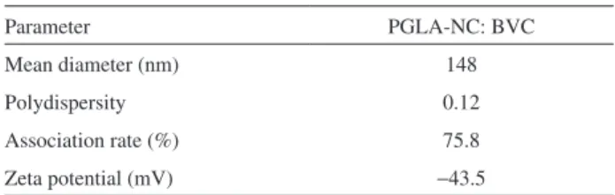
Measurements of the size (hydrodynamic diameter), polydispersity and zeta potential of particles are parameters that indicate the stability of nanocapsules in suspension. Table 1 lists the measured values of these parameters for the PLGA NC suspensions with BVC, as well as the association rate of this drug in the nanocapsules.
The results presented in Table 1 indicate that the PLGA NC suspensions with BVC presented a size (hydrodynamic diameter) and polydispersity compatible
with colloidal suspensions.50 The zeta potential values
of these formulations indicate good stability in solution, because they were lower than –30 mV and aggregation of
particles in suspension was therefore avoided.4,5,52
The BVC association rate in the PLGA nanocapsules was 75.8%, which is a very high value when compared to other studies reported in the literature for local
anesthetics.44-48
The stabilities of the NC polymer suspensions containing BVC were evaluated based on the physicochemical properties of size, polydispersity, zeta potential and association eficiency as a function of time (initial, 15, 60 and 120 days), with all the samples stored in amber glass lasks at room temperature.
The graph in Figure 1(a) shows the particle size
distribution by intensities obtained with the DTS nano
software from light-scattering measurements of the PLGA NC containing BVC.
The results depicted in Figure 1(a) indicate that the size distribution in the formulation remained constant and unimodal for a period of 120 days, conirming the good conditions for preparation of the NC suspensions. Figure 1(b) also demonstrates that the particle size in the PLGA NC containing BVC was initially 147 nm, and underwent a slight increase up to 60 days, after which the particle size was found to decrease. Similar results for polymer nanoparticle sizes
have been reported in the literature.50,51,53
Another parameter investigated was polydispersity, an index that can be indicative of stability, since it
Table 1. Values of the mean diameter (hydrodynamic diameter, nm),
polydispersity, zeta potential (mV) and association rate of the PLGA NC suspensions with BVC
Parameter PGLA-NC: BVC
Mean diameter (nm) 148
Polydispersity 0.12
Association rate (%) 75.8
represents the particle size distribution range. High polydispersity indices indicate size heterogeneity of particles in suspension, while variations in polydispersity values as a function of time indicate the formation of populations of particles with sizes that did not exist initially, possibly resulting from particle aggregation or breakdown/degradation. Polydispersity indices lower than 0.2 are ideal, because they indicate that the particle size distribution falls within a narrow range of sizes. Figure 2 shows the values of polydispersity as a function of time for the PLGA NC containing BVC.
The polydispersity of the NC suspension with BVC did not present statistically significant differences in polydispersity index as a function of time. All the results indicated good polydispersity indices, since they were all below 0.2.
The zeta potential, which relects the load on the particle surface, was another parameter evaluated here. In the absence of steric mechanisms, the stability of nanoparticles is determined by the balance of repulsion and attraction forces that the particles present. Thus, high repulsion forces tend to prevent aggregation. Nanoparticles with a zeta potential of approximately (±) 30 are more stable in
suspension.4,5 Figure 3 shows the zeta potential graph of
the particles as a function of time.
The PLGA NC suspension containing BVC showed an initial zeta potential of –43.5 mV, which changed progressively, reaching –30.2 mV in 120 days (Figure 3), thus indicating the formulation’s good stability. This observed change in the zeta potential could be due to a series of factors, especially changes in the interactions between pharmaceutical/polymer/surfactant at the surface of the polymeric nanocapsules. Previous published works
have shown that surfactants54 and pharmaceuticals55 can
modify zeta potential values in colloidal systems. However, more work is needed to better understand these changes.
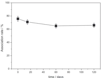
The drug association rates in the nanoparticles were also evaluated as a function of time, as illustrated in Figure 4. The PLGA NC suspension with BVC showed a constant decline in the drug association rate up to 60 days, after which the rate gradually stabilizes by day 120 (Figure 4). The association rate varied from an initial 75.8% to 66.1%
by day 120. According to Müller and co-workers,51 such
a decline in drug association eficiency in NC may be due
Figure 2.Polydispersity of particles as a function of time (0, 15, 60
and 120 days) for PLGA NC suspensions with BVC. The dotted line indicates the maximum value of the polydispersity index for the particle size distribution range (0.2).
Figure 3.Zeta potential of particles as a function of time (0, 15, 60 and 120 days) for PLGA NC suspensions with BVC.
Figure 1.a)Particle size distribution (nm) by intensity (%) of the PLGA
to precipitation of nanocrystals of the drug during the formation of NC stabilized by surfactant agents. It was reported that these nanocrystals present the same size distribution as the NC, and therefore do not pass through the membrane used in the ultrailtration/centrifugation procedure. Over time, these nanocrystals aggregate and precipitate, reducing the drug association rate in the NC.
Guterres et al.50 demonstrated that nanocapsules of
PLA/triglycerides containing dichlofenac showed the formation of nanocrystals that were initially stabilized by surfactants, and over the time formed agglomerates, with consequent precipitation.
Another factor that could explain the reduced association rate of the BVC molecule with time, in the PLGA NC, could be its release from the NC. However, the release proile was not investigated in the present study, because it was not possible to achieve dilution sink conditions due to the quantity of BVC present in the formulation.
Conclusions
This work provided several important pieces of information about the development of formulations containing PLGA NC with BVC, based on the method of interfacial deposition of preformed polymers. The NC polymer suspensions containing this local anesthetic presented good physicochemical stability as a function of time, in terms of size, polydispersity and zeta potential, since these parameters hardly altered with time. Concerning the association rate, a reduction of the amount of BVC in the PLGA NC was observed, probably due to formation of nanocrystals of the pharmaceutical. We therefore suggest that the system
of BVC associated with PLGA nanocapsules could be a useful new formulation, since BVC is a long-lasting local anesthetic.
Acknowledgments
The authors gratefully acknowledge the financial support of the Brazilian research funding agencies FAPESP (06/00121-9, 07/00127-0), CNPq and FUNDUNESP. C.M.M. was the recipient of a fellowship from FAPESP.
References
1. Rawat, M.; Singh, D.; Saraf, S.; Saraf, S. W.; Biol. Pharm. Bull.
2006, 29, 1790.
2. Anton, N.; Benoit, J. P.; Saulnier, P.; J. Control. Release 2008,
128, 185.
3. Soppimath, K. S.; Aminabhavi, T. M.; Kulkarni, A. R.; Rudzinski, W. E.; J. Control. Release 2001, 70, 1.
4. Schaffazick, S. R.; Guterres S. S.; Freitas, L. L.; Pohlmann, A.
R.; Quim. Nova2003, 26, 726.
5. Mohanraj, V. J.; Chen, Y.; Trop. J. Pharm. Res.2006,5, 561.
6. Nurkeeva, Z. S.; Mun, G. A.; Khutoryanskiy, V. V.; Bitekenova, A. B.; Dzhusupbekova, A. B.; J. Biomater. Sci., Polym. Ed.2002,
13, 759.
7. Picos, D. R.; Carril, M. G.; Mena, D. F.; Fuente, L. N.; Rev.
Cubana Farm. 2000, 34, 70.
8. Durán, N.; Mattoso, L. H. C.; Morais, P. C.; Nanotecnologia: Introdução, Preparação e Caracterização de Nanomateriais e
Exemplos de Aplicação, 1a. ed., Artliber: São Paulo, Brasil, 2006.
9. Nair, L. S.; Laurencin, C. T.; Prog. Polym. Sci. 2007, 32, 762. 10. Morales, M. M.; Terapias Avançadas: Células-Tronco, Terapia
Gênica e Nanotecnologia Aplicada à Saúde, 1a. ed., Atheneu:
São Paulo, Brasil, 2007.
11. Covino, B. G.; Vassalo, H. G.; Anestésicos Locais: Mecanismos
de Ação e Uso Clínico, 1st ed., Colina: Rio de Janeiro, 1985.
12. Collins, V. J.; Principles of Anesthesiology: General and
Regional, 3rd ed., Lea&Febiger: New York, 1993.
13. Goodman, A. G.; Gilman, A. G.; As Bases Farmacológicas da
Terapêutica, 9a.ed., McGraw-Hill: Rio de Janeiro, Brasil, 1996.
14. de Jong R. H.; Local Anesthetics, 1st ed., C.C. Thomas: Illinois, 1994.
15. Fraceto, L. F.; de Araújo, D. R.; de Paula, E.; Quim. Nova2008,
31, 1775.
16. Gupta, S. P.; Chem. Rev.1991, 91, 1109.
17. de Araújo, D. R.; Fraceto, L. F.; Braga, A. F. A.; de Paula, E.;
Rev. Bras. Anestesiol. 2005, 55, 316.
18. de Araújo, D. R.; Pinto, L. M. A.; Braga, A. F. A.; de Paula, E.;
Rev. Bras. Anestesiol. 2003,53, 663.
19. Simonetti, M. P. B.; Andrade, M. P.; Rev. Bras. Anestesiol. 1996,
46, 35.
Figure 4. Association rate (%) of the drug in the particles as a function
20. Oliveira, A. G.; Scarpa, M. V.; Correa, M. A.; Cera, L. F. R.; Formariz, T. P.; Quim. Nova2004, 27, 131.
21. Dollo, G.; Le Corre, P.; Chevanne, F.; Le Verge, R.; Int. J.
Pharm.1996, 136, 165.
22. de Araújo, D. R.; Braga, A. F. A.; Moraes, C. M.; Fraceto, L. F.; de Paula, E.; Rev. Bras. Anestesiol.2006, 56, 495.
23. Araújo, D. R.; Tsuneda, S. S.; Cereda, C. M. S.; Carvalho, F. G. F.; Preté, P. S. C; Fernandes, S. A.; Yokaichiya, F.; Franco, M. K. K. D.; Mazzaro, I.; Fraceto, L. F.; Braga, A. F. A.; de Paula,
E.; Eur. J. Pharm. Sci.2008, 33, 60.
24. Moraes, C. M.; Abrami, P.; Araújo, D. R.; Braga, A. F. A.; Issa, M. G.; Ferraz, H. G.; de Paula, E.; Fraceto, L. F.; J. Inclusion
Phenom. Macrocyclic Chem.2007, 57, 313.
25. Moraes, C. M.; Abrami, P.; de Paula, E.; Braga, A. F.; Fraceto, L. F.; Int. J. Pharm.2007, 331, 99.
26. Moraes, C. M.; Abrami, P.; de Paula, E.; Andreo-Filho, N.; Fraceto, L. F.; Quim. Nova2007, 30, 777.
27. Irie, T.; Uekama, K.; Pharm. Sci.1997,86, 147.
28. Loftsson, T.; Masson, M.; Int. J. Pharm.2001,225, 15. 29. Pinto, L. M.; Fraceto, L. F.; Santana, M. H. A.; Pertinhez, T.
A.; Junior, S. O.; de Paula, E.; J. Pharm. Biomed. Anal. 2005,
39, 956.
30. Kuzma, P. J.; Kline, M. D.; Calkins, M. D.; Staats, P. S.; Reg.
Anesth. Pain Med. 1997, 22, 543.
31. Fraceto, L. F.; Pinto, L. M. A., Franzoni, L.; Braga, A. C.; Spisni, A.; Schreier, S.; de Paula, E.; Biophys. Chem.2002, 99, 229.
32. de Araújo, D. R.; Cereda, C. M. S.; Brunetto, G. B.; Pinto, L. M. A.; Santana, M. H. A.; de Paula, E.; Can. J. Anesth. 2004,
51, 566.
33. Cereda, C. M. S.; de Araújo, D. R.; Brunetto, G. B.; de Paula,
E.; J. Pharm. Pharmacol. 2004, 7, 235.
34. Cereda, C. M. S.; Bruschini, G.; De Araújo, D. R.; de Paula,
E.; Reg. Anesth. Pain Med. 2006, 53,1092.
35. Cereda, C. M. S.; Tófoli, G. R.; De Araújo, D. R.; Arana, S., de Paula, E.; Reg. Anesth. Pain Med.2008,33, 62.
36. Araújo, D. R.; Cereda, C. M. S.; Brunetto, G. B.; Vomero, V. U.; Pierucci, A.; Santo Neto, H.; De Oliveira, A. L. R.; Fraceto, L. F.; Braga, A. F. A.; De Paula, E.; J. Pharm. Pharmacol. 2008,
60, 1449.
37. Colombo, G.; Padera, R.; Langer, R.; Kohane, D. S.; J. Biomed.
Mater. Res.,Part A2005, 75, 458.
38. Grant, G. J.; Bansinath, M.; Reg. Anesth. Pain Med.2001, 26, 61.
39. Grant, S. A.; Best Pract. Res. Clin. Anaesth.2002, 16, 345. 40. Le Guévello, P.; Le Corre, P.; Chevanne, F.; Le Verge, R.;
J. Chromatogr. 1993, 622, 284.
41. Rose, J. S.; Neal, J. M.; Kopacz, D. J.; Reg. Anesth. Pain Med.
2005, 30, 275.
42. Kranz, H.; Bodmeier, R.; Eur. J. Pharm. Sci.2008, 34, 164.
43. Holgado, M. A.; Arias, J. L.; Cózar, M. J.; Alvarez-Fuentes, J.; Ganan-Calvo, A. M.; Fernandez-Arevalo, M.; Int. J. Pharm.
2008, 358, 27.
44. Gorner, T.; Gref, R.; Michenot, D.; Sommer, F.; Tran, M. N.; Dellacherie, E.; J. Control. Release1999, 57, 259.
45. Govender, T.; Stolnik, S.; Garnett, M. C.; Illum, L.; Davis, S.
S.; J. Control. Release1999, 57, 171.
46. Govender, T.; Riley, T.; Ehtezazi, T.; Garnett, M. C.; Stolnik, S.; Illum, L.; Davis, S. S.; Int. J. Pharm.2000, 199, 95. 47. Polakovic, M.; Gorner, T.; Gref , R.; Dellacherie, E.;
J. Control. Release 1999, 60, 169.
48. Moraes, C. M.; de Matos, A. P.; de Paula, E.; Rosa, A. H.; Fraceto, L. F.; Mater. Sci. Eng., B2009, 165, 243.
49. Fessi, H., Puiseiux, F.; Devissaguet, J-P.; Ammoury, N.; Benita,
S.; Int. J. Pharm.1989, 55, 1.
50. Guterres, S. S.; Fessi, H.; Barratt, G.; Devissaguet, J. P.; Puisieux, F.; Int. J. Pharm.1995, 113, 57.
51. Muller, C. R.; Haas, S. E.; Bassani, V. L.; Gutterres, S. S.; Fessi, H.; Peralba, M. C. R.; Pohlmann, A. R.; Quim. Nova 2004,27, 555.
52. Couvreur, P.; Barratt, G.; Fattal, E.; Legrand, P.; Vauthier, C.;
Crit. Rev. Ther. Drug Carrier Syst. 2002, 19, 99.
53. Pohlmann, A. R.; Weiss, V.; Mertins, O.; Silveira, N. P.; Guterres, S. S.; Eur. J. Pharm. Sci.2002, 16, 305.
54. Hsu, J. Y.; Nacu, A.; J. Coll. Interf. Sci. 2003, 259, 374. 55. Hsu, J. P.; Lee, H. Y.; Guan, Y. S.; Kim, J. C.; J. Ind. Eng. Chem.
2008, 14, 608.
Received: June 17, 2009 Web Release Date: February 1, 2010